Baatz JE et al. (JUL 2014)
In vivo (Athens,Greece) 28 4 411--423
Cryopreservation of viable human lung tissue for versatile post-thaw analyses and culture.
Clinical trials are currently used to test therapeutic efficacies for lung cancer,infections and diseases. Animal models are also used as surrogates for human disease. Both approaches are expensive and time-consuming. The utility of human biospecimens as models is limited by specialized tissue processing methods that preserve subclasses of analytes (e.g. RNA,protein,morphology) at the expense of others. We present a rapid and reproducible method for the cryopreservation of viable lung tissue from patients undergoing lobectomy or transplant. This method involves the pseudo-diaphragmatic expansion of pieces of fresh lung tissue with cryoprotectant formulation (pseudo-diaphragmatic expansion-cryoprotectant perfusion or PDX-CP) followed by controlled-rate freezing in cryovials. Expansion-perfusion rates,volumes and cryoprotectant formulation were optimized to maintain tissue architecture,decrease crystal formation and increase long-term cell viability. Rates of expansion of 4 cc/min or less and volumes ranging from 0.8-1.2 × tissue volume were well-tolerated by lung tissue obtained from patients with chronic obstructive pulmonary disease or idiopathic pulmonary fibrosis,showing minimal differences compared to standard histopathology. Morphology was greatly improved by the PDX-CP procedure compared to simple fixation. Fresh versus post-thawed lung tissue showed minimal differences in histology,RNA integrity numbers and post-translational modified protein integrity (2-dimensional differential gel electrophoresis). It was possible to derive numerous cell types,including alveolar epithelial cells,fibroblasts and stem cells,from the tissue for at least three months after cryopreservation. This new method should provide a uniform,cost-effective approach to the banking of biospecimens,with versatility to be amenable to any post-acquisition process applicable to fresh tissue samples.
View Publication
Volpe DA and Warren MK (JUN 2003)
Toxicology in vitro : an international journal published in association with BIBRA 17 3 271--7
Myeloid clonogenic assays for comparison of the in vitro toxicity of alkylating agents.
A battery of clonal assays for myeloid progenitor cells (HPP-CFC,CFU-gemm,CFU-gm,CFU-g) was utilized to evaluate the myelotoxicity of a series of alkylating agents representing the spectrum of clinical times to nadir. Bone marrow aspirates from normal volunteers were incubated with mechlorethamine,busulfan,melphalan,carmustine or lomustine for 1 h and then cultured in methylcellulose with 30% serum and cytokines. There was a concentration-dependent inhibition of colony formation and often a differential toxicity to the myeloid progenitors with the alkylators tested. On a molar basis,mechlorethamine and melphalan were the most toxic of the alkylator drugs to the myeloid precursors. The most sensitive progenitor was CFU-gemm with the lowest inhibitory concentration IC(70) concentrations for mechlorethamine,melphalan,carmustine and lomustine. Generally,there was great similarity for drug effects between CFU-g and CFU-gm with overlapping inhibition curves. HPP-CFC proved to be the least sensitive of the progenitors to the toxic actions of the drugs. While there was no correlation between the time to clinical neutropenic nadir and the most sensitive progenitor in the clonal assays,the CFU-gm assay remains a suitable method for determining the myelotoxic potential of cytotoxic agents.
View Publication
Inagi R et al. (NOV 2007)
Nephrology,dialysis,transplantation : official publication of the European Dialysis and Transplant Association - European Renal Association 22 11 3311--7
Establishment of a sandwich ELISA for human megsin, a kidney-specific serine protease inhibitor.
BACKGROUND: We previously identified a novel serine protease inhibitor (serpin),megsin,which is predominantly expressed in the kidney. Megsin expression is up-regulated in human and experimental renal diseases associated with mesangial proliferation and expansion,suggesting that urinary megsin may be a novel diagnostic marker for some renal diseases. METHODS: We established a specific and sensitive sandwich enzyme-linked immunosorbent assay (ELISA) for megsin and measured urinary megsin of patients with various renal diseases. RESULTS: Megsin ELISA specifically detected megsin but not other serpins. The detection limit was 0.04 ng/ml,which allowed detection of urinary megsin in 3.6% of healthy individuals. The antigenic epitope in the urine detected by the ELISA was confirmed as megsin protein by time-of-flight mass spectrometry. Among patients with rapidly progressive glomerulonephritis (n = 18),55.6% were urinary megsin-positive,while 24.1% in IgA nephropathy (n = 112) and 15.1% in chronic non-IgA glomerulonephritis (n = 245) were urinary megsin-positive,respectively. Among patients with chronic renal failure due to unknown causes (n = 74),18.9% were positive for urinary megsin. In diabetic patients with or without nephropathy (n = 1073),12.3% were urinary megsin-positive,while positivity of urinary megsin in patients with non-renal diseases (n = 768) was equivalent (3.3%) to that of healthy individuals. Of note,when urinary megsin-positive patients with diabetic nephropathy (n = 71) were classified into four stages by their proteinuria and estimated glomerular filtration rate,urinary megsin excretion increased as the stage progressed up to stage 3A,suggesting correlation of that with mesangial expansion level. Urinary megsin decreased in the advanced stage,probably reflecting development of glomerulosclerosis. CONCLUSION: We established a high-sensitive megsin ELISA,which detects urinary megsin in some patients with renal diseases and in only a few healthy subjects. Megsin ELISA may be a novel diagnostic tool for renal diseases.
View Publication

 EasySep™小鼠TIL(CD45)正选试剂盒


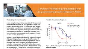 产品手册Services for Predicting Hematotoxicity in Drug Development with HemaTox™ Assays
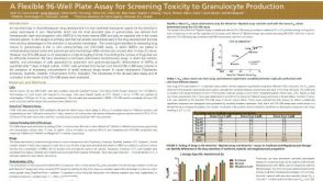 科学海报A Flexible 96-Well Plate Assay for Screening Toxicity to Granulocyte Production
 实验方案How to Prepare and Plate Semi-Solid Methylcellulose Medium for Cell Culture
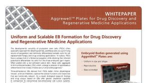 技术公告Uniform and Scalable EB Formation for Drug Discovery and Regenerative Medicine Applications


 沪公网安备31010102008431号