Kuroki MM et al. ( 2005)
Anticancer Research 25 6A 3733--9
Preparation of human IgG and IgM monoclonal antibodies for MK-1/Ep-CAM by using human immunoglobulin gene-transferred mouse and gene cloning of their variable regions.
For antibody-based therapy of cancer,monoclonal antibodies (mAbs) of human origin are superior to mouse,mouse/human chimeric or humanized mAbs,because of their minimum immunogenicity to humans and their efficient collaboration with human effector cells. In the present study,human mAbs were prepared against a pancarcinoma antigen,MK-1 (Ep-CAM),using a genetically-engineered mouse (KM mouse) that contains the human immunoglobulin genes. Spleen cells from KM mice,immunized with recombinant MK-1,were fused with P3-U1 mouse myeloma cells. Of 44 anti-MK-1 clones analyzed,two were of IgG4 and the others of IgM clones. Although the two IgG4 clones were suggested to recognize the same antigenic determinant or two closely located determinants,their VK regions were encoded by different light-chain genes while their VH sequences were identical. The two IgG4 and one of the IgM clones tested revealed antibody-dependent cell-mediated cytotoxicity and complement-dependent cytotoxicity,respectively,against MK-1-expressing cells in vitro,suggesting that these fully human mAbs produced against MK-1 and their V-region genes,which are applicable for the preparation of engineered antibody fragments that may be useful for antibody-based therapy of cancer.
View Publication
Lopez-Izquierdo A et al. (NOV 2014)
American journal of physiology. Heart and circulatory physiology 307 9 H1370--7
A near-infrared fluorescent voltage-sensitive dye allows for moderate-throughput electrophysiological analyses of human induced pluripotent stem cell-derived cardiomyocytes.
Human induced pluripotent stem cell-derived cardiomyocyte (iPSC-CM)-based assays are emerging as a promising tool for the in vitro preclinical screening of QT interval-prolonging side effects of drugs in development. A major impediment to the widespread use of human iPSC-CM assays is the low throughput of the currently available electrophysiological tools. To test the precision and applicability of the near-infrared fluorescent voltage-sensitive dye 1-(4-sulfanatobutyl)-4-β[2-(di-n-butylamino)-6-naphthyl]butadienylquinolinium betaine (di-4-ANBDQBS) for moderate-throughput electrophysiological analyses,we compared simultaneous transmembrane voltage and optical action potential (AP) recordings in human iPSC-CM loaded with di-4-ANBDQBS. Optical AP recordings tracked transmembrane voltage with high precision,generating nearly identical values for AP duration (AP durations at 10%,50%,and 90% repolarization). Human iPSC-CMs tolerated repeated laser exposure,with stable optical AP parameters recorded over a 30-min study period. Optical AP recordings appropriately tracked changes in repolarization induced by pharmacological manipulation. Finally,di-4-ANBDQBS allowed for moderate-throughput analyses,increasing throughput textgreater10-fold over the traditional patch-clamp technique. We conclude that the voltage-sensitive dye di-4-ANBDQBS allows for high-precision optical AP measurements that markedly increase the throughput for electrophysiological characterization of human iPSC-CMs.
View Publication
Xu H et al. (JUL 2016)
Organic & biomolecular chemistry 14 26 6179--83
Cellular thermal shift and clickable chemical probe assays for the determination of drug-target engagement in live cells.
Proof of drug-target engagement in physiologically-relevant contexts is a key pillar of successful therapeutic target validation. We developed two orthogonal technologies,the cellular thermal shift assay (CETSA) and a covalent chemical probe reporter approach (harnessing sulfonyl fluoride tyrosine labeling and subsequent click chemistry) to measure the occupancy of the mRNA-decapping scavenger enzyme DcpS by a small molecule inhibitor in live cells. Enzyme affinity determined using isothermal dose response fingerprinting (ITDRFCETSA) and the concentration required to occupy 50% of the enzyme (OC50) using the chemical probe reporter assay were very similar. In this case,the chemical probe method worked well due to the long offset kinetics of the reversible inhibitor (determined using a fluorescent dye-tagged probe). This work suggests that CETSA could become the first choice assay to determine in-cell target engagement due to its simplicity.
View Publication
Amelioration of murine beta-thalassemia through drug selection of hematopoietic stem cells transduced with a lentiviral vector encoding both gamma-globin and the MGMT drug-resistance gene.
Correction of murine models of beta-thalassemia has been achieved through high-level globin lentiviral vector gene transfer into mouse hematopoietic stem cells (HSCs). However,transduction of human HSCs is less robust and may be inadequate to achieve therapeutic levels of genetically modified erythroid cells. We therefore developed a double gene lentiviral vector encoding both human gamma-globin under the transcriptional control of erythroid regulatory elements and methylguanine methyltransferase (MGMT),driven by a constitutive cellular promoter. MGMT expression provides cellular resistance to alkylator drugs,which can be administered to kill residual untransduced,diseased HSCs,whereas transduced cells are protected. Mice transplanted with beta-thalassemic HSCs transduced with a gamma-globin/MGMT vector initially had subtherapeutic levels of red cells expressing gamma-globin. To enrich gamma-globin-expressing cells,transplanted mice were treated with the alkylator agent 1,3-bis-chloroethyl-1-nitrosourea. This resulted in significant increases in the number of gamma-globin-expressing red cells and the amount of fetal hemoglobin,leading to resolution of anemia. Selection of transduced HSCs was also obtained when cells were drug-treated before transplantation. Mice that received these cells demonstrated reconstitution with therapeutic levels of gamma-globin-expressing cells. These data suggest that MGMT-based drug selection holds promise as a modality to improve gene therapy for beta-thalassemia.
View Publication

 EasySep™小鼠TIL(CD45)正选试剂盒


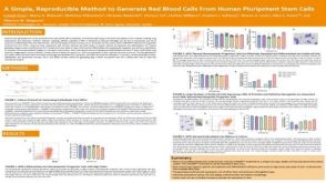 科学海报A Simple, Reproducible Method to Generate Red Blood Cells From Human Pluripotent Stem Cells
 36:59
线上讲座Derivation of Metabolically Active Hepatocytes from Human Pluripotent Stem Cells发布日期: 07/16/2013
36:59
线上讲座Derivation of Metabolically Active Hepatocytes from Human Pluripotent Stem Cells发布日期: 07/16/2013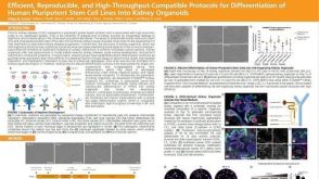 科学海报Efficient, Reproducible and High-Throughput-Compatible Protocols for Differentiation of Human Pluripotent Stem Cell Lines Into Kidney Organoids
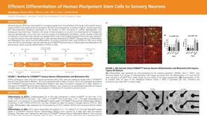 科学海报Efficient Differentiation of Human Pluripotent Stem Cells to Sensory Neurons
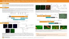 科学海报Rapid, High-Efficiency Differentiation of Motor Neurons from Human Pluripotent Stem Cells
 实验方案How to Culture Human Pluripotent Stem Cell (hPSC)-Derived Forebrain Neurons for MEA Analysis Using the Maestro MEA™ System
 科学海报Generation of Functional 3D Spinal Cord Organoids from Human Pluripotent Stem Cells
 实验方案Gene Editing Human Pluripotent Stem Cells (hPSCs) Using the CellPore™ Transfection System
 沪公网安备31010102008431号