Ucar D et al. (MAR 2009)
Chemico-biological interactions 178 1-3 48--55
Aldehyde dehydrogenase activity as a functional marker for lung cancer.
Aldehyde dehydrogenase (ALDH) activity has been implicated in multiple biological and biochemical pathways and has been used to identify potential cancer stem cells. Our main hypothesis is that ALDH activity may be a lung cancer stem cell marker. Using flow cytometry,we sorted cells with bright (ALDH(br)) and dim (ALDH(lo)) ALDH activity found in H522 lung cancer cell line. We used in vitro proliferation and colony assays as well as a xenograft animal model to test our hypothesis. Cytogenetic analysis demonstrated that the ALDH(br) cells are indeed a different clone,but when left in normal culture conditions will give rise to ALDH(lo) cells. Furthermore,the ALDH(br) cells grow slower,have low clonal efficiency,and give rise to morphologically distinct colonies. The ability to form primary xenografts in NOD/SCID mice by ALDH(br) and ALDH(lo) cells was tested by injecting single cell suspension under the skin in each flank of same animal. Tumor size was calculated weekly. ALDH1A1 and ALDH3A1 immunohistochemistry (IHC) was performed on excised tumors. These tumors were also used to re-establish cell suspension,measure ALDH activity,and re-injection for secondary and tertiary transplants. The results indicate that both cell types can form tumors but the ones from ALDH(br) cells grew much slower in primary recipient mice. Histologically,there was no significant difference in the expression of ALDH in primary tumors originating from ALDH(br) or ALDH(lo) cells. Secondary and tertiary xenografts originating from ALDH(br) grew faster and bigger than those formed by ALDH(lo) cells. In conclusion,ALDH(br) cells may have some of the traditional features of stem cells in terms of being mostly dormant and slow to divide,but require support of other cells (ALDH(lo)) to sustain tumor growth. These observations and the known role of ALDH in drug resistance may have significant therapeutic implications in the treatment of lung cancer.
View Publication
Kakarala M and Wicha MS (JUN 2008)
Journal of clinical oncology : official journal of the American Society of Clinical Oncology 26 17 2813--20
Implications of the cancer stem-cell hypothesis for breast cancer prevention and therapy.
Recent research in breast biology has provided support for the cancer stem-cell hypothesis. Two important components of this hypothesis are that tumors originate in mammary stem or progenitor cells as a result of dysregulation of the normally tightly regulated process of self-renewal. As a result,tumors contain and are driven by a cellular subcomponent that retains key stem-cell properties including self-renewal,which drives tumorigenesis and differentiation that contributes to cellular heterogeneity. Advances in stem-cell technology have led to the identification of stem cells in normal and malignant breast tissue. The study of these stem cells has helped to elucidate the origin of the molecular complexity of human breast cancer. The cancer stem-cell hypothesis has important implications for early detection,prevention,and treatment of breast cancer. Both hereditary and sporadic breast cancers may develop through dysregulation of stem-cell self-renewal pathways. These aberrant stem cells may provide targets for the development of cancer prevention strategies. Furthermore,because breast cancer stem cells may be highly resistant to radiation and chemotherapy,the development of more effective therapies for this disease may require the effective targeting of this cell population.
View Publication
产品类型:
产品号#:
01700
01705
01702
产品名:
ALDEFLUOR™ 试剂盒
ALDEFLUOR™ DEAB试剂, 1.5 mM, 1 mL
ALDEFLUOR™检测缓冲液
Thirukkumaran CM et al. (JUL 2003)
Blood 102 1 377--87
Reovirus oncolysis as a novel purging strategy for autologous stem cell transplantation.
Hematologic stem cell rescue after high-dose cytotoxic therapy is extensively used for the treatment of many hematopoietic and solid cancers. Gene marking studies suggest that occult tumor cells within the autograft may contribute to clinical relapse. To date purging of autografts contaminated with cancer cells has been unsuccessful. The selective oncolytic property of reovirus against myriad malignant histologies in in vitro,in vivo,and ex vivo systems has been previously demonstrated. In the present study we have shown that reovirus can successfully purge cancer cells within autografts. Human monocytic and myeloma cell lines as well as enriched ex vivo lymphoma,myeloma,and Waldenström macroglobulinemia patient tumor specimens were used in an experimental purging model. Viability of the cell lines or purified ex vivo tumor cells of diffuse large B-cell lymphoma,chronic lymphocytic leukemia,Waldenström macroglobulinemia,and small lymphocytic lymphoma was significantly reduced after reovirus treatment. Further,[35S]-methionine labeling and sodium dodecyl sulfate-polyacrylamide gel electrophoresis (SDS-PAGE) of cellular proteins demonstrated reovirus protein synthesis and disruption of host cell protein synthesis as early as 24 hours. Admixtures of apheresis product with the abovementioned tumor cells and cell lines treated with reovirus showed complete purging of disease. In contrast,reovirus purging of enriched ex vivo multiple myeloma,Burkitt lymphoma,and follicular lymphoma was incomplete. The oncolytic action of reovirus did not affect CD34+ stem cells or their long-term colony-forming assays even after granulocyte colony-stimulating factor (G-CSF) stimulation. Our results indicate the ex vivo use of an unattenuated oncolytic virus as an attractive purging strategy for autologous stem cell transplantations.
View Publication
产品类型:
产品号#:
04434
04444
09600
09650
84434
84444
产品名:
MethoCult™ H4434 Classic
MethoCult™ H4434 Classic
StemSpan™ SFEM
StemSpan™ SFEM
Jiang J et al. (SEP 2010)
Cancer research 70 18 7242--52
Crucial roles for protein kinase C isoforms in tumor-specific killing by apoptin.
The chicken anemia virus-derived protein apoptin induces apoptosis in a variety of human malignant and transformed cells but not in normal cells. However,the mechanisms through which apoptin achieves its selective killing effects are not well understood. We developed a lentiviral vector encoding a green fluorescent protein-apoptin fusion gene (LV-GFP-AP) that can efficiently deliver apoptin into hematopoietic cells. Apoptin selectively killed the human multiple myeloma cell lines MM1.R and MM1.S,and the leukemia cell lines K562,HL60,U937,KG1,and NB4. In contrast,normal CD34(+) cells were not killed and maintained their differentiation potential in multilineage colony formation assays. In addition,dexamethasone-resistant MM1.R cells were found to be more susceptible to apoptin-induced cell death than the parental matched MM1.S cells. Death susceptibility correlated with increased phosphorylation and activation of the apoptin protein in MM1.R cells. Expression array profiling identified differential kinase profiles between MM1.R and MM1.S cells. Among these kinases,protein kinase Cβ (PKCβ) was found by immunoprecipitation and in vitro kinase studies to be a candidate kinase responsible for apoptin phosphorylation. Indeed,shRNA knockdown or drug-mediated inhibition of PKCβ significantly reduced apoptin phosphorylation. Furthermore,apoptin-mediated cell death proceeded through the upregulation of PKCβ,activation of caspase-9/3,cleavage of the PKCδ catalytic domain,and downregulation of the MERTK and AKT kinases. Collectively,these results elucidate a novel pathway for apoptin activation involving PKCβ and PKCδ. Further,they highlight the potential of apoptin and its cellular regulators to purge bone marrow used in autologous transplantation for multiple myeloma.
View Publication
产品类型:
产品号#:
04434
04444
09600
09650
产品名:
MethoCult™ H4434 Classic
MethoCult™ H4434 Classic
StemSpan™ SFEM
StemSpan™ SFEM
Jo SY et al. (MAY 2011)
Blood 117 18 4759--68
Requirement for Dot1l in murine postnatal hematopoiesis and leukemogenesis by MLL translocation.
Disruptor of telomeric silencing 1-like (Dot1l) is a histone 3 lysine 79 methyltransferase. Studies of constitutive Dot1l knockout mice show that Dot1l is essential for embryonic development and prenatal hematopoiesis. DOT1L also interacts with translocation partners of Mixed Lineage Leukemia (MLL) gene,which is commonly translocated in human leukemia. However,the requirement of Dot1l in postnatal hematopoiesis and leukemogenesis of MLL translocation proteins has not been conclusively shown. With a conditional Dot1l knockout mouse model,we examined the consequences of Dot1l loss in postnatal hematopoiesis and MLL translocation leukemia. Deletion of Dot1l led to pancytopenia and failure of hematopoietic homeostasis,and Dot1l-deficient cells minimally reconstituted recipient bone marrow in competitive transplantation experiments. In addition,MLL-AF9 cells required Dot1l for oncogenic transformation,whereas cells with other leukemic oncogenes,such as Hoxa9/Meis1 and E2A-HLF,did not. These findings illustrate a crucial role of Dot1l in normal hematopoiesis and leukemogenesis of specific oncogenes.
View Publication
Fischbach NA et al. (FEB 2005)
Blood 105 4 1456--66
HOXB6 overexpression in murine bone marrow immortalizes a myelomonocytic precursor in vitro and causes hematopoietic stem cell expansion and acute myeloid leukemia in vivo.
The HOX family of homeobox genes plays an important role in normal and malignant hematopoiesis. Dysregulated HOX gene expression profoundly effects the proliferation and differentiation of hematopoietic stem cells (HSCs) and committed progenitors,and aberrant activation of HOX genes is a common event in human myeloid leukemia. HOXB6 is frequently overexpressed in human acute myeloid leukemia (AML). To gain further insight into the role of HOXB6 in hematopoiesis,we overexpressed HOXB6 in murine bone marrow using retrovirus-mediated gene transfer. We also explored structure-function relationships using mutant HOXB6 proteins unable to bind to DNA or a key HOX-binding partner,pre-B-cell leukemia transcription factor-1 (PBX1). Additionally,we investigated the potential cooperative interaction with myeloid ecotropic viral integration site 1 homolog (MEIS1). In vivo,HOXB6 expanded HSCs and myeloid precursors while inhibiting erythropoiesis and lymphopoiesis. Overexpression of HOXB6 resulted in AML with a median latency of 223 days. Coexpression of MEIS1 dramatically shortened the onset of AML. Cytogenetic analysis of a subset of HOXB6-induced AMLs revealed recurrent deletions of chromosome bands 2D-E4,a region frequently deleted in HOXA9-induced AMLs. In vitro,HOXB6 immortalized a factor-dependent myelomonocytic precursor capable of granulocytic and monocytic differentiation. These biologic effects of HOXB6 were largely dependent on DNA binding but independent of direct interaction with PBX1.
View Publication

 EasySep™小鼠TIL(CD45)正选试剂盒


 产品手册Highway1™: Fast, Gentle, and Automated Cell Sorting for Every Lab
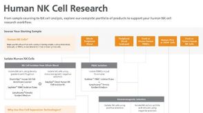
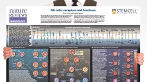 挂图Natural Killer Cells Overview of NK cell receptors, subsets, activation and function
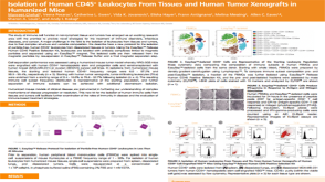 科学海报Isolation of Human CD45+ Leukocytes From Tissues and Human Tumor Xenografts in Humanized Mice

 沪公网安备31010102008431号