Jing J et al. ( 2012)
Molecular cancer therapeutics 11 3 720--729
Comprehensive predictive biomarker analysis for MEK inhibitor GSK1120212.
The MEK1 and MEK2 inhibitor GSK1120212 is currently in phase II/III clinical development. To identify predictive biomarkers,sensitivity to GSK1120212 was profiled for 218 solid tumor cell lines and 81 hematologic malignancy cell lines. For solid tumors,RAF/RAS mutation was a strong predictor of sensitivity. Among RAF/RAS mutant lines,co-occurring PIK3CA/PTEN mutations conferred a cytostatic response instead of a cytotoxic response for colon cancer cells that have the biggest representation of the comutations. Among KRAS mutant cell lines,transcriptomics analysis showed that cell lines with an expression pattern suggestive of epithelial-to-mesenchymal transition were less sensitive to GSK1120212. In addition,a proportion of cell lines from certain tissue types not known to carry frequent RAF/RAS mutations also seemed to be sensitive to GSK1120212. Among these were breast cancer cell lines,with triple negative breast cancer cell lines being more sensitive than cell lines from other breast cancer subtypes. We identified a single gene DUSP6,whose expression was associated with sensitivity to GSK1120212 and lack of expression associated with resistance irrelevant of RAF/RAS status. Among hematologic cell lines,acute myeloid leukemia and chronic myeloid leukemia cell lines were particularly sensitive. Overall,this comprehensive predictive biomarker analysis identified additional efficacy biomarkers for GSK1120212 in RAF/RAS mutant solid tumors and expanded the indication for GSK1120212 to patients who could benefit from this therapy despite the RAF/RAS wild-type status of their tumors.
View Publication
产品类型:
产品号#:
73502
73504
产品名:
Druker BJ (DEC 2008)
Blood 112 13 4808--17
Translation of the Philadelphia chromosome into therapy for CML.
Throughout its history,chronic myeloid leukemia (CML) has set precedents for cancer research and therapy. These range from the identification of the first specific chromosomal abnormality associated with cancer to the development of imatinib as a specific,targeted therapy for the disease. The successful development of imatinib as a therapeutic agent for CML can be attributed directly to decades of scientific discoveries. These discoveries determined that the BCR-ABL tyrosine kinase is the critical pathogenetic event in CML and an ideal target for therapy. This was confirmed in clinical trials of imatinib,with imatinib significantly improving the long-term survival of patients with CML. Continuing in this tradition of scientific discoveries leading to improved therapies,the understanding of resistance to imatinib has rapidly led to strategies to circumvent resistance. Continued studies of hematologic malignancies will allow this paradigm of targeting molecular pathogenetic events to be applied to many additional hematologic cancers.
View Publication
Kurtzberg LS et al. (MAY 2011)
Clinical cancer research : an official journal of the American Association for Cancer Research 17 9 2777--87
Genz-644282, a novel non-camptothecin topoisomerase I inhibitor for cancer treatment.
PURPOSE: Genz-644282 [8,9-dimethoxy-5-(2-N-methylaminoethyl)-2,3-methylenedioxy-5H-dibenzo[c,h][1,6]naphthyridin-6-one] has emerged as a promising candidate for antitumor agents. This report describes the bone marrow colony-forming unit,granulocyte macrophage (CFU-GM) and tumor cell CFU activity of topoisomerase I (Top1) inhibitors,such as Genz-644282,topotecan,irinotecan/SN-38,and ARC-111,and examines their activity in several human tumor xenograft models. EXPERIMENTAL DESIGN: Colony-forming assays were conducted with mouse and human bone marrow and eight human tumor cell lines. In addition,29 human tumor cell lines representing a range of histology and potential resistance mechanisms were assayed for sensitivity to Genz-644282 in a 72-hour exposure assay. The efficacy of Genz-644282 was compared with standard anticancer drugs (i.e.,irinotecan,docetaxel,and dacarbazine) in human tumor xenografts of colon cancer,renal cell carcinoma,non-small cell lung cancer,and melanoma. RESULTS: Human bone marrow CFU-GM was more sensitive to the Top1 inhibitors than was mouse bone marrow CFU-GM. The ratio of mouse to human IC(90) values was more than 10 for the camptothecins and less than 10 for Genz-644282,which had more potency as a cytotoxic agent toward human tumor cells in culture than the camptothecins in the colony-forming and 72-hour proliferation assays. Genz-644282 has superior or equal antitumor activity in the human tumor xenografts than the standard drug comparators. CONCLUSIONS: On the basis of preclinical activity and safety,Genz-644282 was selected for development and is currently undergoing phase 1 clinical trial.
View Publication
Akcakanat A et al. ( 2009)
Molecular Cancer 8 1 75
The rapamycin-regulated gene expression signature determines prognosis for breast cancer
BACKGROUND: Mammalian target of rapamycin (mTOR) is a serine/threonine kinase involved in multiple intracellular signaling pathways promoting tumor growth. mTOR is aberrantly activated in a significant portion of breast cancers and is a promising target for treatment. Rapamycin and its analogues are in clinical trials for breast cancer treatment. Patterns of gene expression (metagenes) may also be used to simulate a biologic process or effects of a drug treatment. In this study,we tested the hypothesis that the gene-expression signature regulated by rapamycin could predict disease outcome for patients with breast cancer. RESULTS: Colony formation and sulforhodamine B (IC50 textless 1 nM) assays,and xenograft animals showed that MDA-MB-468 cells were sensitive to treatment with rapamycin. The comparison of in vitro and in vivo gene expression data identified a signature,termed rapamycin metagene index (RMI),of 31 genes upregulated by rapamycin treatment in vitro as well as in vivo (false discovery rate of 10%). In the Miller dataset,RMI did not correlate with tumor size or lymph node status. High (textgreater75th percentile) RMI was significantly associated with longer survival (P = 0.015). On multivariate analysis,RMI (P = 0.029),tumor size (P = 0.015) and lymph node status (P = 0.001) were prognostic. In van 't Veer study,RMI was not associated with the time to develop distant metastasis (P = 0.41). In the Wang dataset,RMI predicted time to disease relapse (P = 0.009). CONCLUSION: Rapamycin-regulated gene expression signature predicts clinical outcome in breast cancer. This supports the central role of mTOR signaling in breast cancer biology and provides further impetus to pursue mTOR-targeted therapies for breast cancer treatment.
View Publication
产品类型:
产品号#:
73362
73364
100-1050
产品名:
雷帕霉素
雷帕霉素
雷帕霉素
Niu C et al. (SEP 2009)
Blood 114 10 2087--96
c-Myc is a target of RNA-binding motif protein 15 in the regulation of adult hematopoietic stem cell and megakaryocyte development.
RNA-binding motif protein 15 (RBM15) is involved in the RBM15-megakaryoblastic leukemia 1 fusion in acute megakaryoblastic leukemia. Although Rbm15 has been reported to be required for B-cell differentiation and to inhibit myeloid and megakaryocytic expansion,it is not clear what the normal functions of Rbm15 are in the regulation of hematopoietic stem cell (HSC) and megakaryocyte development. In this study,we report that Rbm15 may function in part through regulation of expression of the proto-oncogene c-Myc. Similar to c-Myc knockout (c-Myc-KO) mice,long-term (LT) HSCs are significantly increased in Rbm15-KO mice due to an apparent LT-HSC to short-term HSC differentiation defect associated with abnormal HSC-niche interactions caused by increased N-cadherin and beta(1) integrin expression on mutant HSCs. Both serial transplantation and competitive reconstitution capabilities of Rbm15-KO LT-HSCs are greatly compromised. Rbm15-KO and c-Myc-KO mice also share related abnormalities in megakaryocyte development,with mutant progenitors producing increased,abnormally small low-ploidy megakaryocytes. Consistent with a possible functional interplay between Rbm15 and c-Myc,the megakaryocyte increase in Rbm15-KO mice could be partially reversed by ectopic c-Myc. Thus,Rbm15 appears to be required for normal HSC-niche interactions,for the ability of HSCs to contribute normally to adult hematopoiesis,and for normal megakaryocyte development; these effects of Rbm15 on hematopoiesis may be mediated at least in part by c-Myc.
View Publication

 EasySep™小鼠TIL(CD45)正选试剂盒


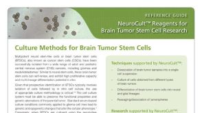 产品手册NeuroCult™: Reagents for Brain Tumor Stem Cell Research
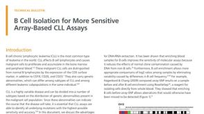 技术公告B Cell Isolation for More Sensitive Array-Based CLL Assays
 产品手册Highway1™: Fast, Gentle, and Automated Cell Sorting for Every Lab
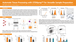 科学海报Automate Tissue Processing with STEMprep™ for Versatile Sample Preparation


 沪公网安备31010102008431号