
 EasySep™小鼠TIL(CD45)正选试剂盒


搜索结果: 'methocult media formulations for human hematopoietic cells serum containing'
-

产品类型:
产品号#:
01700
01705
01702
产品名:
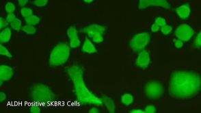
ALDEFLUOR™ 试剂盒
ALDEFLUOR™ DEAB试剂, 1.5 mM, 1 mL
ALDEFLUOR™检测缓冲液
-
产品类型:
产品号#:
15129
15169
产品名:
RosetteSep™人多发性骨髓瘤细胞富集抗体混合物
RosetteSep™人多发性骨髓瘤细胞富集抗体混合物
-
产品类型:
产品号#:
15025
15065
产品名:
RosetteSep™人NK细胞富集抗体混合物
RosetteSep™人NK细胞富集抗体混合物
-
产品类型:
产品号#:
15122
15162
产品名:
RosetteSep™人CD45去除抗体混合物
RosetteSep™人CD45去除抗体混合物
-
产品类型:
产品号#:
09600
09650
产品名:
StemSpan™ SFEM
StemSpan™ SFEM
-
产品类型:
产品号#:
04100
产品名:
MethoCult™ H4100
-
产品类型:
产品号#:
05350
产品名:
-
 产品手册Reliable Antibodies for Your Research
产品类型:
品牌:
EasySep
产品号#:
60100
60100.1
60100AD
60100AD.1
60102
60102FI
60104
60104AD
60104AD.1
60104AZ
60104AZ.1
60104BT
60104BT.1
60104BT.2
60104FI
60104FI.1
60104PE
60104PE.1
60106
60106.1
60106AZ
60106AZ.1
60106BT
60106BT.1
60106FI
60106FI.1
60106PE
60106PE.1
60107
60107.1
601
产品名:
抗β-微管蛋白III抗体,克隆AA10
抗β-微管蛋白III抗体,克隆AA10
抗β-微管蛋白III抗体,克隆AA10,Alexa Fluor® 488
抗β-微管蛋白III抗体,clone AA10,Alexa Fluor® 488
抗小鼠TCR Gamma/Delta抗体,clone GL3
抗小鼠TCR Gamma/Delta抗体,clone GL3,Alexa Fluor® 488
抗小鼠TCR Gamma/Delta抗体,clone GL3,APC
抗小鼠TCR Gamma/Delta抗体,clone GL3,APC
抗小鼠TCR Gamma/Delta抗体,clone GL3,Biotin
抗小鼠TCR Gamma/Delta抗体,clone GL3,Biotin
抗小鼠TCR Gamma/Delta抗体,clone GL3,FITC
抗小鼠TCR Gamma/Delta抗体,clone GL3,PE
抗小鼠TCR Gamma/Delta抗体,clone GL3,PE
抗人CD71(转铁蛋白受体)抗体,clone OKT9
抗人CD71(转铁蛋白受体)抗体,clone OKT9
抗人CD71(转铁蛋白受体)抗体,clone OKT9,APC
抗人CD71(转铁蛋白受体)抗体,clone OKT9,APC
抗人CD71(转铁蛋白受体)抗体,clone OKT9,Biotin
抗人CD71(转铁蛋白受体)抗体,clone OKT9,FITC
抗人CD71(转铁蛋白受体)抗体,clone OKT9,FITC
抗人CD71(转铁蛋白受体)抗体,clone OKT9,PE
抗人CD71(转铁蛋白受体)抗体,clone OKT9,PE
-
产品类型:
产品号#:
02690
09600
09650
产品名:
StemSpan™ CC100
StemSpan™ SFEM
StemSpan™ SFEM
-
产品类型:
产品号#:
09500
09600
09650
04960
04902
04900
04963
04962
04970
04971
04901
产品名:
BIT 9500血清替代物
StemSpan™ SFEM
StemSpan™ SFEM
MegaCult™-C胶原和无细胞因子培养基
胶原蛋白溶液
MegaCult™-C无细胞因子培养基
双室载玻片套件
MegaCult™-C CFU-Mk染色试剂盒
MegaCult™-C无细胞因子全套试剂盒
MegaCult™-C含细胞因子全套试剂盒
MegaCult™-C含细胞因子培养基
-
产品类型:
产品号#:
19055
19055RF
产品名:
EasySep™人NK细胞富集试剂盒
RoboSep™ 人NK细胞富集试剂盒含滤芯吸头

 沪公网安备31010102008431号