Cremona CA and Lloyd AC (SEP 2009)
Journal of cell science 122 Pt 18 3272--81
Loss of anchorage in checkpoint-deficient cells increases genomic instability and promotes oncogenic transformation.
Mammalian cells generally require both mitogens and anchorage signals in order to proliferate. An important characteristic of many tumour cells is that they have lost this anchorage-dependent cell-cycle checkpoint,allowing them to proliferate without signals provided by their normal microenvironment. In the absence of anchorage signals from the extracellular matrix,many cell types arrest cell-cycle progression in G1 phase as a result of Rb-dependent checkpoints. However,despite inactivation of p53 and Rb proteins,SV40LT-expressing cells retain anchorage dependency,suggesting the presence of an uncharacterised cell-cycle checkpoint,which can be overridden by coexpression of oncogenic Ras. We report here that,although cyclin-CDK complexes persisted in suspension,proliferation was inhibited in LT-expressing cells by the CDK inhibitor p27(Kip1) (p27). Interestingly,this did not induce a stable arrest,but aberrant cell-cycle progression associated with stalled DNA replication,rereplication and chromosomal instability,which was sufficient to increase the frequency of oncogenic transformation. These results firstly indicate loss of anchorage in Rb- and p53-deficient cells as a novel mechanism for promotion of genomic instability; secondly suggest that anchorage checkpoints that protect normal cells from inappropriate proliferation act deleteriously in Rb- and p53-deficient cells to promote tumourigenesis; and thirdly indicate caution in the use of CDK inhibitors for cancer treatment.
View Publication
产品类型:
产品号#:
05401
05402
05411
产品名:
MesenCult™ MSC 基础培养基(人)
MesenCult™ MSC刺激添加物(人)
MesenCult™ 增殖试剂盒(人)
Lo J-F et al. (MAR 2011)
Cancer research 71 5 1912--23
The epithelial-mesenchymal transition mediator S100A4 maintains cancer-initiating cells in head and neck cancers.
Cancer-initiating cells (CIC) comprise a rare subpopulation of cells in tumors that are proposed to be responsible for tumor growth. Starting from CICs identified in head and neck squamous cell carcinomas (HNSCC),termed head and neck cancer-initiating cells (HN-CIC),we determined as a candidate stemness-maintaining molecule for HN-CICs the proinflammatory mediator S100A4,which is also known to be an inducer of epithelial-mesenchymal transition. S100A4 knockdown in HN-CICs reduced their self-renewal capability and their stemness and tumorigenic properties,both in vitro and in vivo. Conversely,S100A4 overexpression in HNSCC cells enhanced their stem cell properties. Mechanistic investigations indicated that attenuation of endogenous S100A4 levels in HNSCC cells caused downregulation of Notch2 and PI3K (phosphoinositide 3-kinase)/pAKT along with upregulation of PTEN,consistent with biological findings. Immunohistochemical analysis of HNSCC clinical specimens showed that S100A4 expression was positively correlated with clinical grading,stemness markers,and poorer patient survival. Together,our findings reveal a crucial role for S100A4 signaling pathways in maintaining the stemness properties and tumorigenicity of HN-CICs. Furthermore,our findings suggest that targeting S100A4 signaling may offer a new targeted strategy for HNSCC treatment by eliminating HN-CICs.
View Publication
产品类型:
产品号#:
01700
01705
01702
产品名:
ALDEFLUOR™ 试剂盒
ALDEFLUOR™ DEAB试剂, 1.5 mM, 1 mL
ALDEFLUOR™检测缓冲液
Lelaidier M et al. (OCT 2015)
Oncotarget 6 30 29440--55
TRAIL-mediated killing of acute lymphoblastic leukemia by plasmacytoid dendritic cell-activated natural killer cells.
Acute lymphoblastic leukemia (ALL) still frequently recurs after hematopoietic stem cell transplantation (HSCT),underscoring the need to improve the graft-versus-leukemia (GvL) effect. Natural killer (NK) cells reconstitute in the first months following HSCT when leukemia burden is at its lowest,but ALL cells have been shown to be resistant to NK cell-mediated killing. We show here that this resistance is overcome by NK cell stimulation with TLR-9-activated plasmacytoid dendritic cells (pDCs). NK cell priming with activated pDCs resulted in TRAIL and CD69 up-regulation on NK cells and IFN-γ production. NK cell activation was dependent on IFN-α produced by pDCs,but was not reproduced by IFN-α alone. ALL killing was further enhanced by inhibition of KIR engagement. We showed that ALL lysis was mainly mediated by TRAIL engagement,while the release of cytolytic granules was involved when ALL expressed NK cell activating receptor ligands. Finally,adoptive transfers of activated-pDCs in ALL-bearing humanized mice delayed the leukemia onset and cure 30% of mice. Our data therefore demonstrate that TLR-9 activated pDCs are a powerful tool to overcome ALL resistance to NK cell-mediated killing and to reinforce the GvL effect of HSCT. These results open new therapeutic avenues to prevent relapse in children with ALL.
View Publication
产品类型:
产品号#:
19062
19062RF
19055
19055RF
产品名:
EasySep™人浆细胞样DC富集试剂盒
RoboSep™ 人浆细胞样DC富集试剂盒含滤芯吸头
EasySep™人NK细胞富集试剂盒
RoboSep™ 人NK细胞富集试剂盒含滤芯吸头
Deng S et al. (JAN 2010)
PloS one 5 4 e10277
Distinct expression levels and patterns of stem cell marker, aldehyde dehydrogenase isoform 1 (ALDH1), in human epithelial cancers.
Aldehyde dehydrogenase isoform 1 (ALDH1) has been proved useful for the identification of cancer stem cells. However,our knowledge of the expression and activity of ALDH1 in common epithelial cancers and their corresponding normal tissues is still largely absent. Therefore,we characterized ALDH1 expression in 24 types of normal tissues and a large collection of epithelial tumor specimens (six cancer types,n = 792) by immunohistochemical staining. Using the ALDEFUOR assay,ALDH1 activity was also examined in 16 primary tumor specimens and 43 established epithelial cancer cell lines. In addition,an ovarian cancer transgenic mouse model and 7 murine ovarian cancer cell lines were analyzed. We found that the expression levels and patterns of ALDH1 in epithelial cancers are remarkably distinct,and they correlate with their corresponding normal tissues. ALDH1 protein expression levels are positively correlated with ALDH1 enzymatic activity measured by ALDEFLUOR assay. Long-term in vitro culture doesn't significantly affect ALDH1 activity in epithelial tumor cells. Consistent with research on other cancers,we found that high ALDH1 expression is significantly associated with poor clinical outcomes in serous ovarian cancer patients (n = 439,p = 0.0036). Finally,ALDH(br) tumor cells exhibit cancer stem cell properties and are resistant to chemotherapy. As a novel cancer stem cell marker,ALDH1 can be used for tumors whose corresponding normal tissues express ALDH1 in relatively restricted or limited levels such as breast,lung,ovarian or colon cancer.
View Publication
产品类型:
产品号#:
01700
01705
01701
01702
05620
产品名:
ALDEFLUOR™ 试剂盒
ALDEFLUOR™ DEAB试剂, 1.5 mM, 1 mL
ALDEFLUOR™检测缓冲液
MammoCult™ 人源培养基套装
Morrow M et al. (MAY 2004)
Blood 103 10 3890--6
TEL-AML1 promotes development of specific hematopoietic lineages consistent with preleukemic activity.
The t(12;21)(p13;q22) translocation is the most common chromosomal abnormality yet identified in any pediatric leukemia and gives rise to the TEL-AML1 fusion product. To investigate the effects of TEL-AML1 on hematopoiesis,fetal liver hematopoietic progenitor cells (HPCs) were transduced with retroviral vectors expressing this fusion protein. We show that TEL-AML1 dramatically alters differentiation of HPCs in vitro,preferentially promoting B-lymphocyte development,enhancing self-renewal of B-cell precursors,and leading to the establishment of long-term growth factor-dependent pre-B-cell lines. However,it had no effect on myeloid development in vitro. Further experiments were performed to determine whether TEL-AML1 also demonstrates lineage-specific activity in vivo. TEL-AML1-expressing HPCs displayed a competitive advantage in reconstituting both B-cell and myeloid lineages in vivo but had no effect on reconstitution of the T-cell lineage. Despite promoting these alterations in hematopoiesis,TEL-AML1 did not induce leukemia in transplanted mice. Our study provides a unique insight into the role of TEL-AML1 in leukemia predisposition and a potential model to study the mechanism of leukemogenesis associated with this fusion.
View Publication
产品类型:
产品号#:
03534
03231
产品名:
MethoCult™ GF M3534
MethoCult™ M3231
Pelicano H et al. (DEC 2006)
The Journal of cell biology 175 6 913--23
Mitochondrial respiration defects in cancer cells cause activation of Akt survival pathway through a redox-mediated mechanism.
Cancer cells exhibit increased glycolysis for ATP production due,in part,to respiration injury (the Warburg effect). Because ATP generation through glycolysis is less efficient than through mitochondrial respiration,how cancer cells with this metabolic disadvantage can survive the competition with other cells and eventually develop drug resistance is a long-standing paradox. We report that mitochondrial respiration defects lead to activation of the Akt survival pathway through a novel mechanism mediated by NADH. Respiration-deficient cells (rho(-)) harboring mitochondrial DNA deletion exhibit dependency on glycolysis,increased NADH,and activation of Akt,leading to drug resistance and survival advantage in hypoxia. Similarly,chemical inhibition of mitochondrial respiration and hypoxia also activates Akt. The increase in NADH caused by respiratory deficiency inactivates PTEN through a redox modification mechanism,leading to Akt activation. These findings provide a novel mechanistic insight into the Warburg effect and explain how metabolic alteration in cancer cells may gain a survival advantage and withstand therapeutic agents.
View Publication
产品类型:
产品号#:
04230
产品名:
MethoCult™ H4230
Carlsten M et al. (FEB 2007)
Cancer research 67 3 1317--25
DNAX accessory molecule-1 mediated recognition of freshly isolated ovarian carcinoma by resting natural killer cells.
Although natural killer (NK) cells are well known for their ability to kill tumors,few studies have addressed the interactions between resting (nonactivated) NK cells and freshly isolated human tumors. Here,we show that human leukocyte antigen class I(low) tumor cells isolated directly from patients with advanced ovarian carcinoma trigger degranulation by resting allogeneic NK cells. This was paralleled by induction of granzyme B and caspase-6 activities in the tumor cells and significant tumor cell lysis. Ovarian carcinoma cells displayed ubiquitous expression of the DNAX accessory molecule-1 (DNAM-1) ligand PVR and sparse/heterogeneous expression of the NKG2D ligands MICA/MICB and ULBP1,ULBP2,and ULBP3. In line with the NK receptor ligand expression profiles,antibody-mediated blockade of activating receptor pathways revealed a dominant role for DNAM-1 and a complementary contribution of NKG2D signaling in tumor cell recognition. These results show that resting NK cells are capable of directly recognizing freshly isolated human tumor cells and identify ovarian carcinoma as a potential target for adoptive NK cell-based immunotherapy.
View Publication
产品类型:
产品号#:
18259
18259RF
产品名:
Sloand EM et al. (SEP 2006)
Proceedings of the National Academy of Sciences of the United States of America 103 39 14483--8
Granulocyte colony-stimulating factor preferentially stimulates proliferation of monosomy 7 cells bearing the isoform IV receptor.
Granulocyte colony-stimulating factor (GCSF) administration has been linked to the development of monosomy 7 in severe congenital neutropenia and aplastic anemia. We assessed the effect of pharmacologic doses of GCSF on monosomy 7 cells to determine whether this chromosomal abnormality developed de novo or arose as a result of favored expansion of a preexisting clone. Fluorescence in situ hybridization (FISH) of chromosome 7 was used to identify small populations of aneuploid cells. When bone marrow mononuclear cells from patients with monosomy 7 were cultured with 400 ng/ml GCSF,all samples showed significant increases in the proportion of monosomy 7 cells. In contrast,bone marrow from karyotypically normal aplastic anemia,myelodysplastic syndrome,or healthy individuals did not show an increase in monosomy 7 cells in culture. In bone marrow CD34 cells of patients with myelodysplastic syndrome and monosomy 7,GCSF receptor (GCSFR) protein was increased. Although no mutation was found in genomic GCSFR DNA,CD34 cells showed increased expression of the GCSFR class IV mRNA isoform,which is defective in signaling cellular differentiation. GCSFR signal transduction via the Jak/Stat system was abnormal in monosomy 7 CD34 cells,with increased phosphorylated signal transducer and activation of transcription protein,STAT1-P,and increased STAT5-P relative to STAT3-P. Our results suggest that pharmacologic doses of GCSF increase the proportion of preexisting monosomy 7 cells. The abnormal response of monosomy 7 cells to GCSF would be explained by the expansion of undifferentiated monosomy 7 clones expressing the class IV GCSFR,which is defective in signaling cell maturation.
View Publication
产品类型:
产品号#:
05150
产品名:
MyeloCult™ H5100
Kanai R et al. (JAN 2012)
Journal of the National Cancer Institute 104 1 42--55
Oncolytic virus-mediated manipulation of DNA damage responses: synergy with chemotherapy in killing glioblastoma stem cells.
BACKGROUND: Although both the alkylating agent temozolomide (TMZ) and oncolytic viruses hold promise for treating glioblastoma,which remains uniformly lethal,the effectiveness of combining the two treatments and the mechanism of their interaction on cancer stem cells are unknown. METHODS: We investigated the efficacy of combining TMZ and the oncolytic herpes simplex virus (oHSV) G47Δ in killing glioblastoma stem cells (GSCs),using Chou-Talalay combination index analysis,immunocytochemistry and fluorescence microscopy,and neutral comet assay. The role of treatment-induced DNA double-strand breaks,activation of DNA damage responses,and virus replication in the cytotoxic interaction between G47Δ and TMZ was examined with a panel of pharmacological inhibitors and short-hairpin RNA (shRNA)-mediated knockdown of DNA repair pathways. Comparisons of cell survival and virus replication were performed using a two-sided t test (unpaired). The survival of athymic mice (n = 6-8 mice per group) bearing GSC-derived glioblastoma tumors treated with the combination of G47Δ and TMZ was analyzed by the Kaplan-Meier method and evaluated with a two-sided log-rank test. RESULTS: The combination of G47Δ and TMZ acted synergistically in killing GSCs but not neurons,with associated robust induction of DNA damage. Pharmacological and shRNA-mediated knockdown studies suggested that activated ataxia telangiectasia mutated (ATM) is a crucial mediator of synergy. Activated ATM relocalized to HSV DNA replication compartments where it likely enhanced oHSV replication and could not participate in repairing TMZ-induced DNA damage. Sensitivity to TMZ and synergy with G47Δ decreased with O(6)-methylguanine-DNA-methyltransferase (MGMT) expression and MSH6 knockdown. Combined G47Δ and TMZ treatment extended survival of mice bearing GSC-derived intracranial tumors,achieving long-term remission in four of eight mice (median survival = 228 days; G47Δ alone vs G47Δ + TMZ,hazard ratio of survival = 7.1,95% confidence interval = 1.9 to 26.1,P = .003) at TMZ doses attainable in patients. CONCLUSIONS: The combination of G47Δ and TMZ acts synergistically in killing GSCs through oHSV-mediated manipulation of DNA damage responses. This strategy is highly efficacious in representative preclinical models and warrants clinical translation.
View Publication
产品类型:
产品号#:
05707
05751
产品名:
NeuroCult™化学解离试剂盒(小鼠)
NeuroCult™ NS-A 扩增试剂盒(人)
Golay J et al. (MAR 2006)
Haematologica 91 3 322--30
The sensitivity of acute lymphoblastic leukemia cells carrying the t(12;21) translocation to campath-1H-mediated cell lysis.
BACKGROUND AND OBJECTIVES: Campath-1H is used in conditioning regimens and more recently as an anti-leukemic therapy in acute lymphoblastic leukemias (ALL). We therefore investigated CD52 expression and campath-1H-mediated lysis of ALL cells in vitro. DESIGN AND METHODS: Complement-mediated cytotoxicity assays were performed on freshly isolated neoplastic cells and cell lines using human serum. Antibody-dependent cellular cytotoxicity (ADCC) was performed by calcein-AM release assays. RESULTS: CD52 was expressed in four out of eight ALL cell lines studied. Among 61 freshly isolated ALL samples CD52 was expressed at varying levels in 87% of cases. Whereas ADCC was equivalent in different CD52+ lines,complement-dependent cytotoxicity (CDC) was variable. The REH cell line bearing the t(12;21) translocation showed 47-60% lysis when treated with 10 microg/mL campath-1H compared to 0-6% for the other cell lines expressing equivalent amounts of CD52. Furthermore all nine ALL samples with t(12;21) showed very high CDC (mean 97%) compared to the other 24 CD52+cases (mean 24%)(ptextless0.0001). In t(12;21) samples,efficient CDC was obtained with as little as 1 microg/mL campath-1H. CDC correlated in part with CD52 levels,suggesting that CD52 expression and other yet undefined factors contribute to the particular sensitivity of t(12;21) cells. The resistance of non t(12;21) ALL cases could be overcome to a limited extent by increasing the concentration of campath-1H,blocking the CD55 and CD59 complement inhibitors,and more effectively by combining campath-1H with fludarabine. INTERPRETATION AND CONCLUSIONS: We conclude that most ALL samples express CD52 to a variable level and that campath-1H has cytotoxic activity against CD52+ALL,alone or in combination with cytotoxic drugs.
View Publication
Charafe-Jauffret E et al. (JAN 2010)
Clinical cancer research : an official journal of the American Association for Cancer Research 16 1 45--55
Aldehyde dehydrogenase 1-positive cancer stem cells mediate metastasis and poor clinical outcome in inflammatory breast cancer.
PURPOSE: To examine the role of cancer stem cells (CSC) in mediating metastasis in inflammatory breast cancer (IBC) and the association of these cells with patient outcome in this aggressive type of breast cancer. EXPERIMENTAL DESIGN: CSCs were isolated from SUM149 and MARY-X,an IBC cell line and primary xenograft,by virtue of increased aldehyde dehydrogenase (ALDH) activity as assessed by the ALDEFLUOR assay. Invasion and metastasis of CSC populations were assessed by in vitro and mouse xenograft assays. Expression of ALDH1 was determined on a retrospective series of 109 IBC patients and this was correlated with histoclinical data. All statistical tests were two sided. Log-rank tests using Kaplan-Meier analysis were used to determine the correlation of ALDH1 expression with development of metastasis and patient outcome. RESULTS: Both in vitro and xenograft assays showed that invasion and metastasis in IBC are mediated by a cellular component that displays ALDH activity. Furthermore,expression of ALDH1 in IBC was an independent predictive factor for early metastasis and decreased survival in this patient population. CONCLUSIONS: These results suggest that the metastatic,aggressive behavior of IBC may be mediated by a CSC component that displays ALDH enzymatic activity. ALDH1 expression represents the first independent prognostic marker to predict metastasis and poor patient outcome in IBC. The results illustrate how stem cell research can translate into clinical practice in the IBC field.
View Publication

 EasySep™小鼠TIL(CD45)正选试剂盒


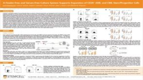 科学海报A Feeder-Free and Serum-Free Culture System Supports Expansion of CD34+ AML and CML Stem/Progenitor Cells
 沪公网安备31010102008431号