McCune K et al. (NOV 2010)
Oncology reports 24 5 1233--9
Loss of ERα and FOXA1 expression in a progression model of luminal type breast cancer: insights from PyMT transgenic mouse model.
The classification of breast cancer into multiple molecular subtypes has necessitated the need for biomarkers that can assess tumor progression and the effects of chemopreventive agents on specific breast cancer subtypes. The goal of this study was to identify biomarkers whose expression are altered along with estrogen receptor α (ERα) in the polyoma middle-T antigen (PyMT) transgenic model of breast cancer and to investigate the chemopreventive activity of phenethyl isothiocyanate (PEITC). The diet of PyMT female mice was fortified with PEITC (8 mmol/kg) and the mammary streak and/or gross tumors and metastases in lungs were subjected to immunohistochemical analyses for ERα,FOXA1,and GATA-3. FOXA1 is associated with luminal type A cancers,while GATA-3 is a marker of luminal progenitor cell differentiation. In both control and PEITC-treated groups,there was a progressive loss of ERα and FOXA1 but persistence of GATA-3 expression indicating that the tumors retain luminal phenotype. Overall,the PyMT induced tumors exhibited the entire gamut of phenotypes from ERα+/FOXA1+/GATA-3+ tumors in the early stage to ERα±/FOXA1-/GATA-3+ in the late stage. Thus,PyMT model serves as an excellent model for studying progression of luminal subtype tumors. PEITC treated animals had multiple small tumors,indicating delay in tumor progression. Although these tumors were histologically similar to those in controls,there was a lower expression of these biomarkers in normal luminal cells indicating delay in tumor initiation. In in vitro studies,PEITC depleted AldeFluor-positive putative stem/progenitor cells,which may partly be responsible for the delay in tumor initiation.
View Publication
Downregulation of miRNA-200c links breast cancer stem cells with normal stem cells.
Human breast tumors contain a breast cancer stem cell (BCSC) population with properties reminiscent of normal stem cells. We found 37 microRNAs that were differentially expressed between human BCSCs and nontumorigenic cancer cells. Three clusters,miR-200c-141,miR-200b-200a-429,and miR-183-96-182 were downregulated in human BCSCs,normal human and murine mammary stem/progenitor cells,and embryonal carcinoma cells. Expression of BMI1,a known regulator of stem cell self-renewal,was modulated by miR-200c. miR-200c inhibited the clonal expansion of breast cancer cells and suppressed the growth of embryonal carcinoma cells in vitro. Most importantly,miR-200c strongly suppressed the ability of normal mammary stem cells to form mammary ducts and tumor formation driven by human BCSCs in vivo. The coordinated downregulation of three microRNA clusters and the similar functional regulation of clonal expansion by miR-200c provide a molecular link that connects BCSCs with normal stem cells.
View Publication
产品类型:
产品号#:
05610
产品名:
EpiCult™-B 小鼠培养基试剂盒
文献
Liu S et al. (JAN 2011)
Cancer research 71 2 614--24
Breast cancer stem cells are regulated by mesenchymal stem cells through cytokine networks.
We have used in vitro and mouse xenograft models to examine the interaction between breast cancer stem cells (CSC) and bone marrow-derived mesenchymal stem cells (MSC). We show that both of these cell populations are organized in a cellular hierarchy in which primitive aldehyde dehydrogenase expressing mesenchymal cells regulate breast CSCs through cytokine loops involving IL6 and CXCL7. In NOD/SCID mice,labeled MSCs introduced into the tibia traffic to sites of growing breast tumor xenografts where they accelerated tumor growth by increasing the breast CSC population. With immunochemistry,we identified MSC-CSC niches in these tumor xenografts as well as in frozen sections from primary human breast cancers. Bone marrow-derived MSCs may accelerate human breast tumor growth by generating cytokine networks that regulate the CSC population.
View Publication
产品类型:
产品号#:
01700
01705
产品名:
ALDEFLUOR™ 试剂盒
ALDEFLUOR™ DEAB试剂
文献
Speen AM et al. ( 2016)
Journal of Biological Chemistry 291 48 25192--25206
Ozone-derived oxysterols affect liver X receptor (LXR) signaling: A potential role for lipid-protein adducts
When inhaled,ozone (O3) interacts with cholesterols of airway epithelial cell membranes or the lung lining fluid,generating chemically reactive oxysterols. The mechanism by which O3-derived oxysterols affect molecular function is unknown. Our data show that in vitro exposure of human bronchial epithelial cells to O3 results in the formation of oxysterols,epoxycholesterol-α and β (α-EpCh,β-EpCh) and Secosterol A and B (Seco A,SecoB),in cell lysates and apical washes. Similarly,bronchoalveolar lavage fluid obtained from human volunteers exposed to O3 contained elevated levels of these oxysterol species. As expected,O3-derived oxysterols have a pro-inflammatory effect and increase NF-κB activity. Interestingly,expression of the cholesterol efflux pump ATP-binding cassette transporter 1 (ABCA1),which is regulated by activation of the liver X receptor (LXR),was suppressed in epithelial cells exposed to O3. Additionally,exposure of LXR knockout mice to O3 enhanced pro-inflammatory cytokine production in the lung,suggesting LXR inhibits O3-induced inflammation. Using alkynyl surrogates of O3-derived oxysterols,our data demonstrate adduction of LXR with Seco A. Similarly,supplementation of epithelial cells with alkynyl-tagged cholesterol followed by O3 exposure causes observable lipid-LXR adduct formation. Experiments using Seco A and the LXR agonist T0901317 (T09) showed reduced expression of ABCA1 as compared to stimulation with T09 alone,indicating that Seco A-LXR protein adduct formation inhibits LXR activation by traditional agonists. Overall,these data demonstrate that O3-derived oxysterols have pro-inflammatory functions and form lipid-protein adducts with LXR,thus leading to suppressed cholesterol regulatory gene expression and providing a biochemical mechanism mediating O3-derived formation of oxidized lipids in the airways and subsequent adverse health effects.
View Publication
产品类型:
产品号#:
05001
05021
05022
产品名:
PneumaCult™-ALI 培养基
PneumaCult™-ALI 培养基含12 mm Transwell®插件
PneumaCult™-ALI 培养基含6.5 mm Transwell®插件
文献
Liu S and Wicha MS (SEP 2010)
Journal of clinical oncology : official journal of the American Society of Clinical Oncology 28 25 4006--12
Targeting breast cancer stem cells.
There is increasing evidence that many cancers,including breast cancer,contain populations of cells that display stem-cell properties. These breast cancer stem cells,by virtue of their relative resistance to radiation and cytotoxic chemotherapy,may contribute to treatment resistance and relapse. The elucidation of pathways that regulate these cells has led to the identification of potential therapeutic targets. A number of agents capable of targeting breast cancer stem cells in preclinical models are currently entering clinical trials. Assessment of the efficacy of the agents will require development of innovative clinical trial designs with appropriate biologic and clinical end points. The effective targeting of breast cancer stem cells has the potential to significantly improve outcome for women with both early-stage and advanced breast cancer.
View Publication
Petersen OW and Polyak K (MAY 2010)
Cold Spring Harbor perspectives in biology 2 5 a003160
Stem cells in the human breast.
The origins of the epithelial cells participating in the development,tissue homeostasis,and cancer of the human breast are poorly understood. However,emerging evidence suggests a role for adult tissue-specific stem cells in these processes. In a hierarchical manner,these generate the two main mammary cell lineages,producing an increasing number of cells with distinct properties. Understanding the biological characteristics of human breast stem cells and their progeny is crucial in attempts to compare the features of normal stem cells and cancer precursor cells and distinguish these from nonprecursor cells and cells from the bulk of a tumor. A historical overview of research on human breast stem cells in primary tissue and in culture reveals the progress that has been made in this area,whereas a focus on the cell-of-origin and reprogramming that occurs during neoplastic conversion provides insight into the enigmatic way in which human breast cancers are skewed toward the luminal epithelial lineage.
View Publication
Cell Symposia: Engineering development and Disease in Organoids 2024
产品号#:
产品名:
文献
E. A. Davis et al. (JUN 2018)
Physiological reports 6 12 e13745
Evidence for a direct effect of the autonomic nervous system on intestinal epithelial stem cell proliferation.
The sympathetic (SNS) and parasympathetic (PNS) branches of the autonomic nervous system have been implicated in the modulation of the renewal of many tissues,including the intestinal epithelium. However,it is not known whether these mechanisms are direct,requiring an interaction between autonomic neurotransmitters and receptors on proliferating epithelial cells. To evaluate the existence of a molecular framework for a direct effect of the SNS or PNS on intestinal epithelial renewal,we measured gene expression for the main autonomic neurotransmitter receptors in this tissue. We separately evaluated intestinal epithelial regions comprised of the stem,progenitor,and mature cells,which allowed us to investigate the distinct contributions of each cell population to this proposed autonomic effect. Notably,we found that the stem cells expressed the receptors for the SNS-associated alpha2A adrenoreceptor and the PNS-associated muscarinic acetylcholine receptors (M1 and M3). In a separate experiment,we found that the application of norepinephrine or acetylcholine decreases the expression of cyclin D1,a gene necessary for cell cycle progression,in intestinal epithelial organoids compared with controls (P {\textless} 0.05). Together,these results provide evidence of a direct mechanism for the autonomic nervous system influence on intestinal epithelial stem cell proliferation.
View Publication

 EasySep™小鼠TIL(CD45)正选试剂盒


 文献
文献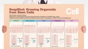 挂图SnapShot: Growing Organoids from Stem Cells Key culture conditions and organoid-forming cells from a variety of epithelial tissues
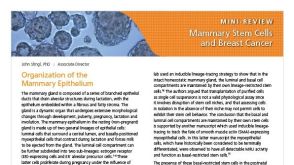
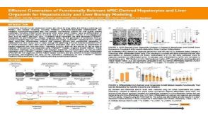 科学海报Efficient Generation of Functionally Relevant hPSC-Derived Hepatocytes and Liver Organoids for Hepatotoxicity and Liver Biology Modeling


 沪公网安备31010102008431号