Menon MP et al. (MAR 2006)
The Journal of clinical investigation 116 3 683--94
Signals for stress erythropoiesis are integrated via an erythropoietin receptor-phosphotyrosine-343-Stat5 axis.
Anemia due to chronic disease or chemotherapy often is ameliorated by erythropoietin (Epo). Present studies reveal that,unlike steady-state erythropoiesis,erythropoiesis during anemia depends sharply on an Epo receptor-phosphotyrosine-343-Stat5 signaling axis. In mice expressing a phosphotyrosine-null (PY-null) Epo receptor allele (EpoR-HM),severe and persistent anemia was induced by hemolysis or 5-fluorouracil. In short-term transplantation experiments,donor EpoR-HM bone marrow cells also failed to efficiently repopulate the erythroid compartment. In each context,stress erythropoiesis was rescued to WT levels upon the selective restoration of an EpoR PY343 Stat5-binding site (EpoR-H allele). As studied using a unique primary culture system,EpoR-HM erythroblasts exhibited marked stage-specific losses in Epo-dependent growth and survival. EpoR-H PY343 signals restored efficient erythroblast expansion,and the selective Epo induction of the Stat5 target genes proviral integration site-1 (Pim-1) and oncostatin-M. Bcl2-like 1 (Bcl-x),in contrast,was not significantly induced via WT-EpoR,EpoR-HM,or EpoR-H alleles. In Kit+ CD71+ erythroblasts,EpoR-PY343 signals furthermore enhanced SCF growth effects,and SCF modulation of Pim-1 kinase and oncostatin-M expression. In maturing Kit- CD71+ erythroblasts,oncostatin-M exerted antiapoptotic effects that likewise depended on EpoR PY343-mediated events. Stress erythropoiesis,therefore,requires stage-specific EpoR-PY343-Stat5 signals,some of which selectively bolster SCF and oncostatin-M action.
View Publication
Vessillier S et al. (SEP 2015)
Journal of immunological methods 424 43--52
Cytokine release assays for the prediction of therapeutic mAb safety in first-in man trials--Whole blood cytokine release assays are poorly predictive for TGN1412 cytokine storm.
The therapeutic monoclonal antibody (mAb) TGN1412 (anti-CD28 superagonist) caused near-fatal cytokine release syndrome (CRS) in all six volunteers during a phase-I clinical trial. Several cytokine release assays (CRAs) with reported predictivity for TGN1412-induced CRS have since been developed for the preclinical safety testing of new therapeutic mAbs. The whole blood (WB) CRA is the most widely used,but its sensitivity for TGN1412-like cytokine release was recently criticized. In a comparative study,using group size required for 90% power with 5% significance as a measure of sensitivity,we found that WB and 10% (v/v) WB CRAs were the least sensitive for TGN1412 as these required the largest group sizes (n = 52 and 79,respectively). In contrast,the peripheral blood mononuclear cell (PBMC) solid phase (SP) CRA was the most sensitive for TGN1412 as it required the smallest group size (n = 4). Similarly,the PBMC SP CRA was more sensitive than the WB CRA for muromonab-CD3 (anti-CD3) which stimulates TGN1412-like cytokine release (n = 4 and 4519,respectively). Conversely,the WB CRA was far more sensitive than the PBMC SP CRA for alemtuzumab (anti-CD52) which stimulates FcγRI-mediated cytokine release (n = 8 and 180,respectively). Investigation of potential factors contributing to the different sensitivities revealed that removal of red blood cells (RBCs) from WB permitted PBMC-like TGN1412 responses in a SP CRA,which in turn could be inhibited by the addition of the RBC membrane protein glycophorin A (GYPA); this observation likely underlies,at least in part,the poor sensitivity of WB CRA for TGN1412. The use of PBMC SP CRA for the detection of TGN1412-like cytokine release is recommended in conjunction with adequately powered group sizes for dependable preclinical safety testing of new therapeutic mAbs.
View Publication
Innate Lymphoid Cells
Overview of innate lymphoid cells (ILCs) development, classification, plasticity and functional diversity
Ramgolam VS et al. (OCT 2009)
Journal of immunology (Baltimore,Md. : 1950) 183 8 5418--27
IFN-beta inhibits human Th17 cell differentiation.
IFN-beta-1a has been used over the past 15 years as a primary therapy for relapsing-remitting multiple sclerosis (MS). However,the immunomodulatory mechanisms that provide a therapeutic effect against this CNS inflammatory disease are not yet completely elucidated. The effect of IFN-beta-1a on Th17 cells,which play a critical role in the development of the autoimmune response,has not been extensively studied in humans. We have investigated the effect of IFN-beta-1a on dendritic cells (DCs) and naive CD4(+)CD45RA(+) T cells derived from untreated MS patients and healthy controls in the context of Th17 cell differentiation. We report that IFN-beta-1a treatment down-regulated the expression of IL-1beta and IL-23p19 in DCs,whereas it induced the gene expression of IL-12p35 and IL-27p28. We propose that IFN-beta-1a-mediated up-regulation of the suppressor of cytokine signaling 3 expression,induced via STAT3 phosphorylation,mediates IL-1beta and IL-23 down-regulation,while IFN-beta-1a-induced STAT1 phosphorylation induces IL-27p28 expression. CD4(+)CD45RA(+) naive T cells cocultured with supernatants from IFN-beta-1a-treated DCs exhibited decreased gene expression of the Th17 cell markers retinoic acid-related orphan nuclear hormone receptor c (RORc),IL-17A,and IL-23R. A direct IFN-beta-1a treatment of CD45RA(+) T cells cultured in Th17-polarizing conditions also down-regulated RORc,IL-17A,and IL-23R,but up-regulated IL-10 gene expression. Studies of the mechanisms involved in the Th17 cell differentiation suggest that IFN-beta-1a inhibits IL-17 and induces IL-10 secretion via activated STAT1 and STAT3,respectively. IFN-beta's suppression of Th17 cell differentiation may represent its most relevant mechanism of selective suppression of the autoimmune response in MS.
View Publication
Dendritic Cells Regulate Extrafollicular Autoreactive B Cells via T Cells Expressing Fas and Fas Ligand.
The extrafollicular (EF) plasmablast response to self-antigens that contain Toll-like receptor (TLR) ligands is prominent in murine lupus models and some bacterial infections,but the inhibitors and activators involved have not been fully delineated. Here,we used two conventional dendritic cell (cDC) depletion systems to investigate the role of cDCs on a classical TLR-dependent autoreactive EF response elicited in rheumatoid-factor B cells by DNA-containing immune complexes. Contrary to our hypothesis,cDC depletion amplified rather than dampened the EF response in Fas-intact but not Fas-deficient mice. Further,we demonstrated that cDC-dependent regulation requires Fas and Fas ligand (FasL) expression by T cells,but not Fas expression by B cells. Thus,cDCs activate FasL-expressing T cells that regulate Fas-expressing extrafollicular helper T (Tefh) cells. These studies reveal a regulatory role for cDCs in B cell plasmablast responses and provide a mechanistic explanation for the excess autoantibody production observed in Fas deficiency.
View Publication
产品类型:
产品号#:
19754
19754RF
产品名:
Schneider E et al. (SEP 2009)
Journal of immunology (Baltimore,Md. : 1950) 183 6 3591--7
IL-33 activates unprimed murine basophils directly in vitro and induces their in vivo expansion indirectly by promoting hematopoietic growth factor production.
IL-33,a new member of the IL-1 family,has been described as an important inducer of Th2 cytokines and mediator of inflammatory responses. In this study,we demonstrate that murine basophils sorted directly from the bone marrow,without prior exposure to IL-3 or Fc(epsilon)R cross-linking,respond to IL-33 alone by producing substantial amounts of histamine,IL-4,and IL-6. These cells express ST2 constitutively and generate a cytokine profile that differs from their IL-3-induced counterpart by a preferential production of IL-6. In vivo,IL-33 promotes basophil expansion in the bone marrow (BM) through an indirect mechanism of action depending on signaling through the beta(c) chain shared by receptors for IL-3,GM-CSF,and IL-5. IL-3 can still signal through its specific beta(IL-3) chain in these mutant mice,which implies that it is not the unique growth-promoting mediator in this setup,but requires IL-5 and/or GMCSF. Our results support a major role of the latter growth factor,which is readily generated by total BM cells as well as sorted basophils in response to IL-33 along with low amounts of IL-3. Furthermore,GM-CSF amplifies IL-3-induced differentiation of basophils from BM cells,whereas IL-5 that is also generated in vivo,affects neither their functions nor their growth in vitro or in vivo. In conclusion,our data provide the first evidence that IL-33 not only activates unprimed basophils directly,but also promotes their expansion in vivo through induction of GM-CSF and IL-3.
View Publication

 EasySep™小鼠TIL(CD45)正选试剂盒


 点播Human Immune Cell Isolation Course Learn how to effectively process human samples and isolate highly pure target cells with EasySep™.
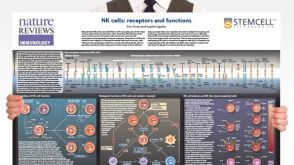 挂图Natural Killer Cells Overview of NK cell receptors, subsets, activation and function
 挂图The Human Leukocyte Antigen (HLA) Complex Provides an overview of the human leukocyte antigen (HLA) complex and nomenclature of HLA alleles
 挂图Innate Lymphoid Cells Overview of innate lymphoid cells (ILCs) development, classification, plasticity and functional diversity
 科学海报Basophil Isolation from Human Peripheral Blood

 沪公网安备31010102008431号