Schneider E et al. (SEP 2009)
Journal of immunology (Baltimore,Md. : 1950) 183 6 3591--7
IL-33 activates unprimed murine basophils directly in vitro and induces their in vivo expansion indirectly by promoting hematopoietic growth factor production.
IL-33,a new member of the IL-1 family,has been described as an important inducer of Th2 cytokines and mediator of inflammatory responses. In this study,we demonstrate that murine basophils sorted directly from the bone marrow,without prior exposure to IL-3 or Fc(epsilon)R cross-linking,respond to IL-33 alone by producing substantial amounts of histamine,IL-4,and IL-6. These cells express ST2 constitutively and generate a cytokine profile that differs from their IL-3-induced counterpart by a preferential production of IL-6. In vivo,IL-33 promotes basophil expansion in the bone marrow (BM) through an indirect mechanism of action depending on signaling through the beta(c) chain shared by receptors for IL-3,GM-CSF,and IL-5. IL-3 can still signal through its specific beta(IL-3) chain in these mutant mice,which implies that it is not the unique growth-promoting mediator in this setup,but requires IL-5 and/or GMCSF. Our results support a major role of the latter growth factor,which is readily generated by total BM cells as well as sorted basophils in response to IL-33 along with low amounts of IL-3. Furthermore,GM-CSF amplifies IL-3-induced differentiation of basophils from BM cells,whereas IL-5 that is also generated in vivo,affects neither their functions nor their growth in vitro or in vivo. In conclusion,our data provide the first evidence that IL-33 not only activates unprimed basophils directly,but also promotes their expansion in vivo through induction of GM-CSF and IL-3.
View Publication
Wulff H et al. (JUL 2004)
Journal of immunology (Baltimore,Md. : 1950) 173 2 776--86
K+ channel expression during B cell differentiation: implications for immunomodulation and autoimmunity.
Using whole-cell patch-clamp,fluorescence microscopy and flow cytometry,we demonstrate a switch in potassium channel expression during differentiation of human B cells from naive to memory cells. Naive and IgD(+)CD27(+) memory B cells express small numbers of the voltage-gated Kv1.3 and the Ca(2+)-activated intermediate-conductance IKCa1 channel when quiescent,and increase IKCa1 expression 45-fold upon activation with no change in Kv1.3 levels. In contrast,quiescent class-switched memory B cells express high levels of Kv1.3 ( approximately 2000 channels/cell) and maintain their Kv1.3(high) expression after activation. Consistent with their channel phenotypes,proliferation of naive and IgD(+)CD27(+) memory B cells is suppressed by the specific IKCa1 inhibitor TRAM-34 but not by the potent Kv1.3 blocker Stichodactyla helianthus toxin,whereas the proliferation of class-switched memory B cells is suppressed by Stichodactyla helianthus toxin but not TRAM-34. These changes parallel those reported for T cells. Therefore,specific Kv1.3 and IKCa1 inhibitors may have use in therapeutic manipulation of selective lymphocyte subsets in immunological disorders.
View Publication
Marwali MR et al. (SEP 2004)
Journal of immunology (Baltimore,Md. : 1950) 173 5 2960--7
Lipid rafts mediate association of LFA-1 and CD3 and formation of the immunological synapse of CTL.
Lipid rafts accumulate in the immunological synapse formed by an organized assembly of the TCR/CD3,LFA-1,and signaling molecules. However,the precise role of lipid rafts in the formation of the immunological synapse is unclear. In this study,we show that LFA-1 on CTL is constitutively active and mediates Ag-independent binding of CTL to target cells expressing its ligands. LFA-1 and CD3 on CTL,but not resting T cells,colocalize in lipid rafts. Binding of LFA-1 on CTL to targets initiates the formation of the immunological synapse,which is formed by LFA-1,CD3,and ganglioside GM1 distributed in the periphery of the cell contact site and cholesterol is more widely distributed. The formation of this synapse is Ag independent,but the recognition of Ag by the TCR induces accumulation of tyrosine phosphorylated proteins in the synapse as well as redistribution of the microtubule organization center toward the cell contact site. Our results suggest that LFA-1 recruits lipid rafts and the TCR/CD3 to the synapse,and facilitates efficient and rapid activation of CTL.
View Publication
产品类型:
产品号#:
18554
18554RF
18564
18564RF
产品名:
Cebo C et al. (JAN 2006)
Journal of immunology (Baltimore,Md. : 1950) 176 2 864--72
The decreased susceptibility of Bcr/Abl targets to NK cell-mediated lysis in response to imatinib mesylate involves modulation of NKG2D ligands, GM1 expression, and synapse formation.
Chronic myeloid leukemia is a clonal multilineage myeloproliferative disease of stem cell origin characterized by the presence of the Bcr/Abl oncoprotein,a constitutively active tyrosine kinase. In previous studies,we have provided evidence that Bcr/Abl overexpression in leukemic cells increased their susceptibility to NK-mediated lysis by different mechanisms. In the present study,using UT-7/9 cells,a high level Bcr/Abl transfectant of UT-7 cells,we show that the treatment of Bcr/Abl target by imatinib mesylate (IM),a specific Abl tyrosine kinase inhibitor,hampers the formation of the NK/target immunological synapse. The main effect of IM involves an induction of surface GM1 ganglioside on Bcr/Abl transfectants that prevents the redistribution of MHC-related Ag molecules in lipid rafts upon interaction with NK cells. IM also affects cell surface glycosylation of targets,as assessed by binding of specific lectins resulting in the subsequent modulation of their binding to lectin type NK receptor,particularly NKG2D. In addition,we demonstrate that the tyrosine kinase activity repression results in a decrease of MHC-related Ags-A/B and UL-16-binding protein expression on Bcr/Abl transfectants UT-7/9. We show that NKG2D controls the NK-mediated lysis of UT-7/9 cells,and IM treatment inhibits this activating pathway. Taken together,our results show that the high expression of Bcr/Abl in leukemic cells controls the expression of NKG2D receptor ligands and membrane GM1 via a tyrosine kinase-dependent mechanism and that the modulation of these molecules by IM interferes with NK cell recognition and cytolysis of the transfectants.
View Publication
产品类型:
产品号#:
15025
15065
产品名:
RosetteSep™人NK细胞富集抗体混合物
RosetteSep™人NK细胞富集抗体混合物
Mace EM et al. ( 2016)
Nature communications 7 12171
Human NK cell development requires CD56-mediated motility and formation of the developmental synapse.
While distinct stages of natural killer (NK) cell development have been defined,the molecular interactions that shape human NK cell maturation are poorly understood. Here we define intercellular interactions between developing NK cells and stromal cells which,through contact-dependent mechanisms,promote the generation of mature,functional human NK cells from CD34(+) precursors. We show that developing NK cells undergo unique,developmental stage-specific sustained and transient interactions with developmentally supportive stromal cells,and that the relative motility of NK cells increases as they move through development in vitro and ex vivo. These interactions include the formation of a synapse between developing NK cells and stromal cells,which we term the developmental synapse. Finally,we identify a role for CD56 in developmental synapse structure,NK cell motility and NK cell development. Thus,we define the developmental synapse leading to human NK cell functional maturation.
View Publication
产品类型:
产品号#:
05150
15025
15065
产品名:
MyeloCult™H5100
RosetteSep™人NK细胞富集抗体混合物
RosetteSep™人NK细胞富集抗体混合物
R. Lorenzetti et al. (jul 2019)
Journal of autoimmunity 101 145--152
Abatacept modulates CD80 and CD86 expression and memory formation in human B-cells.
BACKGROUND Cytotoxic T lymphocyte antigen-4 (CTLA-4) limits T-cell activation and is expressed on T-regulatory cells. Human CTLA-4 deficiency results in severe immune dysregulation. Abatacept (CTLA-4 Ig) is approved for the treatment of rheumatoid arthritis (RA) and its mechanism of action is attributed to effects on T-cells. It is known that CTLA-4 modulates the expression of its ligands CD80 and CD86 on antigen presenting cells (APC) by transendocytosis. As B-cells express CD80/CD86 and function as APC,we hypothesize that B-cells are a direct target of abatacept. OBJECTIVES To investigate direct effects of abatacept on human B-lymphocytes in vitro and in RA patients. METHODS The effect of abatacept on healthy donor B-cells' phenotype,activation and CD80/CD86 expression was studied in vitro. Nine abatacept-treated RA patients were studied. Seven of these were followed up to 24 months,and two up to 12 months only and treatment response,immunoglobulins,ACPA,RF concentrations,B-cell phenotype and ACPA-specific switched memory B-cell frequency were assessed. RESULTS B-cell development was unaffected by abatacept. Abatacept treatment resulted in a dose-dependent decrease of CD80/CD86 expression on B-cells in vitro,which was due to dynamin-dependent internalization. RA patients treated with abatacept showed a progressive decrease in plasmablasts and serum IgG. While ACPA-titers only moderately declined,the frequency of ACPA-specific switched memory B-cells significantly decreased. CONCLUSIONS Abatacept directly targets B-cells by reducing CD80/CD86 expression. Impairment of antigen presentation and T-cell activation may result in altered B-cell selection,providing a new therapeutic mechanism and a base for abatacept use in B-cell mediated autoimmunity.
View Publication
产品类型:
产品号#:
17954
17954RF
产品名:
EasySep™人B细胞分选试剂盒
RoboSep™ 人B细胞分选试剂盒
Koka R et al. (SEP 2004)
Journal of immunology (Baltimore,Md. : 1950) 173 6 3594--8
Cutting edge: murine dendritic cells require IL-15R alpha to prime NK cells.
NK cells protect hosts against viral pathogens and transformed cells,and dendritic cells (DCs) play important roles in activating NK cells. We now find that murine IL-15Ralpha-deficient DCs fail to support NK cell cytolytic activity and elaboration of IFN-gamma,despite the fact that these DCs express normal levels of costimulatory molecules and IL-12. By contrast,IL-15Ralpha expression on NK cells is entirely dispensable for their activation by DCs. In addition,blockade with anti-IL-15Ralpha and anti-IL-2Rbeta but not anti-IL-2Ralpha-specific Abs prevents NK cell activation by wild-type DCs. Finally,presentation of IL-15 by purified IL-15Ralpha/Fc in trans synergizes with IL-12 to support NK cell priming. These findings suggest that murine DCs require IL-15Ralpha to present IL-15 in trans to NK cells during NK cell priming.
View Publication
产品类型:
产品号#:
18755
18755RF
产品名:
EasySep™ 小鼠CD49b正选试剂盒
RoboSep™ 小鼠CD49b正选试剂盒含滤芯吸头
Li Y et al. (FEB 2016)
Journal of Immunology 196 4 1617--25
Hepatic Stellate Cells Directly Inhibit B Cells via Programmed Death-Ligand 1.
We demonstrated previously that mouse hepatic stellate cells (HSCs) suppress T cells via programmed death-ligand 1 (PD-L1),but it remains unknown whether they exert any effects on B cells,the other component of the adaptive immune system. In this study,we found that mouse HSCs directly inhibited B cells and that PD-L1 was also integrally involved. We found that HSCs inhibited the upregulation of activation markers on activated B cells,as well as the proliferation of activated B cells and their cytokine/Ig production in vitro,and that pharmaceutically or genetically blocking the interaction of PD-L1 with programmed cell death protein 1 impaired the ability of HSCs to inhibit B cells. To test the newly discovered B cell-inhibitory activity of HSCs in vivo,we developed a protocol of intrasplenic artery injection to directly deliver HSCs into the spleen. We found that local delivery of wild-type HSCs into the spleens of mice that had been immunized with 4-hydroxy-3-nitrophenylacetyl-Ficoll,a T cell-independent Ag,significantly suppressed Ag-specific IgM and IgG production in vivo,whereas splenic artery delivery of PD-L1-deficient HSCs failed to do so. In conclusion,in addition to inhibiting T cells,mouse HSCs concurrently inhibit B cells via PD-L1. This direct B cell-inhibitory activity of HSCs should contribute to the mechanism by which HSCs maintain the liver's immune homeostasis.
View Publication
产品类型:
产品号#:
19854
19854RF
产品名:
EasySep™小鼠B细胞分选试剂盒
RoboSep™ 小鼠B细胞分选试剂盒
Aflaki E et al. (JUN 2014)
Science translational medicine 6 240 240ra73
Macrophage models of Gaucher disease for evaluating disease pathogenesis and candidate drugs.
Gaucher disease is caused by an inherited deficiency of glucocerebrosidase that manifests with storage of glycolipids in lysosomes,particularly in macrophages. Available cell lines modeling Gaucher disease do not demonstrate lysosomal storage of glycolipids; therefore,we set out to develop two macrophage models of Gaucher disease that exhibit appropriate substrate accumulation. We used these cellular models both to investigate altered macrophage biology in Gaucher disease and to evaluate candidate drugs for its treatment. We generated and characterized monocyte-derived macrophages from 20 patients carrying different Gaucher disease mutations. In addition,we created induced pluripotent stem cell (iPSC)-derived macrophages from five fibroblast lines taken from patients with type 1 or type 2 Gaucher disease. Macrophages derived from patient monocytes or iPSCs showed reduced glucocerebrosidase activity and increased storage of glucocerebroside and glucosylsphingosine in lysosomes. These macrophages showed efficient phagocytosis of bacteria but reduced production of intracellular reactive oxygen species and impaired chemotaxis. The disease phenotype was reversed with a noninhibitory small-molecule chaperone drug that enhanced glucocerebrosidase activity in the macrophages,reduced glycolipid storage,and normalized chemotaxis and production of reactive oxygen species. Macrophages differentiated from patient monocytes or patient-derived iPSCs provide cellular models that can be used to investigate disease pathogenesis and facilitate drug development.
View Publication

 EasySep™小鼠TIL(CD45)正选试剂盒


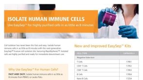 产品手册Isolate Human Immune Cells
 产品手册Human Primary Cells: It All Starts with the Right Cells

 沪公网安备31010102008431号