Francis KR et al. (APR 2016)
Nature medicine 22 4 388--396
Modeling Smith-Lemli-Opitz syndrome with induced pluripotent stem cells reveals a causal role for Wnt/$$-catenin defects in neuronal cholesterol synthesis phenotypes.
Smith-Lemli-Opitz syndrome (SLOS) is a malformation disorder caused by mutations in DHCR7,which impair the reduction of 7-dehydrocholesterol (7DHC) to cholesterol. SLOS results in cognitive impairment,behavioral abnormalities and nervous system defects,though neither affected cell types nor impaired signaling pathways are fully understood. Whether 7DHC accumulation or cholesterol loss is primarily responsible for disease pathogenesis is also unclear. Using induced pluripotent stem cells (iPSCs) from subjects with SLOS,we identified cellular defects that lead to precocious neuronal specification within SLOS derived neural progenitors. We also demonstrated that 7DHC accumulation,not cholesterol deficiency,is critical for SLOS-associated defects. We further identified downregulation of Wnt/$$-catenin signaling as a key initiator of aberrant SLOS iPSC differentiation through the direct inhibitory effects of 7DHC on the formation of an active Wnt receptor complex. Activation of canonical Wnt signaling prevented the neural phenotypes observed in SLOS iPSCs,suggesting that Wnt signaling may be a promising therapeutic target for SLOS.
View Publication
Androgenetic embryonic stem cells form neural progenitor cells in vivo and in vitro.
Uniparental zygotes with two paternal (androgenetic [AG]) or two maternal (gynogenetic [GG]; parthenogenetic [PG]) genomes are not able to develop into viable offspring but can form blastocysts from which embryonic stem cells (ESCs) can be derived. Although some aspects of the in vitro and in vivo differentiation potential of PG and GG ESCs of several species have been studied,the developmental capacity of AG ESCs is much less clear. Here,we investigate the potential of murine AG ESCs to undergo neural differentiation. We observed that AG ESCs differentiate in vitro into pan-neural progenitor cells (pnPCs) that further give rise to cells that express neuronal- and astroglial-specific markers. Neural progeny of in vitro-differentiated AG ESCs exhibited fidelity of expression of six imprinted genes analyzed,with the exception of Ube3a. Bisulfite sequencing for two imprinting control regions suggested that pnPCs predominantly maintained their methylation pattern. Following blastocyst injection of AG and biparental (normal fertilized [N]) ESCs,we found widespread and evenly distributed contribution of ESC-derived cells in both AG and N chimeric early fetal brains. AG and N ESC-derived cells isolated from chimeric fetal brains by fluorescence-activated cell sorting exhibited similar neurosphere-initiating cell frequencies and neural multilineage differentiation potential. Our results indicate that AG ESC-derived neural progenitor/stem cells do not differ from N neural progenitor/stem cells in their self-renewal and neural multilineage differentiation potential. Disclosure of potential conflicts of interest is found at the end of this article.
View Publication
产品类型:
产品号#:
05703
产品名:
NeuroCult™ 分化添加物(小鼠和大鼠)
Matthews TA et al. (JAN 2014)
Brain Research 1543 28--37
Expression of the CHOP-inducible carbonic anhydrase CAVI-b is required for BDNF-mediated protection from hypoxia
Carbonic anhydrases (CAs) comprise a family of zinc-containing enzymes that catalyze the reversible hydration of carbon dioxide. CAs contribute to a myriad of physiological processes,including pH regulation,anion transport and water balance. To date,16 known members of the mammalian alpha-CA family have been identified. Given that the catalytic family members share identical reaction chemistry,their physiologic roles are influenced greatly by their tissue and sub-cellular locations. CAVI is the lone secreted CA and exists in both saliva and the gastrointestinal mucosa. An alternative,stress-inducible isoform of CAVI (CAVI-b) has been shown to be expressed from a cryptic promoter that is activated by the CCAAT/Enhancer-Binding Protein Homologous Protein (CHOP). The CAVI-b isoform is not secreted and is currently of unknown physiological function. Here we use neuronal models,including a model derived using Car6 and CHOP gene ablations,to delineate a role for CAVI-b in ischemic protection. Our results demonstrate that CAVI-b expression,which is increased through CHOP-signaling in response to unfolded protein stress,is also increased by oxygen-glucose deprivation (OGD). While enforced expression of CAVI-b is not sufficient to protect against ischemia,CHOP regulation of CAVI-b is necessary for adaptive changes mediated by BDNF that reduce subsequent ischemic damage. These results suggest that CAVI-b comprises a necessary component of a larger adaptive signaling pathway downstream of CHOP.
View Publication
产品类型:
产品号#:
05700
05701
05702
05703
05704
产品名:
NeuroCult™ 基础培养基(小鼠和大鼠)
NeuroCult™ 扩增添加物(小鼠和大鼠)
NeuroCult™扩增试剂盒(小鼠和大鼠)
NeuroCult™ 分化添加物(小鼠和大鼠)
NeuroCult™ 分化试剂盒(小鼠和大鼠)
Alessandrini F et al. ( 2016)
Journal of Cancer 7 13 1791--1797
Noninvasive Monitoring of Glioma Growth in the Mouse.
Malignant gliomas are the most common and deadly primary malignant brain tumors. In vivo orthotopic models could doubtless represent an appropriate tool to test novel treatment for gliomas. However,methods commonly used to monitor the growth of glioma inside the mouse brain are time consuming and invasive. We tested the reliability of a minimally invasive procedure,based on a secreted luciferase (Gaussia luciferase),to frequently monitor the changes of glioma size. Gluc activity was evaluated from blood samples collected from the tail tip of mice twice a week,allowing to make a growth curve for the tumors. We validated the correlation between Gluc activity and tumor size by analysing the tumor after brain dissection. We found that this method is reliable for monitoring human glioma transplanted in immunodeficient mice,but it has strong limitation in immunocompetent models,where an immune response against the luciferase is developed during the first weeks after transplant.
View Publication
产品类型:
产品号#:
05750
05751
产品名:
NeuroCult™ NS-A 基础培养基(人)
NeuroCult™ NS-A 扩增试剂盒(人)
Wang L et al. (NOV 2008)
PLoS Biology 6 11 e289
Gamma-Secretase Represents a Therapeutic Target for the Treatment of Invasive Glioma Mediated by the p75 Neurotrophin Receptor
The multifunctional signaling protein p75 neurotrophin receptor (p75(NTR)) is a central regulator and major contributor to the highly invasive nature of malignant gliomas. Here,we show that neurotrophin-dependent regulated intramembrane proteolysis (RIP) of p75(NTR) is required for p75(NTR)-mediated glioma invasion,and identify a previously unnamed process for targeted glioma therapy. Expression of cleavage-resistant chimeras of p75(NTR) or treatment of animals bearing p75(NTR)-positive intracranial tumors with clinically applicable gamma-secretase inhibitors resulted in dramatically decreased glioma invasion and prolonged survival. Importantly,proteolytic processing of p75(NTR) was observed in p75(NTR)-positive patient tumor specimens and brain tumor initiating cells. This work highlights the importance of p75(NTR) as a therapeutic target,suggesting that gamma-secretase inhibitors may have direct clinical application for the treatment of malignant glioma.
View Publication
产品类型:
产品号#:
05750
05751
产品名:
NeuroCult™ NS-A 基础培养基(人)
NeuroCult™ NS-A 扩增试剂盒(人)
Azari H et al. (JAN 2011)
Journal of visualized experiments : JoVE 49
Neural-colony forming cell assay: an assay to discriminate bona fide neural stem cells from neural progenitor cells.
The neurosphere assay (NSA) is one of the most frequently used methods to isolate,expand and also calculate the frequency of neural stem cells (NSCs). Furthermore,this serum-free culture system has also been employed to expand stem cells and determine their frequency from a variety of tumors and normal tissues. It has been shown recently that a one-to-one relationship does not exist between neurosphere formation and NSCs. This suggests that the NSA as currently applied,overestimates the frequency of NSCs in a mixed population of neural precursor cells isolated from both the embryonic and adult mammalian brain. This video practically demonstrates a novel collagen based semi- solid assay,the neural-colony forming cell assay (N-CFCA),which has the ability to discriminate stem from progenitor cells based on their long-term proliferative potential,and thus provides a method to enumerate NSC frequency. In the N-CFCA,colonies ≥2 mm in diameter are derived from cells that meet all the functional criteria of a NSC,while colonies textless 2mm are derived from progenitors. The N-CFCA procedure can be used for cells prepared from different sources including primary and cultured adult or embryonic mouse CNS cells. Here we use cells prepared from passage one neurospheres generated from embryonic day 14 mice brain to perform N-CFCA. The cultures are replenished with proliferation medium every seven days for three weeks to allow the plated cells to exhibit their full proliferative potential and then the frequency of neural progenitor and bona fide neural stem cells is calculated respectively by counting the number of colonies that are textless 2mm and the ones that are ≥2mm in reference to the number of cells that were initially plated.
View Publication
产品类型:
产品号#:
05740
产品名:
Saito T et al. (JUL 2013)
PLoS ONE 8 7 e70010
Metformin, a Diabetes Drug, Eliminates Tumor-Initiating Hepatocellular Carcinoma Cells
Metformin has been widely used as an oral drug for diabetes mellitus for approximately 60 years. Interestingly,recent reports showed that metformin exhibited an anti-tumor action in a wide range of malignancies including hepatocellular carcinoma (HCC). In the present study,we investigated its impact on tumor-initiating HCC cells. Metformin suppressed cell growth and induced apoptosis in a dose-dependent manner. Flow cytometric analysis showed that metformin treatment markedly reduced the number of tumor-initiating epithelial cell adhesion molecule (EpCAM)(+) HCC cells. Non-adherent sphere formation assays of EpCAM(+) cells showed that metformin impaired not only their sphere-forming ability,but also their self-renewal capability. Consistent with this,immunostaining of spheres revealed that metformin significantly decreased the number of component cells positive for hepatic stem cell markers such as EpCAM and α-fetoprotein. In a xenograft transplantation model using non-obese diabetic/severe combined immunodeficient mice,metformin and/or sorafenib treatment suppressed the growth of tumors derived from transplanted HCC cells. Notably,the administration of metformin but not sorafenib decreased the number of EpCAM(+) cells and impaired their self-renewal capability. As reported,metformin activated AMP-activated protein kinase (AMPK) through phosphorylation; however its inhibitory effect on the mammalian target of rapamycin (mTOR) pathway did not necessarily correlate with its anti-tumor activity toward EpCAM(+) tumor-initiating HCC cells. These results indicate that metformin is a promising therapeutic agent for the elimination of tumor-initiating HCC cells and suggest as-yet-unknown functions other than its inhibitory effect on the AMPK/mTOR pathway.
View Publication
Beliveau A et al. (MAY 2016)
Scientific reports 6 26143
Aligned Nanotopography Promotes a Migratory State in Glioblastoma Multiforme Tumor Cells.
Glioblastoma multiforme (GBM) is an aggressive,Grade IV astrocytoma with a poor survival rate,primarily due to the GBM tumor cells migrating away from the primary tumor site along the nanotopography of white matter tracts and blood vessels. It is unclear whether this nanotopography influences the biomechanical properties (i.e. cytoskeletal stiffness) of GBM tumor cells. Although GBM tumor cells have an innate propensity to migrate,we believe this capability is enhanced due to the influence of nanotopography on the tumor cells' biomechanical properties. In this study,we used an aligned nanofiber film that mimics the nanotopography in the tumor microenvironment to investigate the mechanical properties of GBM tumor cells in vitro. The data demonstrate that the cytoskeletal stiffness,cell traction stress,and focal adhesion area were significantly lower in the GBM tumor cells compared to healthy astrocytes. Moreover,the cytoskeletal stiffness was significantly reduced when cultured on aligned nanofiber films compared to smooth and randomly aligned nanofiber films. Gene expression analysis showed that tumor cells cultured on the aligned nanotopography upregulated key migratory genes and downregulated key proliferative genes. Therefore,our data suggest that the migratory potential is elevated when GBM tumor cells are migrating along aligned nanotopographical substrates.
View Publication
产品类型:
产品号#:
05750
05751
产品名:
NeuroCult™ NS-A 基础培养基(人)
NeuroCult™ NS-A 扩增试剂盒(人)
Blackmore DG et al. (JAN 2012)
PloS one 7 11 e49912
GH mediates exercise-dependent activation of SVZ neural precursor cells in aged mice.
Here we demonstrate,both in vivo and in vitro,that growth hormone (GH) mediates precursor cell activation in the subventricular zone (SVZ) of the aged (12-month-old) brain following exercise,and that GH signaling stimulates precursor activation to a similar extent to exercise. Our results reveal that both addition of GH in culture and direct intracerebroventricular infusion of GH stimulate neural precursor cells in the aged brain. In contrast,no increase in neurosphere numbers was observed in GH receptor null animals following exercise. Continuous infusion of a GH antagonist into the lateral ventricle of wild-type animals completely abolished the exercise-induced increase in neural precursor cell number. Given that the aged brain does not recover well after injury,we investigated the direct effect of exercise and GH on neural precursor cell activation following irradiation. This revealed that physical exercise as well as infusion of GH promoted repopulation of neural precursor cells in irradiated aged animals. Conversely,infusion of a GH antagonist during exercise prevented recovery of precursor cells in the SVZ following irradiation.
View Publication

 EasySep™小鼠TIL(CD45)正选试剂盒


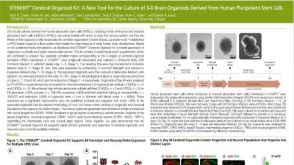 科学海报STEMdiff Cerebral Organoid Kit: A New Tool for the Culture of 3D Brain Organoids Derived from Human Pluripotent Stem Cells

 沪公网安备31010102008431号