Liu Y et al. (MAY 2011)
Nature protocols 6 5 640--55
OLIG gene targeting in human pluripotent stem cells for motor neuron and oligodendrocyte differentiation.
Pluripotent stem cells can be genetically labeled to facilitate differentiation studies. In this paper,we describe a gene-targeting protocol to knock in a GFP cassette into key gene loci in human pluripotent stem cells (hPSCs),and then use the genetically tagged hPSCs to guide in vitro differentiation,immunocytochemical and electrophysiological profiling and in vivo characterization after cell transplantation. The Olig transcription factors have key roles in the transcription regulatory pathways for the genesis of motor neurons (MNs) and oligodendrocytes (OLs). We have generated OLIG2-GFP hPSC reporter lines that reliably mark MNs and OLs for monitoring their sequential differentiation from hPSCs. The expression of the GFP reporter recapitulates the endogenous expression of OLIG genes. The in vitro characterization of fluorescence-activated cell sorting-purified cells is consistent with cells of the MN or OL lineages,depending on the stages at which they are collected. This protocol is efficient and reliable and usually takes 5-7 months to complete. The genetic tagging-differentiation methodology used herein provides a general framework for similar work for differentiation of hPSCs into other lineages.
View Publication
产品类型:
产品号#:
05850
05857
05870
05875
85850
85857
85870
85875
产品名:
mTeSR™1
mTeSR™1
Vukovic J et al. (AUG 2013)
Stem Cells and Development 22 16 2341--2345
A Novel Fluorescent Reporter CDy1 Enriches for Neural Stem Cells Derived from the Murine Brain
Neurogenesis occurs continuously in two brain regions of adult mammals,underpinned by a pool of resident neural stem cells (NSCs) that can differentiate into all neural cell types. To advance our understanding of NSC function and to develop therapeutic and diagnostic approaches,it is important to accurately identify and enrich for NSCs. There are no definitive markers for the identification and enrichment of NSCs present in the mouse brain. Recently,a fluorescent rosamine dye,CDy1,has been identified as a label for pluripotency in cultured human embryonic and induced pluripotent stem cells. As similar cellular characteristics may enable the uptake and retention of CDy1 by other stem cell populations,we hypothesized that this dye may also enrich for primary NSCs from the mouse brain. Because the subventricular zone (SVZ) and the hippocampus represent brain regions that are highly enriched for NSCs in adult mammals,we sampled cells from these areas to test this hypothesis. These experiments revealed that CDy1 staining indeed allows for enrichment and selection of all neurosphere-forming cells from both the SVZ and the hippocampus. We next examined the effectiveness of CDy1 to select for NSCs derived from the SVZ of aged animals,where the total pool of NSCs present is significantly lower than in young animals. We found that CDy1 effectively labels the NSCs in adult and aged animals as assessed by the neurosphere assay and reflects the numbers of NSCs present in aged animals. CDy1,therefore,appears to be a novel marker for enrichment of NSCs in primary brain tissue preparations.
View Publication
产品类型:
产品号#:
05700
05701
05702
产品名:
NeuroCult™ 基础培养基(小鼠和大鼠)
NeuroCult™ 扩增添加物(小鼠和大鼠)
NeuroCult™扩增试剂盒(小鼠和大鼠)
Pollak J et al. (MAR 2017)
PLOS ONE 12 3 e0172884
Ion channel expression patterns in glioblastoma stem cells with functional and therapeutic implications for malignancy
Ion channels and transporters have increasingly recognized roles in cancer progression through the regulation of cell proliferation,migration,and death. Glioblastoma stem-like cells (GSCs) are a source of tumor formation and recurrence in glioblastoma multiforme,a highly aggressive brain cancer,suggesting that ion channel expression may be perturbed in this population. However,little is known about the expression and functional relevance of ion channels that may contribute to GSC malignancy. Using RNA sequencing,we assessed the enrichment of ion channels in GSC isolates and non-tumor neural cell types. We identified a unique set of GSC-enriched ion channels using differential expression analysis that is also associated with distinct gene mutation signatures. In support of potential clinical relevance,expression of selected GSC-enriched ion channels evaluated in human glioblastoma databases of The Cancer Genome Atlas and Ivy Glioblastoma Atlas Project correlated with patient survival times. Finally,genetic knockdown as well as pharmacological inhibition of individual or classes of GSC-enriched ion channels constrained growth of GSCs compared to normal neural stem cells. This first-in-kind global examination characterizes ion channels enriched in GSCs and explores their potential clinical relevance to glioblastoma molecular subtypes,gene mutations,survival outcomes,regional tumor expression,and experimental responses to loss-of-function. Together,the data support the potential biological and therapeutic impact of ion channels on GSC malignancy and provide strong rationale for further examination of their mechanistic and therapeutic importance.
View Publication
产品类型:
产品号#:
05751
70913
产品名:
NeuroCult™ NS-A 扩增试剂盒(人)
Nie S et al. (FEB 2015)
Journal of proteome research 14 2 814--22
Tenascin-C: a novel candidate marker for cancer stem cells in glioblastoma identified by tissue microarrays.
Glioblastoma multiforme (GBM) is a highly aggressive brain tumor,with dismal survival outcomes. Recently,cancer stem cells (CSCs) have been demonstrated to play a role in therapeutic resistance and are considered to be the most likely cause of cancer relapse. The identification of CSCs is an important step toward finding new and effective ways to treat GBM. Tenascin-C (TNC) protein has been identified as a potential marker for CSCs in gliomas based on previous work. Here,we have investigated the expression of TNC in tissue microarrays including 17 GBMs,18 WHO grade III astrocytomas,15 WHO grade II astrocytomas,4 WHO grade I astrocytomas,and 7 normal brain tissue samples by immunohistochemical staining. TNC expression was found to be highly associated with the grade of astrocytoma. It has a high expression level in most of the grade III astrocytomas and GBMs analyzed and a very low expression in most grade II astrocytomas,whereas it is undetectable in grade I astrocytomas and normal brain tissues. Double-immunofluorescence staining for TNC and CD133 in GBM tissues revealed that there was a high overlap between theses two positive populations. The results were further confirmed by flow cytometry analysis of TNC and CD133 in GBM-derived stem-like neurospheres in vitro. A limiting dilution assay demonstrated that the sphere formation ability of CD133(+)/TNC(+) and CD133(-)/TNC(+) cell populations is much higher than that of the CD133(+)/TNC(-) and CD133(-)/TNC(-) populations. These results suggest that TNC is not only a potential prognostic marker for GBM but also a potential marker for glioma CSCs,where the TNC(+) population is identified as a CSC population overlapping with part of the CD133(-) cell population.
View Publication
产品类型:
产品号#:
05750
05751
05752
产品名:
NeuroCult™ NS-A 基础培养基(人)
NeuroCult™ NS-A 扩增试剂盒(人)
NeuroCult™ NS-A 分化试剂盒(人)
Fitzgerald DP et al. (OCT 2006)
Neuroscience 142 3 703--16
Characterization of neogenin-expressing neural progenitor populations and migrating neuroblasts in the embryonic mouse forebrain.
Many studies have demonstrated a role for netrin-1-deleted in colorectal cancer (DCC) interactions in both axon guidance and neuronal migration. Neogenin,a member of the DCC receptor family,has recently been shown to be a chemorepulsive axon guidance receptor for the repulsive guidance molecule (RGM) family of guidance cues [Rajagopalan S,Deitinghoff L,Davis D,Conrad S,Skutella T,Chedotal A,Mueller B,Strittmatter S (2004) Neogenin mediates the action of repulsive guidance molecule. Nat Cell Biol 6:755-762]. Here we show that neogenin is present on neural progenitors,including neurogenic radial glia,in the embryonic mouse forebrain suggesting that neogenin expression is a hallmark of neural progenitor populations. Neogenin-positive progenitors were isolated from embryonic day 14.5 forebrain using flow cytometry and cultured as neurospheres. Neogenin-positive progenitors gave rise to neurospheres displaying a high proliferative and neurogenic potential. In contrast,neogenin-negative forebrain cells did not produce long-term neurosphere cultures and did not possess a significant neurogenic potential. These observations argue strongly for a role for neogenin in neural progenitor biology. In addition,we also observed neogenin on parvalbumin- and calbindin-positive interneuron neuroblasts that were migrating through the medial and lateral ganglionic eminences,suggesting a role for neogenin in tangential migration. Therefore,neogenin may be a multi-functional receptor regulating both progenitor activity and neuroblast migration in the embryonic forebrain.
View Publication
产品类型:
产品号#:
05701
产品名:
NeuroCult™ 扩增添加物(小鼠和大鼠)
Gonzalez-Velasquez FJ and Moss MA (JAN 2008)
Journal of neurochemistry 104 2 500--13
Soluble aggregates of the amyloid-beta protein activate endothelial monolayers for adhesion and subsequent transmigration of monocyte cells.
Increasing evidence suggests that the deposition of amyloid plaques,composed primarily of the amyloid-beta protein (Abeta),within the cerebrovasculature is a frequent occurrence in Alzheimer's disease and may play a significant role in disease progression. Accordingly,the pathogenic mechanisms by which Abeta can alter vascular function may have therapeutic implications. Despite observations that Abeta elicits a number of physiological responses in endothelial cells,ranging from alteration of protein expression to cell death,the Abeta species accountable for these responses remains unexplored. In the current study,we show that isolated soluble Abeta aggregation intermediates activate human brain microvascular endothelial cells for both adhesion and subsequent transmigration of monocyte cells in the absence of endothelial cell death and monolayer disruption. In contrast,unaggregated Abeta monomer and mature Abeta fibril fail to induce any change in endothelial adhesion or transmigration. Correlations between average Abeta aggregate size and observed increases in adhesion illustrate that smaller soluble aggregates are more potent activators of endothelium. These results support previous studies demonstrating heightened neuronal activity of soluble Abeta aggregates,including Abeta-derived diffusible ligands,oligomers,and protofibrils,and further show that soluble aggregates also selectively exhibit activity in a vascular cell model.
View Publication
产品类型:
产品号#:
70034
200-0167
200-0166
产品名:
冻存的人外周血单核细胞
人外周血单核细胞,冷冻
人外周血单核细胞,冷冻
Swartz EW et al. (NOV 2016)
STEM CELLS Translational Medicine 5 11 1461--1472
A Novel Protocol for Directed Differentiation of C9orf72-Associated Human Induced Pluripotent Stem Cells Into Contractile Skeletal Myotubes
Induced pluripotent stem cells (iPSCs) offer an unlimited resource of cells to be used for the study of underlying molecular biology of disease,therapeutic drug screening,and transplant-based regenerative medicine. However,methods for the directed differentiation of skeletal muscle for these purposes remain scarce and incomplete. Here,we present a novel,small molecule-based protocol for the generation of multinucleated skeletal myotubes using eight independent iPSC lines. Through combinatorial inhibition of phosphoinositide 3-kinase (PI3K) and glycogen synthase kinase 3β (GSK3β) with addition of bone morphogenic protein 4 (BMP4) and fibroblast growth factor 2 (FGF2),we report up to 64% conversion of iPSCs into the myogenic program by day 36 as indicated by MYOG+ cell populations. These cells began to exhibit spontaneous contractions as early as 34 days in vitro in the presence of a serum-free medium formulation. We used this protocol to obtain iPSC-derived muscle cells from frontotemporal dementia (FTD) patients harboring C9orf72 hexanucleotide repeat expansions (rGGGGCC),sporadic FTD,and unaffected controls. iPSCs derived from rGGGGCC carriers contained RNA foci but did not vary in differentiation efficiency when compared to unaffected controls nor display mislocalized TDP-43 after as many as 120 days in vitro. This study presents a rapid,efficient,and transgene-free method for generating multinucleated skeletal myotubes from iPSCs and a resource for further modeling the role of skeletal muscle in amyotrophic lateral sclerosis and other motor neuron diseases. SIGNIFICANCE Protocols to produce skeletal myotubes for disease modeling or therapy are scarce and incomplete. The present study efficiently generates functional skeletal myotubes from human induced pluripotent stem cells using a small molecule-based approach. Using this strategy,terminal myogenic induction of up to 64% in 36 days and spontaneously contractile myotubes within 34 days were achieved. Myotubes derived from patients carrying the C9orf72 repeat expansion show no change in differentiation efficiency and normal TDP-43 localization after as many as 120 days in vitro when compared to unaffected controls. This study provides an efficient,novel protocol for the generation of skeletal myotubes from human induced pluripotent stem cells that may serve as a valuable tool in drug discovery and modeling of musculoskeletal and neuromuscular diseases.
View Publication
产品类型:
产品号#:
05832
72302
72304
72307
72308
78006
78006.1
78006.2
78005
78005.1
78005.2
78005.3
34811
34815
34850
34821
34825
34860
05835
05839
100-1044
产品名:
STEMdiff™ 神经花环选择试剂
Y-27632(二盐酸盐)
Y-27632(二盐酸盐)
Y-27632(二盐酸盐)
Y-27632(二盐酸盐)
重组人EGF
重组人EGF
重组人EGF
重组人BDNF
重组人BDNF
重组人BDNF
重组人BDNF
AggreWell™ 800 24孔板,1个
AggreWell™ 800 24孔板,5个
AggreWell™ 800 24孔板启动套装
AggreWell™ 800 6孔板,1个
AggreWell™ 800 6孔板,5个
AggreWell™ 800 6孔板启动套装
STEMdiff™ 神经诱导培养基
STEMdiff™ 神经诱导培养基
Y-27632(二盐酸盐)
Harlow DE et al. (JAN 2014)
Journal of Neuroscience 34 4 1333--1343
Expression of Proteolipid Protein Gene in Spinal Cord Stem Cells and Early Oligodendrocyte Progenitor Cells Is Dispensable for Normal Cell Migration and Myelination
Plp1 gene expression occurs very early in development,well before the onset of myelination,creating a conundrum with regard to the function of myelin proteolipid protein (PLP),one of the major proteins in compact myelin. Using PLP-EGFP mice to investigate Plp1 promoter activity,we found that,at very early time points,PLP-EGFP was expressed in Sox2+ undifferentiated precursors in the spinal cord ventricular zone (VZ),as well as in the progenitors of both neuronal and glial lineages. As development progressed,most PLP-EGFP-expressing cells gave rise to oligodendrocyte progenitor cells (OPCs). The expression of PLP-EGFP in the spinal cord was quite dynamic during development. PLP-EGFP was highly expressed as cells delaminated from the VZ. Expression was downregulated as cells moved laterally through the cord,and then robustly upregulated as OPCs differentiated into mature myelinating oligodendrocytes. The presence of PLP-EGFP expression in OPCs raises the question of its role in this migratory population. We crossed PLP-EGFP reporter mice into a Plp1-null background to investigate the role of PLP in early OPC development. In the absence of PLP,normal numbers of OPCs were generated and their distribution throughout the spinal cord was unaffected. However,the orientation and length of OPC processes during migration was abnormal in Plp1-null mice,suggesting that PLP plays a role either in the structural integrity of OPC processes or in their response to extracellular cues that orient process outgrowth.
View Publication
Robinson M et al. (AUG 2016)
Stem Cell Reviews and Reports 12 4 476--483
Functionalizing Ascl1 with Novel Intracellular Protein Delivery Technology for Promoting Neuronal Differentiation of Human Induced Pluripotent Stem Cells
Pluripotent stem cells can become any cell type found in the body. Accordingly,one of the major challenges when working with pluripotent stem cells is producing a highly homogenous population of differentiated cells,which can then be used for downstream applications such as cell therapies or drug screening. The transcription factor Ascl1 plays a key role in neural development and previous work has shown that Ascl1 overexpression using viral vectors can reprogram fibroblasts directly into neurons. Here we report on how a recombinant version of the Ascl1 protein functionalized with intracellular protein delivery technology (Ascl1-IPTD) can be used to rapidly differentiate human induced pluripotent stem cells (hiPSCs) into neurons. We first evaluated a range of Ascl1-IPTD concentrations to determine the most effective amount for generating neurons from hiPSCs cultured in serum free media. Next,we looked at the frequency of Ascl1-IPTD supplementation in the media on differentiation and found that one time supplementation is sufficient enough to trigger the neural differentiation process. Ascl1-IPTD was efficiently taken up by the hiPSCs and enabled rapid differentiation into TUJ1-positive and NeuN-positive populations with neuronal morphology after 8 days. After 12 days of culture,hiPSC-derived neurons produced by Ascl1-IPTD treatment exhibited greater neurite length and higher numbers of branch points compared to neurons derived using a standard neural progenitor differentiation protocol. This work validates Ascl1-IPTD as a powerful tool for engineering neural tissue from pluripotent stem cells.
View Publication
产品类型:
产品号#:
05832
05833
05838
05872
05873
05940
07174
07180
07183
07190
27147
07191
36254
07930
07931
07940
07955
07956
07959
07954
27845
27945
27840
27865
27940
27965
05835
05839
08581
08582
100-0483
100-0484
100-1061
07952
100-0763
100-0485
100-1077
产品名:
STEMdiff™ 神经花环选择试剂
STEMdiff™神经前体细胞培养基
STEMdiff™神经祖细胞冻存液
Vitronectin XF™
CellAdhere™ 稀释缓冲液
DMEM/F-12 with 15 mM HEPES
CryoStor® CS10
CryoStor® CS10
CryoStor® CS10
CryoStor® CS10
CryoStor® CS10
STEMdiff™ 神经诱导培养基
STEMdiff™ 神经诱导培养基
STEMdiff™SMADi神经诱导试剂盒
STEMdiff™SMADi神经诱导试剂盒,2套
Hausser Scientificᵀᴹ 明线血球计数板
ReLeSR™
CryoStor® CS10
CryoStor® CS10
Vitronectin XF™
温和细胞解离试剂
ReLeSR™
Kim J et al. (NOV 2013)
Stem Cell Research 11 3 978--989
Alginate microcapsule as a 3D platform for the efficient differentiation of human embryonic stem cells to dopamine neurons
Human embryonic stem cells (hESCs) are emerging as an attractive alternative source for cell replacement therapy since the cells can be expanded in culture indefinitely and differentiated into any cell types in the body. In order to optimize cell-to-cell interaction,cell proliferation and differentiation into specific lineages as well as tissue organization,it is important to provide a microenvironment for the hESCs which mimics the stem cell niche. One approach is to provide a three-dimensional (3D) environment such as encapsulation. We present an approach to culture and differentiate hESCs into midbrain dopamine (mdDA) neurons in a 3D microenvironment using alginate microcapsules for the first time. A detailed gene and protein expression analysis during neuronal differentiation showed an increased gene and protein expression of various specific DA neuronal markers,particularly tyrosine hydroxylase (TH) by textgreater100 folds after 2weeks and at least 50% higher expression after 4weeks respectively,compared to cells differentiated under conventional two-dimensional (2D) platform. The encapsulated TH+ cells co-expressed mdDA neuronal markers,forkhead box protein A-2 (FOXA2) and pituitary homeobox-3 (PITX3) after 4weeks and secreted approximately 60pg/ml/106 cells higher DA level when induced. We propose that the 3D platform facilitated an early onset of DA neuronal generation compared to that with conventional 2D system which also secretes more DA under potassium-induction. It is a very useful model to study the proliferation and directed differentiation of hESCs to various lineages,particularly to mdDA neurons. This 3D system also allows the separation of feeder cells from hESCs during the process of differentiation and also has potential for immune-isolation during transplantation studies. ?? 2013 Elsevier B.V.
View Publication
产品类型:
产品号#:
05850
05857
05870
05875
07923
85850
85857
85870
85875
产品名:
Dispase (1 U/mL)
mTeSR™1
mTeSR™1
Rushkevich YN et al. (AUG 2015)
Bulletin of experimental biology and medicine 159 4 576--81
The Use of Autologous Mesenchymal Stem Cells for Cell Therapy of Patients with Amyotrophic Lateral Sclerosis in Belarus.
We studied a new method of treatment of amyotrophic lateral sclerosis with autologous mesenchymal stem cells. Autologous mesenchymal stem cells were injected intravenously (intact cells) or via lumbar puncture (cells committed to neuronal differentiation). Evaluation of the results of cell therapy after 12-month follow-up revealed slowing down of the disease progression in 10 patients in comparison with the control group consisting of 15 patients. The cell therapy was safe for the patients.
View Publication

 EasySep™小鼠TIL(CD45)正选试剂盒


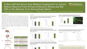 科学海报A New Defined Serum-Free Medium Supplement to Culture Mature Neurons from Primary Embryonic Mouse and Rat CNS
 沪公网安备31010102008431号