Kim S-J et al. (AUG 2010)
Neuroscience letters 479 3 292--6
Omega-3 and omega-6 fatty acids suppress ER- and oxidative stress in cultured neurons and neuronal progenitor cells from mice lacking PPT1.
Reactive oxygen species (ROS) damage brain lipids,carbohydrates,proteins,as well as DNA and may contribute to neurodegeneration. We previously reported that ER- and oxidative stress cause neuronal apoptosis in infantile neuronal ceroid lipofuscinosis (INCL),a lethal neurodegenerative storage disease,caused by palmitoyl-protein thioesterase-1 (PPT1) deficiency. Polyunsaturated fatty acids (PUFA) are essential components of cell membrane phospholipids in the brain and excessive ROS may cause oxidative damage of PUFA leading to neuronal death. Using cultured neurons and neuroprogenitor cells from mice lacking Ppt1,which mimic INCL,we demonstrate that Ppt1-deficient neurons and neuroprogenitor cells contain high levels of ROS,which may cause peroxidation of PUFA and render them incapable of providing protection against oxidative stress. We tested whether treatment of these cells with omega-3 or omega-6 PUFA protects the neurons and neuroprogenitor cells from oxidative stress and suppress apoptosis. We report here that both omega-3 and omega-6 fatty acids protect the Ppt1-deficient cells from ER- as well as oxidative stress and suppress apoptosis. Our results suggest that PUFA supplementation may have neuroprotective effects in INCL.
View Publication
SFN 2016,Advances in Drug Discovery 2017,iForum 2017
产品号#:
05790
05792
05793
05794
05795
R1061
R1034
R1088
R1032
产品名:
BrainPhys™神经元培养基
BrainPhys™神经元培养基和SM1试剂盒
BrainPhys™ 神经元培养基N2-A和SM1试剂盒
BrainPhys™原代神经元试剂盒
BrainPhys™ hPSC 神经元试剂盒
Jin HK et al. (MAY 2002)
The Journal of clinical investigation 109 9 1183--91
Intracerebral transplantation of mesenchymal stem cells into acid sphingomyelinase-deficient mice delays the onset of neurological abnormalities and extends their life span.
Types A and B Niemann-Pick disease (NPD) are lysosomal storage disorders resulting from loss of acid sphingomyelinase (ASM) activity. We have used a knockout mouse model of NPD (ASMKO mice) to evaluate the effects of direct intracerebral transplantation of bone marrow-derived mesenchymal stem cells (MSCs) on the progression of neurological disease in this disorder. MSCs were transduced with a retroviral vector to overexpress ASM and were injected into the hippocampus and cerebellum of 3-week-old ASMKO pups. Transplanted cells migrated away from the injection sites and survived at least 6 months after transplantation. Seven of 8 treated mice,but none of the untreated controls,survived for textgreater or = 7 months after transplant. Survival times were greater in sex-matched than in sex-mismatched transplants. Transplantation significantly delayed the Purkinje cell loss that is characteristic of NPD,although the protective effect declined with distance from the injection site. Overall ASM activity in brain homogenates was low,but surviving Purkinje cells contained the retrovirally expressed human enzyme,and transplanted animals showed a reduction in cerebral sphingomyelin. These results reveal the potential of treating neurodegenerative lysosomal storage disorders by intracerebral transplantation of bone marrow-derived MSCs.
View Publication
产品类型:
产品号#:
05350
产品名:
Zhou Q et al. (FEB 2016)
Molecular biology of the cell 27 4 627--39
Inhibition of the histone demethylase Kdm5b promotes neurogenesis and derepresses Reln (reelin) in neural stem cells from the adult subventricular zone of mice.
The role of epigenetic regulators in the control of adult neurogenesis is largely undefined. We show that the histone demethylase enzyme Kdm5b (Jarid1b) negatively regulates neurogenesis from adult subventricular zone (SVZ) neural stem cells (NSCs) in culture. shRNA-mediated depletion of Kdm5b in proliferating adult NSCs decreased proliferation rates and reduced neurosphere formation in culture. When transferred to differentiation culture conditions,Kdm5b-depleted adult NSCs migrated from neurospheres with increased velocity. Whole-genome expression screening revealed widespread transcriptional changes with Kdm5b depletion,notably the up-regulation of reelin (Reln),the inhibition of steroid biosynthetic pathway component genes and the activation of genes with intracellular transport functions in cultured adult NSCs. Kdm5b depletion increased extracellular reelin concentration in the culture medium and increased phosphorylation of the downstream reelin signaling target Disabled-1 (Dab1). Sequestration of extracellular reelin with CR-50 reelin-blocking antibodies suppressed the increase in migratory velocity of Kdm5b-depleted adult NSCs. Chromatin immunoprecipitation revealed that Kdm5b is present at the proximal promoter of Reln,and H3K4me3 methylation was increased at this locus with Kdm5b depletion in differentiating adult NSCs. Combined the data suggest Kdm5b negatively regulates neurogenesis and represses Reln in neural stem cells from the adult SVZ.
View Publication
Belzile J-P et al. (APR 2014)
Journal of virology 88 8 4021--4039
Human cytomegalovirus infection of human embryonic stem cell-derived primitive neural stem cells is restricted at several steps but leads to the persistence of viral DNA.
UNLABELLED Congenital human cytomegalovirus (HCMV) infection is a major cause of central nervous system structural anomalies and sensory impairments. It is likely that the stage of fetal development,as well as the state of differentiation of susceptible cells at the time of infection,affects the severity of the disease. We used human embryonic stem (ES) cell-derived primitive prerosette neural stem cells (pNSCs) and neural progenitor cells (NPCs) maintained in chemically defined conditions to study HCMV replication in cells at the early stages of neural development. In contrast to what was observed previously using fetus-derived NPCs,infection of ES cell-derived pNSCs with HCMV was nonprogressive. At a low multiplicity of infection,we observed only a small percentage of cells expressing immediate-early genes (IE) and early genes. IE expression was found to be restricted to cells negative for the anterior marker FORSE-1,and treatment of pNSCs with retinoic acid restored IE expression. Differentiation of pNSCs into NPCs restored IE expression but not the transactivation of early genes. Virions produced in NPCs and pNSCs were exclusively cell associated and were mostly non-neural tropic. Finally,we found that viral genomes could persist in pNSC cultures for up to a month after infection despite the absence of detectable IE expression by immunofluorescence,and infectious virus could be produced upon differentiation of pNSCs to neurons. In conclusion,our results highlight the complex array of hurdles that HCMV must overcome in order to infect primitive neural stem cells and suggest that these cells might act as a reservoir for the virus. IMPORTANCE Human cytomegalovirus (HCMV) is a betaherpesvirus that is highly prevalent in the population. HCMV infection is usually asymptomatic but can lead to severe consequences in immunosuppressed individuals. HCMV is also the most important infectious cause of congenital developmental birth defects. Manifestations of fetal HCMV disease range from deafness and learning disabilities to more severe symptoms such as microcephaly. In this study,we have used embryonic stem cells to generate primitive neural stem cells and have used these to model HCMV infection of the fetal central nervous system (CNS) in vitro. Our results reveal that these cells,which are similar to those present in the developing neural tube,do not support viral replication but instead likely constitute a viral reservoir. Future work will define the effect of viral persistence on cellular functions as well as the exogenous signals leading to the reactivation of viral replication in the CNS.
View Publication
产品类型:
产品号#:
05850
05857
05870
05875
85850
85857
85870
85875
产品名:
mTeSR™1
mTeSR™1
Abeysinghe HCS et al. (SEP 2015)
Stem cell research & therapy 6 1 186
Pre-differentiation of human neural stem cells into GABAergic neurons prior to transplant results in greater repopulation of the damaged brain and accelerates functional recovery after transient ischemic stroke.
INTRODUCTION Despite attempts to prevent brain injury during the hyperacute phase of stroke,most sufferers end up with significant neuronal loss and functional deficits. The use of cell-based therapies to recover the injured brain offers new hope. In the current study,we employed human neural stem cells (hNSCs) isolated from subventricular zone (SVZ),and directed their differentiation into GABAergic neurons followed by transplantation to ischemic brain. METHODS Pre-differentiated GABAergic neurons,undifferentiated SVZ-hNSCs or media alone were stereotaxically transplanted into the rat brain (n=7/group) 7 days after endothelin-1 induced stroke. Neurological outcome was assessed by neurological deficit scores and the cylinder test. Transplanted cell survival,cellular phenotype and maturation were assessed using immunohistochemistry and confocal microscopy. RESULTS Behavioral assessments revealed accelerated improvements in motor function 7 days post-transplant in rats treated with pre-differentiated GABAergic cells in comparison to media alone and undifferentiated hNSC treated groups. Histopathology 28 days-post transplant indicated that pre-differentiated cells maintained their GABAergic neuronal phenotype,showed evidence of synaptogenesis and up-regulated expression of both GABA and calcium signaling proteins associated with neurotransmission. Rats treated with pre-differentiated cells also showed increased neurogenic activity within the SVZ at 28 days,suggesting an additional trophic role of these GABAergic cells. In contrast,undifferentiated SVZ-hNSCs predominantly differentiated into GFAP-positive astrocytes and appeared to be incorporated into the glial scar. CONCLUSION Our study is the first to show enhanced exogenous repopulation of a neuronal phenotype after stroke using techniques aimed at GABAergic cell induction prior to delivery that resulted in accelerated and improved functional recovery.
View Publication
产品类型:
产品号#:
05750
05751
产品名:
NeuroCult™ NS-A 基础培养基(人)
NeuroCult™ NS-A 扩增试剂盒(人)
Akizu N et al. (MAY 2015)
Nature genetics 47 5 528--34
Biallelic mutations in SNX14 cause a syndromic form of cerebellar atrophy and lysosome-autophagosome dysfunction.
Pediatric-onset ataxias often present clinically as developmental delay and intellectual disability,with prominent cerebellar atrophy as a key neuroradiographic finding. Here we describe a new clinically distinguishable recessive syndrome in 12 families with cerebellar atrophy together with ataxia,coarsened facial features and intellectual disability,due to truncating mutations in the sorting nexin gene SNX14,encoding a ubiquitously expressed modular PX domain-containing sorting factor. We found SNX14 localized to lysosomes and associated with phosphatidylinositol (3,5)-bisphosphate,a key component of late endosomes/lysosomes. Patient-derived cells showed engorged lysosomes and a slower autophagosome clearance rate upon autophagy induction by starvation. Zebrafish morphants for snx14 showed dramatic loss of cerebellar parenchyma,accumulation of autophagosomes and activation of apoptosis. Our results characterize a unique ataxia syndrome due to biallelic SNX14 mutations leading to lysosome-autophagosome dysfunction.
View Publication
产品类型:
产品号#:
05850
05857
05870
05875
85850
85857
85870
85875
产品名:
mTeSR™1
mTeSR™1
Wang P et al. (DEC 2015)
Molecular autism 6 1 55
CRISPR/Cas9-mediated heterozygous knockout of the autism gene CHD8 and characterization of its transcriptional networks in neurodevelopment.
BACKGROUND Disruptive mutation in the CHD8 gene is one of the top genetic risk factors in autism spectrum disorders (ASDs). Previous analyses of genome-wide CHD8 occupancy and reduced expression of CHD8 by shRNA knockdown in committed neural cells showed that CHD8 regulates multiple cell processes critical for neural functions,and its targets are enriched with ASD-associated genes. METHODS To further understand the molecular links between CHD8 functions and ASD,we have applied the CRISPR/Cas9 technology to knockout one copy of CHD8 in induced pluripotent stem cells (iPSCs) to better mimic the loss-of-function status that would exist in the developing human embryo prior to neuronal differentiation. We then carried out transcriptomic and bioinformatic analyses of neural progenitors and neurons derived from the CHD8 mutant iPSCs. RESULTS Transcriptome profiling revealed that CHD8 hemizygosity (CHD8 (+/-)) affected the expression of several thousands of genes in neural progenitors and early differentiating neurons. The differentially expressed genes were enriched for functions of neural development,$$-catenin/Wnt signaling,extracellular matrix,and skeletal system development. They also exhibited significant overlap with genes previously associated with autism and schizophrenia,as well as the downstream transcriptional targets of multiple genes implicated in autism. Providing important insight into how CHD8 mutations might give rise to macrocephaly,we found that seven of the twelve genes associated with human brain volume or head size by genome-wide association studies (e.g.,HGMA2) were dysregulated in CHD8 (+/-) neural progenitors or neurons. CONCLUSIONS We have established a renewable source of CHD8 (+/-) iPSC lines that would be valuable for investigating the molecular and cellular functions of CHD8. Transcriptomic profiling showed that CHD8 regulates multiple genes implicated in ASD pathogenesis and genes associated with brain volume.
View Publication
产品类型:
产品号#:
05850
05857
05870
05875
85850
85857
85870
85875
产品名:
mTeSR™1
mTeSR™1
Dafinca R et al. (APR 2016)
Stem cells (Dayton,Ohio) 34 8 2016
C9orf72 Hexanucleotide Expansions are Associated with Altered ER Calcium Homeostasis and Stress Granule Formation in iPSC-Derived Neurons from Patients with Amyotrophic Lateral Sclerosis and Frontotemporal Dementia.
An expanded hexanucleotide repeat in a noncoding region of the C9orf72 gene is a major cause of amyotrophic lateral sclerosis (ALS),accounting for up to 40% of familial cases and 7% of sporadic ALS in European populations. We have generated induced pluripotent stem cells (iPSCs) from fibroblasts of patients carrying C9orf72 hexanucleotide expansions,differentiated these to functional motor and cortical neurons and performed an extensive phenotypic characterization. In C9orf72 iPSC-derived motor neurons,decreased cell survival is correlated with dysfunction in Ca(2+) homeostasis,reduced levels of the anti-apoptotic protein Bcl-2,increased endoplasmic reticulum (ER) stress and reduced mitochondrial membrane potential. Furthermore,C9orf72 motor neurons,and also cortical neurons,show evidence of abnormal protein aggregation and stress granule formation. This study is an extensive characterization of iPSC-derived motor neurons as cellular models of ALS carrying C9orf72 hexanucleotide repeats,which describes a novel pathogenic link between C9orf72 mutations,dysregulation of calcium signalling and altered proteostasis and provides a potential pharmacological target for the treatment of ALS and the related neurodegenerative disease frontotemporal dementia (FTD). This article is protected by copyright. All rights reserved.
View Publication
产品类型:
产品号#:
05850
05857
05870
05875
85850
85857
85870
85875
产品名:
mTeSR™1
mTeSR™1
Bai R-Y et al. (SEP 2011)
Neuro-oncology 13 9 974--82
Antiparasitic mebendazole shows survival benefit in 2 preclinical models of glioblastoma multiforme.
Glioblastoma multiforme (GBM) is the most common and aggressive brain cancer,and despite treatment advances,patient prognosis remains poor. During routine animal studies,we serendipitously observed that fenbendazole,a benzimidazole antihelminthic used to treat pinworm infection,inhibited brain tumor engraftment. Subsequent in vitro and in vivo experiments with benzimidazoles identified mebendazole as the more promising drug for GBM therapy. In GBM cell lines,mebendazole displayed cytotoxicity,with half-maximal inhibitory concentrations ranging from 0.1 to 0.3 µM. Mebendazole disrupted microtubule formation in GBM cells,and in vitro activity was correlated with reduced tubulin polymerization. Subsequently,we showed that mebendazole significantly extended mean survival up to 63% in syngeneic and xenograft orthotopic mouse glioma models. Mebendazole has been approved by the US Food and Drug Administration for parasitic infections,has a long track-record of safe human use,and was effective in our animal models with doses documented as safe in humans. Our findings indicate that mebendazole is a possible novel anti-brain tumor therapeutic that could be further tested in clinical trials.
View Publication
产品类型:
产品号#:
05751
07980
产品名:
NeuroCult™ NS-A 扩增试剂盒(人)
肝素溶液
Bilican B et al. (APR 2012)
Proceedings of the National Academy of Sciences of the United States of America 109 15 5803--8
Mutant induced pluripotent stem cell lines recapitulate aspects of TDP-43 proteinopathies and reveal cell-specific vulnerability.
Transactive response DNA-binding (TDP-43) protein is the dominant disease protein in amyotrophic lateral sclerosis (ALS) and a subgroup of frontotemporal lobar degeneration (FTLD-TDP). Identification of mutations in the gene encoding TDP-43 (TARDBP) in familial ALS confirms a mechanistic link between misaccumulation of TDP-43 and neurodegeneration and provides an opportunity to study TDP-43 proteinopathies in human neurons generated from patient fibroblasts by using induced pluripotent stem cells (iPSCs). Here,we report the generation of iPSCs that carry the TDP-43 M337V mutation and their differentiation into neurons and functional motor neurons. Mutant neurons had elevated levels of soluble and detergent-resistant TDP-43 protein,decreased survival in longitudinal studies,and increased vulnerability to antagonism of the PI3K pathway. We conclude that expression of physiological levels of TDP-43 in human neurons is sufficient to reveal a mutation-specific cell-autonomous phenotype and strongly supports this approach for the study of disease mechanisms and for drug screening.
View Publication

 EasySep™小鼠TIL(CD45)正选试剂盒


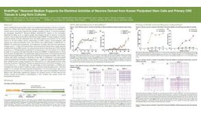 科学海报BrainPhys™ Neuronal Medium Supports the Electrical Activities of Neurons Derived from Human Pluripotent Stem Cells and Primary CNS Tissues in Long-Term Cultures
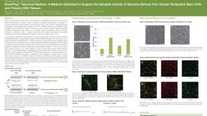 科学海报BrainPhys™ Neuronal Medium: A Medium Optimized to Support the Synaptic Activity of Neurons Derived from Human Pluripotent Stem Cells and Primary CNS Tissues
 沪公网安备31010102008431号