Meco D et al. (AUG 2014)
Neuro-Oncology 16 8 1067--1077
Ependymoma stem cells are highly sensitive to temozolomide in vitro and in orthotopic models
BACKGROUND Ependymoma management remains challenging because of the inherent chemoresistance of this tumor. To determine whether ependymoma stem cells (SCs) might contribute to therapy resistance,we investigated the sensitivity of ependymoma SCs to temozolomide and etoposide. METHODS The efficacies of the two DNA damaging agents were explored in two ependymoma SC lines in vitro and in vivo models. RESULTS Ependymoma SC lines were highly sensitive to temozolomide and etoposide in vitro,but only temozolomide impaired tumor-initiation properties. Consistently,temozolomide but not etoposide showed significant antitumoral activity on ependymoma SC-driven subcutaneous and orthotopic xenografts by reducing the mitotic fraction. In vitro temozolomide at the EC50 (10 µM) induced accumulation of cells in the G2/M phase that was unexpectedly accompanied by downregulation of p27 and p21 without modulation of full-length p53 (FLp53). Differentiation-committed ependymoma SCs acquired resistance to temozolomide. Inhibition of proliferation was partly due to apoptosis,that occurred earlier in differentiated cells as compared to neurospheres. The activation of apoptosis correlated with an increase in p53β/γ isoforms without modulation of FLp53 under both serum-free and differentiation-promoting media. Incubation of cells in both conditions with temozolomide resulted in increased glioneuronal differentiation exhibiting elevated glial fibrillary acidic protein,galactosylceramidase,and βIII-tubulin expression compared to untreated controls. O(6)-methylguanine DNA methyltransferase (MGMT) transcript levels were very low in SCs,and were increased by treatment and,epigenetically,by differentiation through MGMT promoter unmethylation. CONCLUSION Ependymoma growth might be impaired by temozolomide through preferential depletion of a less differentiated,more tumorigenic,MGMT-negative cell population with stem-like properties.
View Publication
产品类型:
产品号#:
05750
05751
产品名:
NeuroCult™ NS-A 基础培养基(人)
NeuroCult™ NS-A 扩增试剂盒(人)
Daynac M et al. (JUL 2013)
Stem Cell Research 11 1 516--528
Quiescent neural stem cells exit dormancy upon alteration of GABAAR signaling following radiation damage
Quiescent neural stem cells (NSCs) are considered the reservoir for adult neurogenesis,generating new neurons throughout life. Until now,their isolation has not been reported,which has hampered studies of their regulatory mechanisms. We sorted by FACS quiescent NSCs and their progeny from the subventricular zone (SVZ) of adult mice according to the expression of the NSC marker LeX/CD15,the EGF receptor (EGFR) and the CD24 in combination with the vital DNA marker Hoechst 33342. Characterization of sorted cells showed that the LeX(bright)/EGFR-negative population was enriched in quiescent cells having an NSC phenotype. In contrast to proliferating NSCs and progenitors,the LeX(bright)/EGFR-negative cells,i.e. quiescent NSCs,resisted to a moderate dose of gamma-radiation (4Gy),entered the cell cycle two days after irradiation prior to EGFR acquisition and ultimately repopulated the SVZ. We further show that the GABAAR signaling regulates their cell cycle entry by using specific GABAAR agonists/antagonists and that the radiation-induced depletion of neuroblasts,the major GABA source,provoked their proliferation in the irradiated SVZ. Our study demonstrates that quiescent NSCs are specifically enriched in the LeX(bright)/EGFR-negative population,and identifies the GABAAR signaling as a regulator of the SVZ niche size by modulating the quiescence of NSCs.
View Publication
产品类型:
产品号#:
05700
05701
05702
产品名:
NeuroCult™ 基础培养基(小鼠和大鼠)
NeuroCult™ 扩增添加物(小鼠和大鼠)
NeuroCult™扩增试剂盒(小鼠和大鼠)
Bagci-Onder T et al. (JUN 2015)
Brain 138 6 1710--1721
Targeting breast to brain metastatic tumours with death receptor ligand expressing therapeutic stem cells
Characterizing clinically relevant brain metastasis models and assessing the therapeutic efficacy in such models are fundamental for the development of novel therapies for metastatic brain cancers. In this study,we have developed an in vivo imageable breast-to-brain metastasis mouse model. Using real time in vivo imaging and subsequent composite fluorescence imaging,we show a widespread distribution of micro- and macro-metastasis in different stages of metastatic progression. We also show extravasation of tumour cells and the close association of tumour cells with blood vessels in the brain thus mimicking the multi-foci metastases observed in the clinics. Next,we explored the ability of engineered adult stem cells to track metastatic deposits in this model and show that engineered stem cells either implanted or injected via circulation efficiently home to metastatic tumour deposits in the brain. Based on the recent findings that metastatic tumour cells adopt unique mechanisms of evading apoptosis to successfully colonize in the brain,we reasoned that TNF receptor superfamily member 10A/10B apoptosis-inducing ligand (TRAIL) based pro-apoptotic therapies that induce death receptor signalling within the metastatic tumour cells might be a favourable therapeutic approach. We engineered stem cells to express a tumour selective,potent and secretable variant of a TRAIL,S-TRAIL,and show that these cells significantly suppressed metastatic tumour growth and prolonged the survival of mice bearing metastatic breast tumours. Furthermore,the incorporation of pro-drug converting enzyme,herpes simplex virus thymidine kinase,into therapeutic S-TRAIL secreting stem cells allowed their eradication post-tumour treatment. These studies are the first of their kind that provide insight into targeting brain metastasis with stem-cell mediated delivery of pro-apoptotic ligands and have important clinical implications.
View Publication
产品类型:
产品号#:
05700
05701
产品名:
NeuroCult™ 基础培养基(小鼠和大鼠)
NeuroCult™ 扩增添加物(小鼠和大鼠)
Yang Z-J et al. (AUG 2008)
Cancer cell 14 2 135--45
Medulloblastoma can be initiated by deletion of Patched in lineage-restricted progenitors or stem cells.
Medulloblastoma is the most common malignant brain tumor in children,but the cells from which it arises remain unclear. Here we examine the origin of medulloblastoma resulting from mutations in the Sonic hedgehog (Shh) pathway. We show that activation of Shh signaling in neuronal progenitors causes medulloblastoma by 3 months of age. Shh pathway activation in stem cells promotes stem cell proliferation but only causes tumors after commitment to-and expansion of-the neuronal lineage. Notably,tumors initiated in stem cells develop more rapidly than those initiated in progenitors,with all animals succumbing by 3-4 weeks. These studies suggest that medulloblastoma can be initiated in progenitors or stem cells but that Shh-induced tumorigenesis is associated with neuronal lineage commitment.
View Publication
产品类型:
产品号#:
05700
05701
05702
05703
05704
产品名:
NeuroCult™ 基础培养基(小鼠和大鼠)
NeuroCult™ 扩增添加物(小鼠和大鼠)
NeuroCult™扩增试剂盒(小鼠和大鼠)
NeuroCult™ 分化添加物(小鼠和大鼠)
NeuroCult™ 分化试剂盒(小鼠和大鼠)
Ishii Y et al. (MAR 2008)
Molecular and cellular neurosciences 37 3 507--18
Characterization of neuroprogenitor cells expressing the PDGF beta-receptor within the subventricular zone of postnatal mice.
We report a considerable number of cells in the ventricular and the subventricular zones (SVZ) of newborn mice to stain positive for the PDGF beta-receptor (PDGFRB). Many of them also stained for nestin and/or GFAP but less frequently for the neuroblast marker doublecortin and for the mitotic marker Ki-67. The SVZ of mice with nestin-Cre conditional deletion of PDGFRB expressed the receptor only on blood vessels and was devoid of any morphological abnormality. PDGFRB(-/-) neurospheres showed a higher rate of apoptosis without any significant decrease in proliferation. They demonstrated reduced capacities of migration and neuronal differentiation in response to not only PDGF-BB but also bFGF. Furthermore,the PDGFR kinase inhibitor STI571 blocked the effects of bFGF in control neurosphere cultures. bFGF increased the activity of the PDGFRB promoter as well as the expression and phosphorylation of PDGFRB. These results suggest the presence of the signaling convergence between PDGF and FGF. PDGFRB is needed for survival,and the effects of bFGF in migration and neural differentiation of the cells may be potentiated by induction of PDGFRB.
View Publication
Mitochondrially targeted vitamin E succinate efficiently kills breast tumour-initiating cells in a complex II-dependent manner.
BACKGROUND Accumulating evidence suggests that breast cancer involves tumour-initiating cells (TICs),which play a role in initiation,metastasis,therapeutic resistance and relapse of the disease. Emerging drugs that target TICs are becoming a focus of contemporary research. Mitocans,a group of compounds that induce apoptosis of cancer cells by destabilising their mitochondria,are showing their potential in killing TICs. In this project,we investigated mitochondrially targeted vitamin E succinate (MitoVES),a recently developed mitocan,for its in vitro and in vivo efficacy against TICs. METHODS The mammosphere model of breast TICs was established by culturing murine NeuTL and human MCF7 cells as spheres. This model was verified by stem cell marker expression,tumour initiation capacity and chemotherapeutic resistance. Cell susceptibility to MitoVES was assessed and the cell death pathway investigated. In vivo efficacy was studied by grafting NeuTL TICs to form syngeneic tumours. RESULTS Mammospheres derived from NeuTL and MCF7 breast cancer cells were enriched in the level of stemness,and the sphere cells featured altered mitochondrial function. Sphere cultures were resistant to several established anti-cancer agents while they were susceptible to MitoVES. Killing of mammospheres was suppressed when the mitochondrial complex II,the molecular target of MitoVES,was knocked down. Importantly,MitoVES inhibited progression of syngeneic HER2(high) tumours derived from breast TICs by inducing apoptosis in tumour cells. CONCLUSIONS These results demonstrate that using mammospheres,a plausible model for studying TICs,drugs that target mitochondria efficiently kill breast tumour-initiating cells.
View Publication
产品类型:
产品号#:
05701
产品名:
NeuroCult™ 扩增添加物(小鼠和大鼠)
A. Odawara et al. (JUL 2018)
Scientific reports 8 1 10416
Toxicological evaluation of convulsant and anticonvulsant drugs in human induced pluripotent stem cell-derived cortical neuronal networks using an MEA system.
Functional evaluation assays using human induced pluripotent stem cell (hiPSC)-derived neurons can predict the convulsion toxicity of new drugs and the neurological effects of antiepileptic drugs. However,differences in responsiveness depending on convulsant type and antiepileptic drugs,and an evaluation index capable of comparing in vitro responses with in vivo responses are not well known. We observed the difference in synchronized burst patterns in the epileptiform activities induced by pentylentetrazole (PTZ) and 4-aminopryridine (4-AP) with different action mechanisms using multi-electrode arrays (MEAs); we also observed that 100 µM of the antiepileptic drug phenytoin suppressed epileptiform activities induced by PTZ,but increased those induced by 4-AP. To compare in vitro results with in vivo convulsive responses,frequency analysis of below 250 Hz,excluding the spike component,was performed. The in vivo convulsive firing enhancement of the high gamma$ wave and beta$ wave component were observed remarkably in in vitro hiPSC-derived neurons with astrocytes in co-culture. MEA measurement of hiPSC-derived neurons in co-culture with astrocytes and our analysis methods,including frequency analysis,appear effective for predicting convulsion toxicity,side effects,and their mechanism of action as well as the comparison of convulsions induced in vivo.
View Publication
Maire C et al. (JAN 2014)
STEM CELLS 32 1 313--326
Pten Loss in Olig2 Expressing Neural Progenitor Cells and Oligodendrocytes Leads to Interneuron Dysplasia and Leukodystrophy
Therapeutic modulation of phosphatidylinositol 3-kinase (PI3K)/PTEN signaling is currently being explored for multiple neurological indications including brain tumors and seizure disorders associated with cortical malformations. The effects of PI3K/PTEN signaling are highly cell context dependent but the function of this pathway in specific subsets of neural stem/progenitor cells generating oligodendroglial lineage cells has not been fully studied. To address this,we created Olig2-cre:Pten(fl/fl) mice that showed a unique pattern of Pten loss and PI3K activation in Olig2-lineage cells. Olig2-cre:Pten(fl/fl) animals progressively developed central nervous system white matter hypermyelination by 3 weeks of age leading to later onset leukodystrophy,chronic neurodegeneration,and death by 9 months. In contrast,during immediate postnatal development,oligodendroglia were unaffected but abnormal and accelerated differentiation of lateral subventricular zone stem cells produced calretinin-positive interneuron dysplasia. Neural stem cells isolated from Olig2-cre:Pten(fl/fl) mice also exhibited accelerated differentiation and proliferation into calretinin-positive interneurons and oligodendrocytes indicating such effects are cell autonomous. Opposition of the pathway by treatment of human primary neural progenitor cells (NPCs) with the PI3K inhibitor,NVP-BKM120,blocked in vitro differentiation of neurons and oligodendroglia indicating PI3K/PTEN effects on NPCs can be bidirectional. In summary,our results suggest Pten is a developmental rheostat regulating interneuron and oligodendroglial differentiation and support testing of PI3K modulating drugs as treatment for developmental and myelination disorders. However,such agents may need to be administered at ages that minimize potential effects on early stem/progenitor cell development.
View Publication
产品类型:
产品号#:
05700
05701
05702
05750
05751
产品名:
NeuroCult™ 基础培养基(小鼠和大鼠)
NeuroCult™ 扩增添加物(小鼠和大鼠)
NeuroCult™扩增试剂盒(小鼠和大鼠)
NeuroCult™ NS-A 基础培养基(人)
NeuroCult™ NS-A 扩增试剂盒(人)
Maricque BB et al. (FEB 2017)
Nucleic acids research 45 4 e16
A genome-integrated massively parallel reporter assay reveals DNA sequence determinants of cis-regulatory activity in neural cells.
Recent large-scale genomics efforts to characterize the cis-regulatory sequences that orchestrate genome-wide expression patterns have produced impressive catalogues of putative regulatory elements. Most of these sequences have not been functionally tested,and our limited understanding of the non-coding genome prevents us from predicting which sequences are bona fide cis-regulatory elements. Recently,massively parallel reporter assays (MPRAs) have been deployed to measure the activity of putative cis-regulatory sequences in several biological contexts,each with specific advantages and distinct limitations. We developed LV-MPRA,a novel lentiviral-based,massively parallel reporter gene assay,to study the function of genome-integrated regulatory elements in any mammalian cell type; thus,making it possible to apply MPRAs in more biologically relevant contexts. We measured the activity of 2,600 sequences in U87 glioblastoma cells and human neural progenitor cells (hNPCs) and explored how regulatory activity is encoded in DNA sequence. We demonstrate that LV-MPRA can be applied to estimate the effects of local DNA sequence and regional chromatin on regulatory activity. Our data reveal that primary DNA sequence features,such as GC content and dinucleotide composition,accurately distinguish sequences with high activity from sequences with low activity in a full chromosomal context,and may also function in combination with different transcription factor binding sites to determine cell type specificity. We conclude that LV-MPRA will be an important tool for identifying cis-regulatory elements and stimulating new understanding about how the non-coding genome encodes information.
View Publication
产品类型:
产品号#:
05833
05710
05835
05839
产品名:
STEMdiff™神经前体细胞培养基
STEMdiff™ 神经诱导培养基
STEMdiff™ 神经诱导培养基
Niu H et al. (MAR 2017)
Neuroscience Letters 642 71--76
Recombinant insulin-like growth factor binding protein-4 inhibits proliferation and promotes differentiation of neural progenitor cells
Insulin-like growth factor (IGF) is involved in regulating many processes during neural development,and IGF binding protein-4 (IGFBP4) functions as a modulator of IGF actions or in an IGF-independent manner (e.g.,via inhibiting Wnt/β-catenin signaling). In the present study,neural progenitor cells (NPCs) were isolated from the forebrain of newborn mice to investigate effects of IGFBP4 on the proliferation and differentiation of NPCs. The proliferation of NPCs was evaluated using Cell Counting Kit-8 (CCK-8) after treatment with or without IGFBP4 as well as blockers of IGF-IR and β-catenin. Phosphorylation levels of Akt,Erk1,2 and p38 were analyzed by Western blotting. The differentiation of NPCs was evaluated using immunofluorescence and Western blotting. It was shown that exogenous IGFBP4 significantly inhibited the proliferation of NPCs and it did not induce a more pronounced inhibition of cell proliferation after blockade of IGF-IR but it did after antagonism of β-catenin. Akt phosphorylation was significantly decreased and phosphorylation levels of Erk1,2 and p38 were not significantly changed in IGFBP4-treated NPCs. Excessive IGFBP4 significantly promoted NPCs to differentiate into astrocytes and neurons. These data suggested that exogenous IGFBP4 inhibits proliferation and promotes differentiation of neural progenitor cells mainly through IGF-IR signaling pathway.
View Publication

 EasySep™小鼠TIL(CD45)正选试剂盒


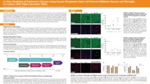 科学海报In Vitro Modeling of Parkinson’s Disease Using Human Pluripotent Stem Cell-Derived Midbrain Neuron and Microglia Co-Culture with Alpha-Synuclein Fibrils
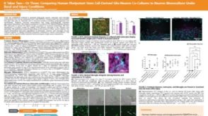 科学海报It Takes Two—Or Three: Comparing Human Pluripotent Stem Cell Derived Glia-Neuron Co-Cultures to Neuron Monoculture Under Basal and Injury Conditions
 沪公网安备31010102008431号