Galavotti S et al. (FEB 2013)
Oncogene 32 6 699--712
The autophagy-associated factors DRAM1 and p62 regulate cell migration and invasion in glioblastoma stem cells.
The aggressiveness of glioblastoma multiforme (GBM) is defined by local invasion and resistance to therapy. Within established GBM,a subpopulation of tumor-initiating cells with stem-like properties (GBM stem cells,GSCs) is believed to underlie resistance to therapy. The metabolic pathway autophagy has been implicated in the regulation of survival in GBM. However,the status of autophagy in GBM and its role in the cancer stem cell fraction is currently unclear. We found that a number of autophagy regulators are highly expressed in GBM tumors carrying a mesenchymal signature,which defines aggressiveness and invasion,and are associated with components of the MAPK pathway. This autophagy signature included the autophagy-associated genes DRAM1 and SQSTM1,which encode a key regulator of selective autophagy,p62. High levels of DRAM1 were associated with shorter overall survival in GBM patients. In GSCs,DRAM1 and SQSTM1 expression correlated with activation of MAPK and expression of the mesenchymal marker c-MET. DRAM1 knockdown decreased p62 localization to autophagosomes and its autophagy-mediated degradation,thus suggesting a role for DRAM1 in p62-mediated autophagy. In contrast,autophagy induced by starvation or inhibition of mTOR/PI-3K was not affected by either DRAM1 or p62 downregulation. Functionally,DRAM1 and p62 regulate cell motility and invasion in GSCs. This was associated with alterations of energy metabolism,in particular reduced ATP and lactate levels. Taken together,these findings shed new light on the role of autophagy in GBM and reveal a novel function of the autophagy regulators DRAM1 and p62 in control of migration/invasion in cancer stem cells.
View Publication
Pei Y et al. (MAY 2012)
Development (Cambridge,England) 139 10 1724--33
WNT signaling increases proliferation and impairs differentiation of stem cells in the developing cerebellum.
The WNT pathway plays multiple roles in neural development and is crucial for establishment of the embryonic cerebellum. In addition,WNT pathway mutations are associated with medulloblastoma,the most common malignant brain tumor in children. However,the cell types within the cerebellum that are responsive to WNT signaling remain unknown. Here we investigate the effects of canonical WNT signaling on two important classes of progenitors in the developing cerebellum: multipotent neural stem cells (NSCs) and granule neuron precursors (GNPs). We show that WNT pathway activation in vitro promotes proliferation of NSCs but not GNPs. Moreover,mice that express activated β-catenin in the cerebellar ventricular zone exhibit increased proliferation of NSCs in that region,whereas expression of the same protein in GNPs impairs proliferation. Although β-catenin-expressing NSCs proliferate they do not undergo prolonged expansion or neoplastic growth; rather,WNT signaling markedly interferes with their capacity for self-renewal and differentiation. At a molecular level,mutant NSCs exhibit increased expression of c-Myc,which might account for their transient proliferation,but also express high levels of bone morphogenetic proteins and the cyclin-dependent kinase inhibitor p21,which might contribute to their altered self-renewal and differentiation. These studies suggest that the WNT pathway is a potent regulator of cerebellar stem cell growth and differentiation.
View Publication
产品类型:
产品号#:
05700
05701
05702
产品名:
NeuroCult™ 基础培养基(小鼠和大鼠)
NeuroCult™ 扩增添加物(小鼠和大鼠)
NeuroCult™扩增试剂盒(小鼠和大鼠)
Katikireddy KR et al. (OCT 2016)
The American Journal of Pathology 186 10 2736--2750
Existence of Neural CrestDerived Progenitor Cells in Normal and Fuchs Endothelial Dystrophy Corneal Endothelium
Human corneal endothelial cells are derived from neural crest and because of postmitotic arrest lack competence to repair cell loss from trauma,aging,and degenerative disorders such as Fuchs endothelial corneal dystrophy (FECD). Herein,we identified a rapidly proliferating subpopulation of cells from the corneal endothelium of adult normal and FECD donors that exhibited features of neural crest-derived progenitor (NCDP) cells by showing absence of senescence with passaging,propensity to form spheres,and increased colony forming efficacy compared with the primary cells. The collective expression of stem cell-related genes SOX2,OCT4,LGR5,TP63 (p63),as well as neural crest marker genes PSIP1 (p75(NTR)),PAX3,SOX9,AP2B1 (AP-2β),and NES,generated a phenotypic footprint of endothelial NCDPs. NCDPs displayed multipotency by differentiating into microtubule-associated protein 2,β-III tubulin,and glial fibrillary acidic protein positive neurons and into p75(NTR)-positive human corneal endothelial cells that exhibited transendothelial resistance of functional endothelium. In conclusion,we found that mitotically incompetent ocular tissue cells contain adult NCDPs that exhibit a profile of transcription factors regulating multipotency and neural crest progenitor characteristics. Identification of normal NCDPs in FECD-affected endothelium holds promise for potential autologous cell therapies.
View Publication
产品类型:
产品号#:
05835
05839
产品名:
STEMdiff™ 神经诱导培养基
STEMdiff™ 神经诱导培养基
Meco D et al. (AUG 2014)
Neuro-Oncology 16 8 1067--1077
Ependymoma stem cells are highly sensitive to temozolomide in vitro and in orthotopic models
BACKGROUND Ependymoma management remains challenging because of the inherent chemoresistance of this tumor. To determine whether ependymoma stem cells (SCs) might contribute to therapy resistance,we investigated the sensitivity of ependymoma SCs to temozolomide and etoposide. METHODS The efficacies of the two DNA damaging agents were explored in two ependymoma SC lines in vitro and in vivo models. RESULTS Ependymoma SC lines were highly sensitive to temozolomide and etoposide in vitro,but only temozolomide impaired tumor-initiation properties. Consistently,temozolomide but not etoposide showed significant antitumoral activity on ependymoma SC-driven subcutaneous and orthotopic xenografts by reducing the mitotic fraction. In vitro temozolomide at the EC50 (10 µM) induced accumulation of cells in the G2/M phase that was unexpectedly accompanied by downregulation of p27 and p21 without modulation of full-length p53 (FLp53). Differentiation-committed ependymoma SCs acquired resistance to temozolomide. Inhibition of proliferation was partly due to apoptosis,that occurred earlier in differentiated cells as compared to neurospheres. The activation of apoptosis correlated with an increase in p53β/γ isoforms without modulation of FLp53 under both serum-free and differentiation-promoting media. Incubation of cells in both conditions with temozolomide resulted in increased glioneuronal differentiation exhibiting elevated glial fibrillary acidic protein,galactosylceramidase,and βIII-tubulin expression compared to untreated controls. O(6)-methylguanine DNA methyltransferase (MGMT) transcript levels were very low in SCs,and were increased by treatment and,epigenetically,by differentiation through MGMT promoter unmethylation. CONCLUSION Ependymoma growth might be impaired by temozolomide through preferential depletion of a less differentiated,more tumorigenic,MGMT-negative cell population with stem-like properties.
View Publication
产品类型:
产品号#:
05750
05751
产品名:
NeuroCult™ NS-A 基础培养基(人)
NeuroCult™ NS-A 扩增试剂盒(人)
Daynac M et al. (JUL 2013)
Stem Cell Research 11 1 516--528
Quiescent neural stem cells exit dormancy upon alteration of GABAAR signaling following radiation damage
Quiescent neural stem cells (NSCs) are considered the reservoir for adult neurogenesis,generating new neurons throughout life. Until now,their isolation has not been reported,which has hampered studies of their regulatory mechanisms. We sorted by FACS quiescent NSCs and their progeny from the subventricular zone (SVZ) of adult mice according to the expression of the NSC marker LeX/CD15,the EGF receptor (EGFR) and the CD24 in combination with the vital DNA marker Hoechst 33342. Characterization of sorted cells showed that the LeX(bright)/EGFR-negative population was enriched in quiescent cells having an NSC phenotype. In contrast to proliferating NSCs and progenitors,the LeX(bright)/EGFR-negative cells,i.e. quiescent NSCs,resisted to a moderate dose of gamma-radiation (4Gy),entered the cell cycle two days after irradiation prior to EGFR acquisition and ultimately repopulated the SVZ. We further show that the GABAAR signaling regulates their cell cycle entry by using specific GABAAR agonists/antagonists and that the radiation-induced depletion of neuroblasts,the major GABA source,provoked their proliferation in the irradiated SVZ. Our study demonstrates that quiescent NSCs are specifically enriched in the LeX(bright)/EGFR-negative population,and identifies the GABAAR signaling as a regulator of the SVZ niche size by modulating the quiescence of NSCs.
View Publication
产品类型:
产品号#:
05700
05701
05702
产品名:
NeuroCult™ 基础培养基(小鼠和大鼠)
NeuroCult™ 扩增添加物(小鼠和大鼠)
NeuroCult™扩增试剂盒(小鼠和大鼠)
Bagci-Onder T et al. (JUN 2015)
Brain 138 6 1710--1721
Targeting breast to brain metastatic tumours with death receptor ligand expressing therapeutic stem cells
Characterizing clinically relevant brain metastasis models and assessing the therapeutic efficacy in such models are fundamental for the development of novel therapies for metastatic brain cancers. In this study,we have developed an in vivo imageable breast-to-brain metastasis mouse model. Using real time in vivo imaging and subsequent composite fluorescence imaging,we show a widespread distribution of micro- and macro-metastasis in different stages of metastatic progression. We also show extravasation of tumour cells and the close association of tumour cells with blood vessels in the brain thus mimicking the multi-foci metastases observed in the clinics. Next,we explored the ability of engineered adult stem cells to track metastatic deposits in this model and show that engineered stem cells either implanted or injected via circulation efficiently home to metastatic tumour deposits in the brain. Based on the recent findings that metastatic tumour cells adopt unique mechanisms of evading apoptosis to successfully colonize in the brain,we reasoned that TNF receptor superfamily member 10A/10B apoptosis-inducing ligand (TRAIL) based pro-apoptotic therapies that induce death receptor signalling within the metastatic tumour cells might be a favourable therapeutic approach. We engineered stem cells to express a tumour selective,potent and secretable variant of a TRAIL,S-TRAIL,and show that these cells significantly suppressed metastatic tumour growth and prolonged the survival of mice bearing metastatic breast tumours. Furthermore,the incorporation of pro-drug converting enzyme,herpes simplex virus thymidine kinase,into therapeutic S-TRAIL secreting stem cells allowed their eradication post-tumour treatment. These studies are the first of their kind that provide insight into targeting brain metastasis with stem-cell mediated delivery of pro-apoptotic ligands and have important clinical implications.
View Publication
产品类型:
产品号#:
05700
05701
产品名:
NeuroCult™ 基础培养基(小鼠和大鼠)
NeuroCult™ 扩增添加物(小鼠和大鼠)
Yang Z-J et al. (AUG 2008)
Cancer cell 14 2 135--45
Medulloblastoma can be initiated by deletion of Patched in lineage-restricted progenitors or stem cells.
Medulloblastoma is the most common malignant brain tumor in children,but the cells from which it arises remain unclear. Here we examine the origin of medulloblastoma resulting from mutations in the Sonic hedgehog (Shh) pathway. We show that activation of Shh signaling in neuronal progenitors causes medulloblastoma by 3 months of age. Shh pathway activation in stem cells promotes stem cell proliferation but only causes tumors after commitment to-and expansion of-the neuronal lineage. Notably,tumors initiated in stem cells develop more rapidly than those initiated in progenitors,with all animals succumbing by 3-4 weeks. These studies suggest that medulloblastoma can be initiated in progenitors or stem cells but that Shh-induced tumorigenesis is associated with neuronal lineage commitment.
View Publication
产品类型:
产品号#:
05700
05701
05702
05703
05704
产品名:
NeuroCult™ 基础培养基(小鼠和大鼠)
NeuroCult™ 扩增添加物(小鼠和大鼠)
NeuroCult™扩增试剂盒(小鼠和大鼠)
NeuroCult™ 分化添加物(小鼠和大鼠)
NeuroCult™ 分化试剂盒(小鼠和大鼠)
Chambers SM et al. (JUL 2012)
Nature biotechnology 30 7 715--20
Combined small-molecule inhibition accelerates developmental timing and converts human pluripotent stem cells into nociceptors.
Considerable progress has been made in identifying signaling pathways that direct the differentiation of human pluripotent stem cells (hPSCs) into specialized cell types,including neurons. However,differentiation of hPSCs with extrinsic factors is a slow,step-wise process,mimicking the protracted timing of human development. Using a small-molecule screen,we identified a combination of five small-molecule pathway inhibitors that yield hPSC-derived neurons at textgreater75% efficiency within 10 d of differentiation. The resulting neurons express canonical markers and functional properties of human nociceptors,including tetrodotoxin (TTX)-resistant,SCN10A-dependent sodium currents and response to nociceptive stimuli such as ATP and capsaicin. Neuronal fate acquisition occurs about threefold faster than during in vivo development,suggesting that use of small-molecule pathway inhibitors could become a general strategy for accelerating developmental timing in vitro. The quick and high-efficiency derivation of nociceptors offers unprecedented access to this medically relevant cell type for studies of human pain.
View Publication
产品类型:
产品号#:
05850
05857
05870
05875
72082
72142
72144
72232
72234
85850
85857
85870
85875
100-1051
产品名:
DAPT
SB431542(水合物)
SB431542(水合物)
mTeSR™1
mTeSR™1
SB431542(水合物)
D. Alvarez-Carbonell et al. (JUL 2018)
Journal of neuroimmune pharmacology : the official journal of the Society on NeuroImmune Pharmacology
The Glucocorticoid Receptor Is a Critical Regulator of HIV Latency in Human Microglial Cells.
We have developed models of HIV latency using microglia derived from adult human patient brain cortex and transformed with the SV40 T large and hTERT antigens. Latent clones infected by HIV reporter viruses display high levels of spontaneous HIV reactivation in culture. BrainPhys,a medium highly representative of the CNS extracellular environment,containing low glucose and 1{\%} FBS,reduced,but did not prevent,HIV reactivation. We hypothesized that spontaneous HIV reactivation in culture was due to the expression of pro-inflammatory genes,such as TNF-alpha$,taking place in the absence of the natural inhibitory signals from astrocytes and neurons. Indeed,expression and secretion of TNF-alpha$ is strongly reduced in HIV-latently infected microglia compared to the subset of cells that have undergone spontaneous HIV reactivation. Whereas inhibitors of NF-kappa$B or of macrophage activation only had a short-term silencing effect,addition of dexamethasone (DEXA),a glucocorticoid receptor (GR) agonist and mediator of anti-inflammation,silenced the HIV provirus in a long-term,and shRNA-mediated knock-down of GR activated HIV. DEXA also decreased secretion of a number of cytokines,including TNF-alpha$. Chromatin immunoprecipitation analysis revealed that DEXA strongly increased GR occupancy at the HIV promoter,and reduced histone 3 acetylated levels. Moreover,TNF-alpha$ expression inhibitors in combination with DEXA induced further HIV silencing and increased the histone 3 lysine 27 tri-methylated epigenetic mark of repression at the HIV promoter region. We conclude that GR is a critical repressor of HIV transcription in microglia,and a novel potential pharmacological target to restrict HIV expression in the CNS.
View Publication
产品类型:
产品号#:
05790
05792
05793
05794
05795
产品名:
BrainPhys™神经元培养基
BrainPhys™神经元培养基和SM1试剂盒
BrainPhys™ 神经元培养基N2-A和SM1试剂盒
BrainPhys™原代神经元试剂盒
BrainPhys™ hPSC 神经元试剂盒
Ishii Y et al. (MAR 2008)
Molecular and cellular neurosciences 37 3 507--18
Characterization of neuroprogenitor cells expressing the PDGF beta-receptor within the subventricular zone of postnatal mice.
We report a considerable number of cells in the ventricular and the subventricular zones (SVZ) of newborn mice to stain positive for the PDGF beta-receptor (PDGFRB). Many of them also stained for nestin and/or GFAP but less frequently for the neuroblast marker doublecortin and for the mitotic marker Ki-67. The SVZ of mice with nestin-Cre conditional deletion of PDGFRB expressed the receptor only on blood vessels and was devoid of any morphological abnormality. PDGFRB(-/-) neurospheres showed a higher rate of apoptosis without any significant decrease in proliferation. They demonstrated reduced capacities of migration and neuronal differentiation in response to not only PDGF-BB but also bFGF. Furthermore,the PDGFR kinase inhibitor STI571 blocked the effects of bFGF in control neurosphere cultures. bFGF increased the activity of the PDGFRB promoter as well as the expression and phosphorylation of PDGFRB. These results suggest the presence of the signaling convergence between PDGF and FGF. PDGFRB is needed for survival,and the effects of bFGF in migration and neural differentiation of the cells may be potentiated by induction of PDGFRB.
View Publication
产品类型:
产品号#:
05700
05701
05702
产品名:
NeuroCult™ 基础培养基(小鼠和大鼠)
NeuroCult™ 扩增添加物(小鼠和大鼠)
NeuroCult™扩增试剂盒(小鼠和大鼠)
B. S. Souza et al. (dec 2016)
Scientific Reports 6 1 39775
Zika virus infection induces mitosis abnormalities and apoptotic cell death of human neural progenitor cells
Zika virus (ZIKV) infection has been associated with severe complications both in the developing and adult nervous system. To investigate the deleterious effects of ZIKV infection,we used human neural progenitor cells (NPC),derived from induced pluripotent stem cells (iPSC). We found that NPC are highly susceptible to ZIKV and the infection results in cell death. ZIKV infection led to a marked reduction in cell proliferation,ultrastructural alterations and induction of autophagy. Induction of apoptosis of Sox2 + cells was demonstrated by activation of caspases 3/7,8 and 9,and by ultrastructural and flow cytometry analyses. ZIKV-induced death of Sox2 + cells was prevented by incubation with the pan-caspase inhibitor,Z-VAD-FMK. By confocal microscopy analysis we found an increased number of cells with supernumerary centrosomes. Live imaging showed a significant increase in mitosis abnormalities,including multipolar spindle,chromosome laggards,micronuclei and death of progeny after cell division. FISH analysis for chromosomes 12 and 17 showed increased frequency of aneuploidy,such as monosomy,trisomy and polyploidy. Our study reinforces the link between ZIKV and abnormalities in the developing human brain,including microcephaly.
View Publication


 EasySep™小鼠TIL(CD45)正选试剂盒





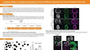 科学海报A Reliable, Efficient, and Matrix-Free Method to Generate Midbrain Organoids from Human Pluripotent Stem Cells
 沪公网安备31010102008431号