Tomov ML et al. (DEC 2016)
Scientific Reports 6 1 37637
Distinct and Shared Determinants of Cardiomyocyte Contractility in Multi-Lineage Competent Ethnically Diverse Human iPSCs
The realization of personalized medicine through human induced pluripotent stem cell (iPSC) technology can be advanced by transcriptomics,epigenomics,and bioinformatics that inform on genetic pathways directing tissue development and function. When possible,population diversity should be included in new studies as resources become available. Previously we derived replicate iPSC lines of African American,Hispanic-Latino and Asian self-designated ethnically diverse (ED) origins with normal karyotype,verified teratoma formation,pluripotency biomarkers,and tri-lineage in vitro commitment. Here we perform bioinformatics of RNA-Seq and ChIP-seq pluripotency data sets for two replicate Asian and Hispanic-Latino ED-iPSC lines that reveal differences in generation of contractile cardiomyocytes but similar and robust differentiation to multiple neural,pancreatic,and smooth muscle cell types. We identify shared and distinct genes and contributing pathways in the replicate ED-iPSC lines to enhance our ability to understand how reprogramming to iPSC impacts genes and pathways contributing to cardiomyocyte contractility potential.
View Publication
产品类型:
产品号#:
05835
05839
08581
08582
产品名:
STEMdiff™ 神经诱导培养基
STEMdiff™ 神经诱导培养基
STEMdiff™SMADi神经诱导试剂盒
STEMdiff™SMADi神经诱导试剂盒,2套
Paulsen BdS et al. (APR 2014)
Schizophrenia Research 154 1-3 30--35
Valproate reverts zinc and potassium imbalance in schizophrenia-derived reprogrammed cells
Schizophrenia has been considered a devastating clinical syndrome rather than a single disease. Nevertheless,the mechanisms behind the onset of schizophrenia have been only partially elucidated. Several studies propose that levels of trace elements are abnormal in schizophrenia; however,conflicting data generated from different biological sources prevent conclusions being drawn. In this work,we used synchrotron radiation X-ray microfluorescence spectroscopy to compare trace element levels in neural progenitor cells (NPCs) derived from two clones of induced pluripotent stem cell lines of a clozapine-resistant schizophrenic patient and two controls. Our data reveal the presence of elevated levels of potassium and zinc in schizophrenic NPCs. Neural cells treated with valproate,an adjunctive medication for schizophrenia,brought potassium and zinc content back to control levels. These results expand the understanding of atomic element imbalance related to schizophrenia and may provide novel insights for the screening of drugs to treat mental disorders. ?? 2014 Elsevier B.V.
View Publication
产品类型:
产品号#:
05850
05857
05870
05875
85850
85857
85870
85875
产品名:
mTeSR™1
mTeSR™1
Y. Kim et al. (MAY 2018)
Cell reports 23 9 2550--2558
Mitochondrial Aging Defects Emerge in Directly Reprogrammed Human Neurons due to Their Metabolic Profile.
Mitochondria are a major target for aging and are instrumental in the age-dependent deterioration of the human brain,but studying mitochondria in aging human neurons has been challenging. Direct fibroblast-to-induced neuron (iN) conversion yields functional neurons that retain important signs of aging,in contrast to iPSC differentiation. Here,we analyzed mitochondrial features in iNs from individuals of different ages. iNs from old donors display decreased oxidative phosphorylation (OXPHOS)-related gene expression,impaired axonal mitochondrial morphologies,lower mitochondrial membrane potentials,reduced energy production,and increased oxidized proteins levels. In contrast,the fibroblasts from which iNs were generated show only mild age-dependent changes,consistent with a metabolic shift from glycolysis-dependent fibroblasts to OXPHOS-dependent iNs. Indeed,OXPHOS-induced old fibroblasts show increased mitochondrial aging features similar to iNs. Our data indicate that iNs are a valuable tool for studying mitochondrial aging and support a bioenergetic explanation for the high susceptibility of the brain to aging.
View Publication
Kang HS et al. (DEC 2015)
Journal of Korean medical science 30 12 1764--76
Advanced Properties of Urine Derived Stem Cells Compared to Adipose Tissue Derived Stem Cells in Terms of Cell Proliferation, Immune Modulation and Multi Differentiation.
Adipose tissue stem cells (ADSCs) would be an attractive autologous cell source. However,ADSCs require invasive procedures,and has potential complications. Recently,urine stem cells (USCs) have been proposed as an alternative stem cell source. In this study,we compared USCs and ADSCs collected from the same patients on stem cell characteristics and capacity to differentiate into various cell lineages to provide a useful guideline for selecting the appropriate type of cell source for use in clinical application. The urine samples were collected via urethral catheterization,and adipose tissue was obtained from subcutaneous fat tissue during elective laparoscopic kidney surgery from the same patient (n = 10). Both cells were plated for primary culture. Cell proliferation,colony formation,cell surface markers,immune modulation,chromosome stability and multi-lineage differentiation were analyzed for each USCs and ADSCs at cell passage 3,5,and 7. USCs showed high cell proliferation rate,enhanced colony forming ability,strong positive for stem cell markers expression,high efficiency for inhibition of immune cell activation compared to ADSCs at cell passage 3,5,and 7. In chromosome stability analysis,both cells showed normal karyotype through all passages. In analysis of multi-lineage capability,USCs showed higher myogenic,neurogenic,and endogenic differentiation rate,and lower osteogenic,adipogenic,and chondrogenic differentiation rate compared to ADSCs. Therefore,we expect that USC can be an alternative autologous stem cell source for muscle,neuron and endothelial tissue reconstruction instead of ADSCs.
View Publication
产品类型:
产品号#:
05752
产品名:
NeuroCult™ NS-A 分化试剂盒(人)
Kayama T et al. (JAN 2018)
Biochemical and Biophysical Research Communications 495 1 1028--1033
Temporally coordinated spiking activity of human induced pluripotent stem cell-derived neurons co-cultured with astrocytes
In culture conditions,human induced-pluripotent stem cells (hiPSC)-derived neurons form synaptic connections with other cells and establish neuronal networks,which are expected to be an in vitro model system for drug discovery screening and toxicity testing. While early studies demonstrated effects of co-culture of hiPSC-derived neurons with astroglial cells on survival and maturation of hiPSC-derived neurons,the population spiking patterns of such hiPSC-derived neurons have not been fully characterized. In this study,we analyzed temporal spiking patterns of hiPSC-derived neurons recorded by a multi-electrode array system. We discovered that specific sets of hiPSC-derived neurons co-cultured with astrocytes showed more frequent and highly coherent non-random synchronized spike trains and more dynamic changes in overall spike patterns over time. These temporally coordinated spiking patterns are physiological signs of organized circuits of hiPSC-derived neurons and suggest benefits of co-culture of hiPSC-derived neurons with astrocytes.
View Publication
产品类型:
产品号#:
05790
05792
05793
05794
05795
产品名:
BrainPhys™神经元培养基
BrainPhys™神经元培养基和SM1试剂盒
BrainPhys™ 神经元培养基N2-A和SM1试剂盒
BrainPhys™原代神经元试剂盒
BrainPhys™ hPSC 神经元试剂盒
Gupta S et al. (DEC 2017)
Journal of Neurochemistry
Fibroblast growth factor 2 regulates activity and gene expression of human post-mitotic excitatory neurons
Many neuropsychiatric disorders are thought to result from subtle changes in neural circuit formation. We used human embryonic stem cells and induced pluripotent stem cells (hiPSCs) to model mature,post-mitotic excitatory neurons and examine effects of fibroblast growth factor 2 (FGF2). FGF2 gene expression is known to be altered in brain regions of major depressive disorder (MDD) patients and FGF2 has anti-depressive effects in animal models of depression. We generated stable inducible neurons (siNeurons) conditionally expressing human neurogenin-2 (NEUROG2) to generate a homogenous population of post-mitotic excitatory neurons and study the functional as well as the transcriptional effects of FGF2. Upon induction of NEUROG2 with doxycycline,the vast majority of cells are post-mitotic,and the gene expression profile recapitulates that of excitatory neurons within 6 days. Using hES cell lines that inducibly express NEUROG2 as well as GCaMP6f,we were able to characterize spontaneous calcium activity in these neurons and show that calcium transients increase in the presence of FGF2. The FGF2-responsive genes were determined by RNA-Seq. FGF2-regulated genes previously identified in non-neuronal cell types were up-regulated (EGR1,ETV4,SPRY4,and DUSP6) as a result of chronic FGF2 treatment of siNeurons. Novel neuron-specific genes were also identified that may mediate FGF2-dependent increases in synaptic efficacy including NRXN3,SYT2,and GALR1. Since several of these genes have been implicated in MDD previously,these results will provide the basis for more mechanistic studies of the role of FGF2 in MDD.
View Publication
Scalable Production of Glioblastoma Tumor-initiating Cells in 3 Dimension Thermoreversible Hydrogels.
There is growing interest in developing drugs that specifically target glioblastoma tumor-initiating cells (TICs). Current cell culture methods,however,cannot cost-effectively produce the large numbers of glioblastoma TICs required for drug discovery and development. In this paper we report a new method that encapsulates patient-derived primary glioblastoma TICs and grows them in 3 dimension thermoreversible hydrogels. Our method allows long-term culture (˜50 days,10 passages tested,accumulative ˜>10(10)-fold expansion) with both high growth rate (˜20-fold expansion/7 days) and high volumetric yield (˜2.0%A-%10(7)%cells/ml) without the loss of stemness. The scalable method can be used to produce sufficient,affordable glioblastoma TICs for drug discovery.
View Publication
产品类型:
产品号#:
05750
05751
产品名:
NeuroCult™ NS-A 基础培养基(人)
NeuroCult™ NS-A 扩增试剂盒(人)
Huat T et al. (APR 2015)
International Journal of Molecular Sciences 16 5 9693--9718
MicroRNA Expression Profile of Neural Progenitor-Like Cells Derived from Rat Bone Marrow Mesenchymal Stem Cells under the Influence of IGF-1, bFGF and EGF
Insulin-like growth factor 1 (IGF-1) enhances cellular proliferation and reduces apoptosis during the early differentiation of bone marrow derived mesenchymal stem cells (BMSCs) into neural progenitor-like cells (NPCs) in the presence of epidermal growth factor (EGF) and basic fibroblast growth factor (bFGF). BMSCs were differentiated in three groups of growth factors: (A) EGF + bFGF,(B) EGF + bFGF + IGF-1,and (C) without growth factor. To unravel the molecular mechanisms of the NPCs derivation,microarray analysis using GeneChip miRNA arrays was performed. The profiles were compared among the groups. Annotated microRNA fingerprints (GSE60060) delineated 46 microRNAs temporally up-regulated or down-regulated compared to group C. The expressions of selected microRNAs were validated by real-time PCR. Among the 46 microRNAs,30 were consistently expressed for minimum of two consecutive time intervals. In Group B,only miR-496 was up-regulated and 12 microRNAs,including the let-7 family,miR-1224,miR-125a-3p,miR-214,miR-22,miR-320,miR-708,and miR-93,were down-regulated. Bioinformatics analysis reveals that some of these microRNAs (miR-22,miR-214,miR-125a-3p,miR-320 and let-7 family) are associated with reduction of apoptosis. Here,we summarize the roles of key microRNAs associated with IGF-1 in the differentiation of BMSCs into NPCs. These findings may provide clues to further our understanding of the mechanisms and roles of microRNAs as key regulators of BMSC-derived NPC maintenance.
View Publication
产品类型:
产品号#:
05750
05751
产品名:
NeuroCult™ NS-A 基础培养基(人)
NeuroCult™ NS-A 扩增试剂盒(人)
E. Gabriel et al. (JAN 2017)
Cell stem cell 20 3 397--406.e5
Recent Zika Virus Isolates Induce Premature Differentiation of Neural Progenitors in Human Brain Organoids.
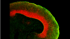
The recent Zika virus (ZIKV) epidemic is associated with microcephaly in newborns. Although the connection between ZIKV and neurodevelopmental defects is widely recognized,the underlying mechanisms are poorly understood. Here we show that two recently isolated strains of ZIKV,an American strain from an infected fetal brain (FB-GWUH-2016) and a closely-related Asian strain (H/PF/2013),productively infect human iPSC-derived brain organoids. Both of these strains readily target to and replicate in proliferating ventricular zone (VZ) apical progenitors. The main phenotypic effect was premature differentiation of neural progenitors associated with centrosome perturbation,even during early stages of infection,leading to progenitor depletion,disruption of the VZ,impaired neurogenesis,and cortical thinning. The infection pattern and cellular outcome differ from those seen with the extensively passaged ZIKV strain MR766. The structural changes we see after infection with these more recently isolated viral strains closely resemble those seen in ZIKV-associated microcephaly.
View Publication

 EasySep™小鼠TIL(CD45)正选试剂盒


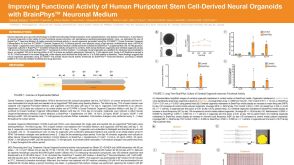 科学海报Improving Functional Activity of Human Pluripotent Stem Cell-Derived Neural Organoids with BrainPhys Neuronal Medium

 沪公网安备31010102008431号