Perez-Campo FM et al. (JUN 2014)
STEM CELLS 32 6 1591--1601
MOZ-Mediated Repression of p16 INK 4 a Is Critical for the Self-Renewal of Neural and Hematopoietic Stem Cells
Although inhibition of p16(INK4a) expression is critical to preserve the proliferative capacity of stem cells,the molecular mechanisms responsible for silencing p16(INK4a) expression remain poorly characterized. Here,we show that the histone acetyltransferase (HAT) monocytic leukemia zinc finger protein (MOZ) controls the proliferation of both hematopoietic and neural stem cells by modulating the transcriptional repression of p16(INK4a) . In the absence of the HAT activity of MOZ,expression of p16(INK4a) is upregulated in progenitor and stem cells,inducing an early entrance into replicative senescence. Genetic deletion of p16(INK4a) reverses the proliferative defect in both Moz(HAT) (-) (/) (-) hematopoietic and neural progenitors. Our results suggest a critical requirement for MOZ HAT activity to silence p16(INK4a) expression and to protect stem cells from early entrance into replicative senescence.
View Publication
产品类型:
产品号#:
05700
05701
05702
05707
产品名:
NeuroCult™ 基础培养基(小鼠和大鼠)
NeuroCult™ 扩增添加物(小鼠和大鼠)
NeuroCult™扩增试剂盒(小鼠和大鼠)
NeuroCult™化学解离试剂盒(小鼠)
Usta S et al. (OCT 2014)
Annals of translational medicine 2 10 97
Chemically defined serum-free and xeno-free media for multiple cell lineages.
Cell culture is one of the most common methods used to recapitulate a human disease environment in a laboratory setting. Cell culture techniques are used to grow and maintain cells of various types including those derived from primary tissues,such as stem cells and cancer tumors. However,a major confounding factor with cell culture is the use of serum and animal (xeno) products in the media. The addition of animal products introduces batch and lot variations that lead to experimental variability,confounds studies with therapeutic outcomes for cultured cells,and represents a major cost associated with cell culture. Here we report a commercially available serum-free,albumin-free,and xeno free (XF) media (Neuro-Pure(TM)) that is more cost-effective than other commercial medias. Neuro-Pure was used to maintain and differentiate various cells of neuronal lineages,fibroblasts,as well as specific cancer cell lines; without the use of contaminants such serum,albumin,and animal products. Neuro-Pure allows for a controlled and reproducible cell culture environment that is applicable to translational medicine and general tissue culture.
View Publication
产品类型:
产品号#:
05761
产品名:
用于小鼠和大鼠神经干细胞和祖细胞分化培养的试剂盒
Zhang L et al. (APR 2016)
Human Reproduction 31 4 832--843
Protein kinase A inhibitor, H89, enhances survival and clonogenicity of dissociated human embryonic stem cells through Rho-associated coiled-coil containing protein kinase (ROCK) inhibition
H89 inhibits the dissociation-induced phosphorylation of PKA and two substrates of Rho-associated coiled-coil containing protein kinase (ROCK),myosin light chain (MLC2) and myosin phosphatase target subunit 1 (MYPT1),significantly increases cell survival and colony formation,and strongly depresses dissociation-induced cell death and cell blebbing without affecting the pluripotency of hESCs and their differentiation in vitro.
View Publication
产品类型:
产品号#:
05835
05839
产品名:
STEMdiff™ 神经诱导培养基
STEMdiff™ 神经诱导培养基
Bagó et al. (FEB 2017)
Science Translational Medicine 9 375 eaah6510
Tumor-homing cytotoxic human induced neural stem cells for cancer therapy
Engineered neural stem cells (NSCs) are a promising approach to treating glioblastoma (GBM). The ideal NSC drug carrier for clinical use should be easily isolated and autologous to avoid immune rejection. We transdifferentiated (TD) human fibroblasts into tumor-homing early-stage induced NSCs (h-iNSC(TE)),engineered them to express optical reporters and different therapeutic gene products,and assessed the tumor-homing migration and therapeutic efficacy of cytotoxic h-iNSC(TE) in patient-derived GBM models of surgical and nonsurgical disease. Molecular and functional analysis revealed that our single-factor SOX2 TD strategy converted human skin fibroblasts into h-iNSC(TE) that were nestin(+) and expressed pathways associated with tumor-homing migration in 4 days. Time-lapse motion analysis showed that h-iNSC(TE) rapidly migrated to human GBM cells and penetrated human GBM spheroids,a process inhibited by blockade of CXCR4. Serial imaging showed that h-iNSC(TE) delivery of the proapoptotic agent tumor necrosis factor-α-related apoptosis-inducing ligand (TRAIL) reduced the size of solid human GBM xenografts 250-fold in 3 weeks and prolonged median survival from 22 to 49 days. Additionally,h-iNSC(TE) thymidine kinase/ganciclovir enzyme/prodrug therapy (h-iNSC(TE)-TK) reduced the size of patient-derived GBM xenografts 20-fold and extended survival from 32 to 62 days. Mimicking clinical NSC therapy,h-iNSC(TE)-TK therapy delivered into the postoperative surgical resection cavity delayed the regrowth of residual GBMs threefold and prolonged survival from 46 to 60 days. These results suggest that TD of human skin into h-iNSC(TE) is a platform for creating tumor-homing cytotoxic cell therapies for cancer,where the potential to avoid carrier rejection could maximize treatment durability in human trials.
View Publication

 EasySep™小鼠TIL(CD45)正选试剂盒


 抗人SOX2 (ANOP3)抗体,clone 2E1 兔Monoclonal 抗体,抗人、小鼠、大鼠SOX2
 抗人突触蛋白(MRX96)抗体,clone 249 兔Monoclonal 抗体,抗人Synaptophysin
 抗人TBR1(IDDAS)Polyclonal抗体 抗人、小鼠、大鼠TBR1的兔Polyclonal抗体
 抗人PAX6(AN)抗体,Polyclonal 兔Polyclonal抗体,抗人、小鼠PAX6
 抗人巢蛋白抗体,clone 10C2 小鼠Monoclonal IgG1抗体,抗人、食蟹猴巢蛋白
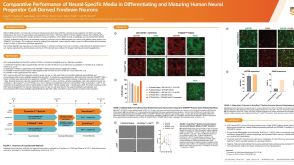 科学海报Comparative Performance of Neural-Specific Media in Differentiating and Maturing Human Neural Progenitor Cell-Derived Forebrain Neurons
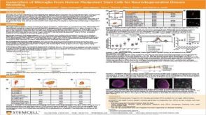 科学海报Generation of Microglia From Human Pluripotent Stem Cells for Neurodegenerative Disease Modeling
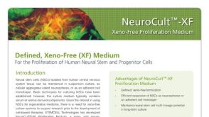 产品手册NeuroCult™-XF: Xeno-Free Culture Medium for the Proliferation of Human Neural Stem Cells
 沪公网安备31010102008431号