Gundemir S et al. (SEP 2016)
Neuro-Oncology now157
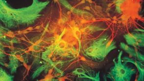
The complex role of transglutaminase 2 in glioblastoma proliferation
BACKGROUND Glioblastomas (GBMs) are a heterogeneous group of primary brain tumors. These tumors are resistant to therapeutic interventions and invariably recur after surgical resection. The multifunctional protein transglutaminase 2 (TG2) has been shown to promote cell survival in a number of different tumors. There is also evidence that TG2 may be a pro-survival factor in GBMs. However,the roles that TG2 plays in facilitating GBM survival and proliferation have not yet been clearly delineated . METHODS The functions of TG2 are often cell- and context-specific. Therefore,in this study we examined the ability of TG2 to facilitate GBM proliferation using colony formation assays and 5-ethynyl-2'-deoxyuridine (EdU) incorporation in several different GBM cell lines as well as neurospheres derived from patient tumors representing the 3 major subtypes of GBM tumors (mesenchymal,proneural,and classical) and maintained in the absence of serum. TG2 knockdown or selective TG2 inhibitors were used to modulate TG2 expression and activity. RESULTS We show that TG2 plays differential roles in the proliferative process depending on the cell type. In most,but not all,GBM models TG2 plays a crucial role in the proliferative process,and some but not all TG2 inhibitors were highly effective at reducing proliferation in a large subset of the GBM models. CONCLUSION Our results show that TG2 plays an important-but notoriously context-specific-role in GBM cell biology. Nonetheless,as future studies unravel the genetic fingerprints" that make TG2 inhibitors effective this information could be exploited to develop TG2 inhibitors into personalized GBM therapies.
View Publication
产品类型:
产品号#:
05750
05751
产品名:
NeuroCult™ NS-A 基础培养基(人)
NeuroCult™ NS-A 扩增试剂盒(人)
Wang F et al. (DEC 2017)
Stem Cell Research & Therapy 8 1 26
CCL11 promotes migration and proliferation of mouse neural progenitor cells
BACKGROUND Neonatal hypoxia-ischemia induces massive brain damage during the perinatal period,resulting in long-term consequences to central nervous system structural and functional maturation. Although neural progenitor cells (NPCs) migrate through the parenchyma and home in to injury sites in the rodent brain,the molecular mechanisms are unknown. We examined the role of chemokines in mediating NPC migration after neonatal hypoxic-ischemic brain injury. METHODS Nine-day-old mice were exposed to a 120-minute hypoxia following unilateral carotid occlusion. Chemokine levels were quantified in mouse brain extract. Migration and proliferation assays were performed using embryonic and infant mouse NPCs. RESULTS The neonatal hypoxic-ischemic brain injury resulted in an ipsilateral lesion,which was extended to the cortical and striatal areas. NPCs migrated toward an injured area,where a marked increase of CC chemokines was detected. In vitro studies showed that incubation of NPCs with recombinant mouse CCL11 promoted migration and proliferation. These effects were partly inhibited by a CCR3 antagonist,SB297006. CONCLUSIONS Our data implicate an important effect of CCL11 for mouse NPCs. The effective activation of NPCs may offer a promising strategy for neuroregeneration in neonatal hypoxic-ischemic brain injury.
View Publication
产品类型:
产品号#:
05700
05701
05702
产品名:
NeuroCult™ 基础培养基(小鼠和大鼠)
NeuroCult™ 扩增添加物(小鼠和大鼠)
NeuroCult™扩增试剂盒(小鼠和大鼠)
Wang Y et al. (MAY 2010)
Neuroscience 167 3 750--7
Erythropoietin (EPO) regulates the proliferation and differentiation of erythroid cells by binding to its specific transmembrane receptor (EPOR). The presence of EPO and its receptor in the CNS suggests a different function for EPO other than erythropoiesis. The purpose of the present study was to examine EPOR expression and the role of EPO in the proliferation of neonatal spinal cord-derived neural progenitor cells. The effect of EPO on cell cycle progression was also examined,as well as the signaling cascades involved in this process. Our results showed that EPOR was present in the neural progenitor cells and EPO significantly enhanced their proliferation. Cell cycle analysis of EPO-treated neural progenitor cells indicated a reduced percentage of cells in G0/G1 phase,whereas the cell proliferation index (S phase plus G2/M phase) was increased. EPO also increased the proportion of 5-bromo-2-deoxyuridine (BrdU)-positive cells. With respect to the cell cycle signaling,we examined the cyclin-dependent kinases D1,D2 and E,and cyclin-dependent kinase inhibitors,p21cip1,p27kip1 and p57kip2. No significant differences were observed in the expression of these transcripts after EPO administration. Interestingly,the anti-apoptotic factors,mcl-1 and bcl-2 were significantly increased twofold. Moreover,these specific effects of EPO were eliminated by incubation of the progenitor cells with anti-EPO neutralizing antibody. Those observations suggested that EPO may play a role in normal spinal cord development by regulating cell proliferation and apoptosis.
View Publication

 EasySep™小鼠TIL(CD45)正选试剂盒


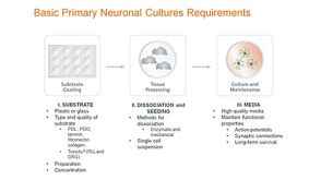
 技术手册In Vitro Proliferation and Differentiation of Human Neural Stem and Progenitor Cells using NeuroCult™ or NeuroCult™-XF
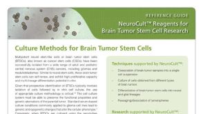 产品手册NeuroCult™: Reagents for Brain Tumor Stem Cell Research
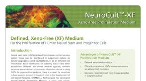 产品手册NeuroCult™-XF: Xeno-Free Culture Medium for the Proliferation of Human Neural Stem Cells

 沪公网安备31010102008431号