Rahman M et al. (MAR 2015)
Anatomy & cell biology 48 1 25--35
Neurosphere and adherent culture conditions are equivalent for malignant glioma stem cell lines.
Certain limitations of the neurosphere assay (NSA) have resulted in a search for alternative culture techniques for brain tumor-initiating cells (TICs). Recently,reports have described growing glioblastoma (GBM) TICs as a monolayer using laminin. We performed a side-by-side analysis of the NSA and laminin (adherent) culture conditions to compare the growth and expansion of GBM TICs. GBM cells were grown using the NSA and adherent culture conditions. Comparisons were made using growth in culture,apoptosis assays,protein expression,limiting dilution clonal frequency assay,genetic affymetrix analysis,and tumorigenicity in vivo. In vitro expansion curves for the NSA and adherent culture conditions were virtually identical (P=0.24) and the clonogenic frequencies (5.2% for NSA vs. 5.0% for laminin,P=0.9) were similar as well. Likewise,markers of differentiation (glial fibrillary acidic protein and beta tubulin III) and proliferation (Ki67 and MCM2) revealed no statistical difference between the sphere and attachment methods. Several different methods were used to determine the numbers of dead or dying cells (trypan blue,DiIC,caspase-3,and annexin V) with none of the assays noting a meaningful variance between the two methods. In addition,genetic expression analysis with microarrays revealed no significant differences between the two groups. Finally,glioma cells derived from both methods of expansion formed large invasive tumors exhibiting GBM features when implanted in immune-compromised animals. A detailed functional,protein and genetic characterization of human GBM cells cultured in serum-free defined conditions demonstrated no statistically meaningful differences when grown using sphere (NSA) or adherent conditions. Hence,both methods are functionally equivalent and remain suitable options for expanding primary high-grade gliomas in tissue culture.
View Publication
产品类型:
产品号#:
05750
05751
产品名:
NeuroCult™ NS-A 基础培养基(人)
NeuroCult™ NS-A 扩增试剂盒(人)
Katori S et al. (JUL 2009)
The Journal of neuroscience : the official journal of the Society for Neuroscience 29 29 9137--47
Protocadherin-alpha family is required for serotonergic projections to appropriately innervate target brain areas.
Serotonergic axons from the raphe nuclei in the brainstem project to every region of the brain,where they make connections through their extensive terminal arborizations. This serotonergic innervation contributes to various normal behaviors and psychiatric disorders. The protocadherin-alpha (Pcdha) family of clustered protocadherins consists of 14 cadherin-related molecules generated from a single gene cluster. We found that the Pcdhas were strongly expressed in the serotonergic neurons. To elucidate their roles,we examined serotonergic fibers in a mouse mutant (Pcdha(Delta CR/Delta CR)) lacking the Pcdha cytoplasmic region-encoding exons,which are common to the gene cluster. In the first week after birth,the distribution pattern of serotonergic fibers in Pcdha(Delta CR/Delta CR) mice was similar to wild-type,but by 3 weeks of age,when the serotonergic axonal termini complete their arborizations,the distribution of the projections was abnormal. In some target regions,notably the globus pallidus and substantia nigra,the normally even distribution of serotonin axonal terminals was,in the mutants,dense at the periphery of each region,but sparse in the center. In the stratum lacunosum-molecular of the hippocampus,the mutants showed denser serotonergic innervation than in wild-type,and in the dentate gyrus of the hippocampus and the caudate-putamen,the innervation was sparser. Together,the abnormalities suggested that Pcdha proteins are important in the late-stage maturation of serotonergic projections. Further examination of alternatively spliced exons encoding the cytoplasmic tail showed that the A-type (but not the B-type) cytoplasmic tail was essential for the normal development of serotonergic projections.
View Publication
产品类型:
产品号#:
03800
03801
03802
03803
03804
03805
03806
产品名:
ClonaCell™-HY杂交瘤试剂盒
ClonaCell™-HY培养基A
ClonaCell™-HY 培养基 B
ClonaCell™-HY 培养基 C
ClonaCell™-HY 培养基 D
ClonaCell™-HY 培养基 E
ClonaCell™-HY PEG
Bravard A et al. (JAN 2015)
Nucleic acids research 43 2 904--16
The prion protein is critical for DNA repair and cell survival after genotoxic stress.
The prion protein (PrP) is highly conserved and ubiquitously expressed,suggesting that it plays an important physiological function. However,despite decades of investigation,this role remains elusive. Here,by using animal and cellular models,we unveil a key role of PrP in the DNA damage response. Exposure of neurons to a genotoxic stress activates PRNP transcription leading to an increased amount of PrP in the nucleus where it interacts with APE1,the major mammalian endonuclease essential for base excision repair,and stimulates its activity. Preventing the induction of PRNP results in accumulation of abasic sites in DNA and impairs cell survival after genotoxic treatment. Brains from Prnp(-/-) mice display a reduced APE1 activity and a defect in the repair of induced DNA damage in vivo. Thus,PrP is required to maintain genomic stability in response to genotoxic stresses.
View Publication
产品类型:
产品号#:
05700
05701
05702
产品名:
NeuroCult™ 基础培养基(小鼠和大鼠)
NeuroCult™ 扩增添加物(小鼠和大鼠)
NeuroCult™扩增试剂盒(小鼠和大鼠)
Mao J et al. (OCT 2015)
Aging Cell 14 5 784--796
A herbal medicine for Alzheimer's disease and its active constituents promote neural progenitor proliferation
Aberrant neural progenitor cell (NPC) proliferation and self-renewal have been linked to age-related neurodegeneration and neurodegenerative disorders including Alzheimer's disease (AD). Rhizoma Acori tatarinowii is a traditional Chinese herbal medicine against cognitive decline. In this study,we found that the extract of Rhizoma Acori tatarinowii (AT) and its active constituents,asarones,promote NPC proliferation. Oral administration of AT enhanced NPC proliferation and neurogenesis in the hippocampi of adult and aged mice as well as that of transgenic AD model mice. AT and its fractions also enhanced the proliferation of NPCs cultured in vitro. Further analysis identified α-asarone and β-asarone as the two active constituents of AT in promoting neurogenesis. Our mechanistic study revealed that AT and asarones activated extracellular signal-regulated kinase (ERK) but not Akt,two critical kinase cascades for neurogenesis. Consistently,the inhibition of ERK activities effectively blocked the enhancement of NPC proliferation by AT or asarones. Our findings suggest that AT and asarones,which can be orally administrated,could serve as preventive and regenerative therapeutic agents to promote neurogenesis against age-related neurodegeneration and neurodegenerative disorders.
View Publication
P. Gonzalez-Sanchez et al. ( 2017)
Frontiers in cellular neuroscience 11 363
Store-Operated Calcium Entry Is Required for mGluR-Dependent Long Term Depression in Cortical Neurons.
Store-operated calcium entry (SOCE) is a Calcium (Ca2+) influx pathway activated by depletion of intracellular stores that occurs in eukaryotic cells. In neurons,the presence and functions of SOCE are still in question. Here,we show evidences for the existence of SOCE in primary mouse cortical neurons. Endoplasmic reticulum (ER)-Ca2+ depletion using thapsigargin (Tg) triggered a maintained cytosolic Ca2+ increase,which rapidly returned to basal level in the presence of the SOCE blockers 2-Aminoethoxydiphenyl borate (2-APB) and YM-58483. Neural SOCE is also engaged by activation of metabotropic glutamate receptors (mGluRs) with (S)-3,5-dihydroxyphenylglycine (DHPG) (agonist of group I mGluRs),being an essential mechanism to maintain the mGluR-driven Ca2+ signal. Activation of group I of mGluRs triggers long-term depression (LTD) in many brain regions,but the underlying mechanism and,specifically,the necessity of Ca2+ increase in the postsynaptic neuron is controversial. In primary cortical neurons,we now show that the inhibition of Ca2+ influx through SOCE impaired DHPG-LTD,pointing out a key function of calcium and SOCE in synaptic plasticity.
View Publication
产品类型:
产品号#:
05790
05792
05793
05794
05795
产品名:
BrainPhys™神经元培养基
BrainPhys™神经元培养基和SM1试剂盒
BrainPhys™ 神经元培养基N2-A和SM1试剂盒
BrainPhys™原代神经元试剂盒
BrainPhys™ hPSC 神经元试剂盒
Andrade LNdS et al. (SEP 2012)
Human Molecular Genetics 21 17 3825--3834
Evidence for premature aging due to oxidative stress in iPSCs from Cockayne syndrome
Cockayne syndrome (CS) is a human premature aging disorder associated with neurological and developmental abnormalities,caused by mutations mainly in the CS group B gene (ERCC6). At the molecular level,CS is characterized by a deficiency in the transcription-couple DNA repair pathway. To understand the role of this molecular pathway in a pluripotent cell and the impact of CSB mutation during human cellular development,we generated induced pluripotent stem cells (iPSCs) from CSB skin fibroblasts (CSB-iPSC). Here,we showed that the lack of functional CSB does not represent a barrier to genetic reprogramming. However,iPSCs derived from CSB patient's fibroblasts exhibited elevated cell death rate and higher reactive oxygen species (ROS) production. Moreover,these cellular phenotypes were accompanied by an up-regulation of TXNIP and TP53 transcriptional expression. Our findings suggest that CSB modulates cell viability in pluripotent stem cells,regulating the expression of TP53 and TXNIP and ROS production.
View Publication
McPherson CA et al. (JUL 2011)
Brain,behavior,and immunity 25 5 850--62
Interleukin (IL)-1 and IL-6 regulation of neural progenitor cell proliferation with hippocampal injury: differential regulatory pathways in the subgranular zone (SGZ) of the adolescent and mature mouse brain.
Current data suggests an association between elevations in interleukin 1 (IL-1)α,IL-1β,and IL-6 and the proliferation of neural progenitor cells (NPCs) following brain injury. A limited amount of work implicates changes in these pro-inflammatory responses with diminished NPC proliferation observed as a function of aging. In the current study,adolescent (21day-old) and 1year-old CD-1 male mice were injected with trimethyltin (TMT,2.3mg/kg,i.p.) to produce acute apoptosis of hippocampal dentate granule cells. In this model,fewer 5-bromo-2'-deoxyuridine (BrdU)+ NPC were observed in both naive and injured adult hippocampus as compared to the corresponding number seen in adolescent mice. At 48h post-TMT,a similar level of neuronal death was observed across ages,yet activated ameboid microglia were observed in the adolescent and hypertrophic process-bearing microglia in the adult. IL-1α mRNA levels were elevated in the adolescent hippocampus; IL-6 mRNA levels were elevated in the adult. In subgranular zone (SGZ) isolated by laser-capture microdissection,IL-1β was detected but not elevated by TMT,IL-1a was elevated at both ages,while IL-6 was elevated only in the adult. Naïve NPCs isolated from the hippocampus expressed transcripts for IL-1R1,IL-6Rα,and gp130 with significantly higher levels of IL-6Rα mRNA in the adult. In vitro,IL-1α (150pg/ml) stimulated proliferation of adolescent NPCs; IL-6 (10ng/ml) inhibited proliferation of adolescent and adult NPCs. Microarray analysis of SGZ post-TMT indicated a prominence of IL-1a/IL-1R1 signaling in the adolescent and IL-6/gp130 signaling in the adult.
View Publication

 EasySep™小鼠TIL(CD45)正选试剂盒


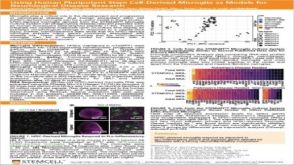 科学海报Using Human Pluripotent Stem Cell-Derived Microglia As Models For Neurological Disease Research
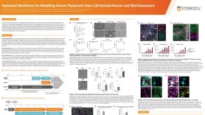 科学海报Optimized Workflows for Modelling Human Pluripotent Stem Cell-Derived Neuron and Glial Interactions
 实验方案How to Culture Primary Rodent Neurons for MEA Analysis Using the Maestro MEA™ System
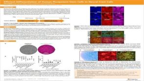 科学海报Efficient Generation of Human Pluripotent Stem Cells to Neural Crest Cells

 沪公网安备31010102008431号