Noormohammadi A et al. (NOV 2016)
Nature Communications 7 13649
Somatic increase of CCT8 mimics proteostasis of human pluripotent stem cells and extends C. elegans lifespan
Human embryonic stem cells can replicate indefinitely while maintaining their undifferentiated state and,therefore,are immortal in culture. This capacity may demand avoidance of any imbalance in protein homeostasis (proteostasis) that would otherwise compromise stem cell identity. Here we show that human pluripotent stem cells exhibit enhanced assembly of the TRiC/CCT complex,a chaperonin that facilitates the folding of 10% of the proteome. We find that ectopic expression of a single subunit (CCT8) is sufficient to increase TRiC/CCT assembly. Moreover,increased TRiC/CCT complex is required to avoid aggregation of mutant Huntingtin protein. We further show that increased expression of CCT8 in somatic tissues extends Caenorhabditis elegans lifespan in a TRiC/CCT-dependent manner. Ectopic expression of CCT8 also ameliorates the age-associated demise of proteostasis and corrects proteostatic deficiencies in worm models of Huntington's disease. Our results suggest proteostasis is a common principle that links organismal longevity with hESC immortality.
View Publication
Jenkins PM et al. (DEC 2015)
Nanoscale research letters 10 1 972
A nerve guidance conduit with topographical and biochemical cues: potential application using human neural stem cells.
Despite major advances in the pathophysiological understanding of peripheral nerve damage,the treatment of nerve injuries still remains an unmet medical need. Nerve guidance conduits present a promising treatment option by providing a growth-permissive environment that 1) promotes neuronal cell survival and axon growth and 2) directs axonal extension. To this end,we designed an electrospun nerve guidance conduit using a blend of polyurea and poly-caprolactone with both biochemical and topographical cues. Biochemical cues were integrated into the conduit by functionalizing the polyurea with RGD to improve cell attachment. Topographical cues that resemble natural nerve tissue were incorporated by introducing intraluminal microchannels aligned with nanofibers. We determined that electrospinning the polymer solution across a two electrode system with dissolvable sucrose fibers produced a polymer conduit with the appropriate biomimetic properties. Human neural stem cells were cultured on the conduit to evaluate its ability to promote neuronal growth and axonal extension. The nerve guidance conduit was shown to enhance cell survival,migration,and guide neurite extension.
View Publication
产品类型:
产品号#:
05850
05857
05870
05875
85850
85857
85870
85875
产品名:
mTeSR™1
mTeSR™1
Belzile J-P et al. (APR 2014)
Journal of virology 88 8 4021--4039
Human cytomegalovirus infection of human embryonic stem cell-derived primitive neural stem cells is restricted at several steps but leads to the persistence of viral DNA.
UNLABELLED Congenital human cytomegalovirus (HCMV) infection is a major cause of central nervous system structural anomalies and sensory impairments. It is likely that the stage of fetal development,as well as the state of differentiation of susceptible cells at the time of infection,affects the severity of the disease. We used human embryonic stem (ES) cell-derived primitive prerosette neural stem cells (pNSCs) and neural progenitor cells (NPCs) maintained in chemically defined conditions to study HCMV replication in cells at the early stages of neural development. In contrast to what was observed previously using fetus-derived NPCs,infection of ES cell-derived pNSCs with HCMV was nonprogressive. At a low multiplicity of infection,we observed only a small percentage of cells expressing immediate-early genes (IE) and early genes. IE expression was found to be restricted to cells negative for the anterior marker FORSE-1,and treatment of pNSCs with retinoic acid restored IE expression. Differentiation of pNSCs into NPCs restored IE expression but not the transactivation of early genes. Virions produced in NPCs and pNSCs were exclusively cell associated and were mostly non-neural tropic. Finally,we found that viral genomes could persist in pNSC cultures for up to a month after infection despite the absence of detectable IE expression by immunofluorescence,and infectious virus could be produced upon differentiation of pNSCs to neurons. In conclusion,our results highlight the complex array of hurdles that HCMV must overcome in order to infect primitive neural stem cells and suggest that these cells might act as a reservoir for the virus. IMPORTANCE Human cytomegalovirus (HCMV) is a betaherpesvirus that is highly prevalent in the population. HCMV infection is usually asymptomatic but can lead to severe consequences in immunosuppressed individuals. HCMV is also the most important infectious cause of congenital developmental birth defects. Manifestations of fetal HCMV disease range from deafness and learning disabilities to more severe symptoms such as microcephaly. In this study,we have used embryonic stem cells to generate primitive neural stem cells and have used these to model HCMV infection of the fetal central nervous system (CNS) in vitro. Our results reveal that these cells,which are similar to those present in the developing neural tube,do not support viral replication but instead likely constitute a viral reservoir. Future work will define the effect of viral persistence on cellular functions as well as the exogenous signals leading to the reactivation of viral replication in the CNS.
View Publication
产品类型:
产品号#:
05850
05857
05870
05875
85850
85857
85870
85875
产品名:
mTeSR™1
mTeSR™1
Sareen D et al. (AUG 2014)
Journal of Comparative Neurology 522 12 2707--2728
Human induced pluripotent stem cells are a novel source of neural progenitor cells (iNPCs) that migrate and integrate in the rodent spinal cord
Transplantation of human neural progenitor cells (NPCs) into the brain or spinal cord to replace lost cells,modulate the injury environment,or create a permissive milieu to protect and regenerate host neurons is a promising therapeutic strategy for neurological diseases. Deriving NPCs from human fetal tissue is feasible,although problematic issues include limited sources and ethical concerns. Here we describe a new and abundant source of NPCs derived from human induced pluripotent stem cells (iPSCs). A novel chopping technique was used to transform adherent iPSCs into free-floating spheres that were easy to maintain and were expandable (EZ spheres) (Ebert et al. [2013] Stem Cell Res 10:417–427). These EZ spheres could be differentiated towards NPC spheres with a spinal cord phenotype using a combination of all-trans retinoic acid (RA) and epidermal growth factor (EGF) and fibroblast growth factor-2 (FGF-2) mitogens. Suspension cultures of NPCs derived from human iPSCs or fetal tissue have similar characteristics,although they were not similar when grown as adherent cells. In addition,iPSC-derived NPCs (iNPCs) survived grafting into the spinal cord of athymic nude rats with no signs of overgrowth and with a very similar profile to human fetal-derived NPCs (fNPCs). These results suggest that human iNPCs behave like fNPCs and could thus be a valuable alternative for cellular regenerative therapies of neurological diseases. J. Comp. Neurol. 522:2707–2728,2014. textcopyright 2014 Wiley Periodicals,Inc.
View Publication
产品类型:
产品号#:
05850
05857
05870
05875
85850
85857
85870
85875
产品名:
mTeSR™1
mTeSR™1
Fè et al. ( 2014)
PloS one 9 3 e91519
Comparative expression study of the endo-G protein coupled receptor (GPCR) repertoire in human glioblastoma cancer stem-like cells, U87-MG cells and non malignant cells of neural origin unveils new potential therapeutic targets.
Glioblastomas (GBMs) are highly aggressive,invasive brain tumors with bad prognosis and unmet medical need. These tumors are heterogeneous being constituted by a variety of cells in different states of differentiation. Among these,cells endowed with stem properties,tumor initiating/propagating properties and particularly resistant to chemo- and radiotherapies are designed as the real culprits for tumor maintenance and relapse after treatment. These cells,termed cancer stem-like cells,have been designed as prominent targets for new and more efficient cancer therapies. G-protein coupled receptors (GPCRs),a family of membrane receptors,play a prominent role in cell signaling,cell communication and crosstalk with the microenvironment. Their role in cancer has been highlighted but remains largely unexplored. Here,we report a descriptive study of the differential expression of the endo-GPCR repertoire in human glioblastoma cancer stem-like cells (GSCs),U-87 MG cells,human astrocytes and fetal neural stem cells (f-NSCs). The endo-GPCR transcriptome has been studied using Taqman Low Density Arrays. Of the 356 GPCRs investigated,138 were retained for comparative studies between the different cell types. At the transcriptomic level,eight GPCRs were specifically expressed/overexpressed in GSCs. Seventeen GPCRs appeared specifically expressed in cells with stem properties (GSCs and f-NSCs). Results of GPCR expression at the protein level using mass spectrometry and proteomic analysis are also presented. The comparative GPCR expression study presented here gives clues for new pathways specifically used by GSCs and unveils novel potential therapeutic targets.
View Publication
Ankam S et al. (JAN 2013)
Acta Biomaterialia 9 1 4535--45
Substrate topography and size determine the fate of human embryonic stem cells to neuronal or glial lineage
Efficient derivation of neural cells from human embryonic stem cells (hESCs) remains an unmet need for the treatment of neurological disorders. The limiting factors for current methods include being labor-intensive,time-consuming and expensive. In this study,we hypothesize that the substrate topography,with optimal geometry and dimension,can modulate the neural fate of hESCs and enhance the efficiency of differentiation. A multi-architectural chip (MARC) containing fields of topographies varying in geometry and dimension was developed to facilitate high-throughput analysis of topography-induced neural differentiation in vitro. The hESCs were subjected to direct differentiation"�
View Publication
产品类型:
产品号#:
05850
05857
05870
05875
07920
85850
85857
85870
85875
07922
产品名:
ACCUTASE™
mTeSR™1
mTeSR™1
ACCUTASE™
Fornara O et al. (FEB 2016)
Cell death and differentiation 23 2 261--9
Cytomegalovirus infection induces a stem cell phenotype in human primary glioblastoma cells: prognostic significance and biological impact.
Glioblastoma (GBM) is associated with poor prognosis despite aggressive surgical resection,chemotherapy,and radiation therapy. Unfortunately,this standard therapy does not target glioma cancer stem cells (GCSCs),a subpopulation of GBM cells that can give rise to recurrent tumors. GBMs express human cytomegalovirus (HCMV) proteins,and previously we found that the level of expression of HCMV immediate-early (IE) protein in GBMs is a prognostic factor for poor patient survival. In this study,we investigated the relation between HCMV infection of GBM cells and the presence of GCSCs. Primary GBMs were characterized by their expression of HCMV-IE and GCSCs marker CD133 and by patient survival. The extent to which HCMV infection of primary GBM cells induced a GCSC phenotype was evaluated in vitro. In primary GBMs,a large fraction of CD133-positive cells expressed HCMV-IE,and higher co-expression of these two proteins predicted poor patient survival. Infection of GBM cells with HCMV led to upregulation of CD133 and other GSCS markers (Notch1,Sox2,Oct4,Nestin). HCMV infection also promoted the growth of GBM cells as neurospheres,a behavior typically displayed by GCSCs,and this phenotype was prevented by either chemical inhibition of the Notch1 pathway or by treatment with the anti-viral drug ganciclovir. GBM cells that maintained expression of HCMV-IE failed to differentiate into neuronal or astrocytic phenotypes. Our findings imply that HCMV infection induces phenotypic plasticity of GBM cells to promote GCSC features and may thereby increase the aggressiveness of this tumor.
View Publication
产品类型:
产品号#:
05752
产品名:
NeuroCult™ NS-A 分化试剂盒(人)
Wakimoto H et al. (APR 2009)
Cancer research 69 8 3472--81
Human glioblastoma-derived cancer stem cells: establishment of invasive glioma models and treatment with oncolytic herpes simplex virus vectors.
Glioblastoma,the most malignant type of primary brain tumor,is one of the solid cancers where cancer stem cells have been isolated,and studies have suggested resistance of those cells to chemotherapy and radiotherapy. Here,we report the establishment of CSC-enriched cultures derived from human glioblastoma specimens. They grew as neurospheres in serum-free medium with epidermal growth factor and fibroblast growth factor 2,varied in the level of CD133 expression and very efficiently formed highly invasive and/or vascular tumors upon intracerebral implantation into immunodeficient mice. As a novel therapeutic strategy for glioblastoma-derived cancer stem-like cells (GBM-SC),we have tested oncolytic herpes simplex virus (oHSV) vectors. We show that although ICP6 (UL39)-deleted mutants kill GBM-SCs as efficiently as wild-type HSV,the deletion of gamma34.5 significantly attenuated the vectors due to poor replication. However,this was significantly reversed by the additional deletion of alpha47. Infection with oHSV G47Delta (ICP6(-),gamma34.5(-),alpha47(-)) not only killed GBM-SCs but also inhibited their self-renewal as evidenced by the inability of viable cells to form secondary tumor spheres. Importantly,despite the highly invasive nature of the intracerebral tumors generated by GBM-SCs,intratumoral injection of G47Delta significantly prolonged survival. These results for the first time show the efficacy of oHSV against human GBM-SCs,and correlate this cytotoxic property with specific oHSV mutations. This is important for designing new oHSV vectors and clinical trials. Moreover,the new glioma models described in this study provide powerful tools for testing experimental therapeutics and studying invasion and angiogenesis.
View Publication
Vector-Free and Transgene-Free Human iPS Cells Differentiate into Functional Neurons and Enhance Functional Recovery after Ischemic Stroke in Mice
Stroke is a leading cause of human death and disability in the adult population in the United States and around the world. While stroke treatment is limited,stem cell transplantation has emerged as a promising regenerative therapy to replace or repair damaged tissues and enhance functional recovery after stroke. Recently,the creation of induced pluripotent stem (iPS) cells through reprogramming of somatic cells has revolutionized cell therapy by providing an unlimited source of autologous cells for transplantation. In addition,the creation of vector-free and transgene-free human iPS (hiPS) cells provides a new generation of stem cells with a reduced risk of tumor formation that was associated with the random integration of viral vectors seen with previous techniques. However,the potential use of these cells in the treatment of ischemic stroke has not been explored. In the present investigation,we examined the neuronal differentiation of vector-free and transgene-free hiPS cells and the transplantation of hiPS cell-derived neural progenitor cells (hiPS-NPCs) in an ischemic stroke model in mice. Vector-free hiPS cells were maintained in feeder-free and serum-free conditions and differentiated into functional neurons in vitro using a newly developed differentiation protocol. Twenty eight days after transplantation in stroke mice,hiPS-NPCs showed mature neuronal markers in vivo. No tumor formation was seen up to 12 months after transplantation. Transplantation of hiPS-NPCs restored neurovascular coupling,increased trophic support and promoted behavioral recovery after stroke. These data suggest that using vector-free and transgene-free hiPS cells in stem cell therapy are safe and efficacious in enhancing recovery after focal ischemic stroke in mice.
View Publication

 EasySep™小鼠TIL(CD45)正选试剂盒


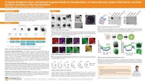 科学海报A Human Pluripotent Stem Cell-Derived Organoid Model for Recapitulation of Central Nervous System (CNS) Barrier and Fluid Secretion Functions of the Choroid Plexus

 沪公网安备31010102008431号