Murphy SV et al. (JAN 2013)
Journal of biomedical materials research. Part A 101 1 272--84
Evaluation of hydrogels for bio-printing applications.
In the United States alone,there are approximately 500,000 burn injuries that require medical treatment every year. Limitations of current treatments necessitate the development of new methods that can be applied quicker,result in faster wound regeneration,and yield skin that is cosmetically similar to undamaged skin. The development of new hydrogel biomaterials and bioprinting deposition technologies has provided a platform to address this need. Herein we evaluated characteristics of twelve hydrogels to determine their suitability for bioprinting applications. We chose hydrogels that are either commercially available,or are commonly used for research purposes. We evaluated specific hydrogel properties relevant to bioprinting applications,specifically; gelation time,swelling or contraction,stability,biocompatibility and printability. Further,we described regulatory,commercial and financial aspects of each of the hydrogels. While many of the hydrogels screened may exhibit characteristics suitable for other applications,UV-crosslinked Extracel,a hyaluronic acid-based hydrogel,had many of the desired properties for our bioprinting application. Taken together with commercial availability,shelf life,potential for regulatory approval and ease of use,these materials hold the potential to be further developed into fast and effective wound healing treatments.
View Publication
Smith Sa et al. (MAR 2012)
Journal of Virology 86 5 2665--75
Persistence of circulating memory B cell clones with potential for Dengue virus disease enhancement for decades following infection
Symptomatic dengue virus infection ranges in disease severity from an influenza-like illness to life-threatening shock. One model of the mechanism underlying severe disease proposes that weakly neutralizing,dengue serotype cross-reactive antibodies induced during a primary infection facilitate virus entry into Fc receptor-bearing cells during a subsequent secondary infection,increasing viral replication and the release of cytokines and vasoactive mediators,culminating in shock. This process has been termed antibody-dependent enhancement of infection and has significantly hindered vaccine development. Much of our understanding of this process has come from studies using mouse monoclonal antibodies (MAbs); however,antibody responses in mice typically exhibit less complexity than those in humans. A better understanding of the humoral immune response to natural dengue virus infection in humans is sorely needed. Using a high-efficiency human hybridoma technology,we isolated 37 hybridomas secreting human MAbs to dengue viruses from 12 subjects years or even decades following primary or secondary infection. The majority of the human antibodies recovered were broadly cross-reactive,directed against either envelope or premembrane proteins,and capable of enhancement of infection in vitro; few exhibited serotype-specific binding or potent neutralizing activity. Memory B cells encoding enhancing antibodies predominated in the circulation,even two or more decades following infection. Mapping the epitopes and activity of naturally occurring dengue antibodies should prove valuable in determining whether the enhancing and neutralizing activity of antibodies can be separated. Such principles could be used in the rational design of vaccines that enhance the induction of neutralizing antibodies,while lowering the risk of dengue shock syndrome.
View Publication
Meng A et al. (DEC 2003)
Experimental hematology 31 12 1348--56
Ionizing radiation and busulfan inhibit murine bone marrow cell hematopoietic function via apoptosis-dependent and -independent mechanisms.
OBJECTIVE: Ionizing radiation (IR) and busulfan (BU) are commonly used as preconditioning regimens for bone marrow transplantation (BMT). We examined whether induction of apoptosis in murine bone marrow (BM) hematopoietic cells contributes to IR- and BU-induced suppression of their hematopoietic function. METHODS: The hematopoietic functions of hematopoietic stem cells (HSCs) and progenitors were analyzed by the cobblestone area-forming cell (CAFC) assay. Apoptosis was determined by measuring 3,3'-dihexyloxacarbocyanine iodide (DiCO6) uptake,annexin V staining,and/or sub-G(0/1) cells. Four cell types were studied: murine BM mononuclear cells (BM-MNCs),linage-negative hematopoietic cells (Lin-) cells),Lin- Scal+ c-kit+ cells,and Lin- Scal- c-kit+ cells by flow cytometry. RESULTS: Exposure of BM-MNCs to IR (4 Gy) or incubation of the cells with BU (30 microM) resulted in a significant reduction in CAFC frequency (ptextless0.001). The survival fractions of various day-types of CAFC for the irradiated cells were less than 10%,while that for BU-treated cells was 71.3% on day 7 and progressively declined to 5.3% on day 35. Interestingly,IR significantly induced apoptosis in BM-MNCs,Lin- cells,HSCs,and progenitors,whereas BU failed to increase apoptosis in these cells. In addition,preincubation of BM-MNCs with z-Val-Ala-Asp (OCH3)-fluoromethylketone,methyl ester (z-VAD) attenuated IR-induced reduction in CAFC but not that induced by BU. CONCLUSION: IR and BU differentially suppress the hematopoietic function of HSCs and progenitors by fundamentally different mechanisms. IR inhibits the function primarily by the induction of HSC and progenitor apoptosis. In contrast,BU suppresses HSC and progenitor function via an apoptosis-independent mechanism.
View Publication

 EasySep™小鼠TIL(CD45)正选试剂盒


 产品手册High Affinity Anti-EPO Reagents for Consistent Results
 产品手册NeuroCult™: Reagents for Brain Tumor Stem Cell Research
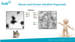
 点播Human Intestinal Organoid Course Learn the basics of culturing and applying human intestinal organoids in your research.


 沪公网安备31010102008431号