Katori S et al. (JUL 2009)
The Journal of neuroscience : the official journal of the Society for Neuroscience 29 29 9137--47
Protocadherin-alpha family is required for serotonergic projections to appropriately innervate target brain areas.
Serotonergic axons from the raphe nuclei in the brainstem project to every region of the brain,where they make connections through their extensive terminal arborizations. This serotonergic innervation contributes to various normal behaviors and psychiatric disorders. The protocadherin-alpha (Pcdha) family of clustered protocadherins consists of 14 cadherin-related molecules generated from a single gene cluster. We found that the Pcdhas were strongly expressed in the serotonergic neurons. To elucidate their roles,we examined serotonergic fibers in a mouse mutant (Pcdha(Delta CR/Delta CR)) lacking the Pcdha cytoplasmic region-encoding exons,which are common to the gene cluster. In the first week after birth,the distribution pattern of serotonergic fibers in Pcdha(Delta CR/Delta CR) mice was similar to wild-type,but by 3 weeks of age,when the serotonergic axonal termini complete their arborizations,the distribution of the projections was abnormal. In some target regions,notably the globus pallidus and substantia nigra,the normally even distribution of serotonin axonal terminals was,in the mutants,dense at the periphery of each region,but sparse in the center. In the stratum lacunosum-molecular of the hippocampus,the mutants showed denser serotonergic innervation than in wild-type,and in the dentate gyrus of the hippocampus and the caudate-putamen,the innervation was sparser. Together,the abnormalities suggested that Pcdha proteins are important in the late-stage maturation of serotonergic projections. Further examination of alternatively spliced exons encoding the cytoplasmic tail showed that the A-type (but not the B-type) cytoplasmic tail was essential for the normal development of serotonergic projections.
View Publication
产品类型:
产品号#:
03800
03801
03802
03803
03804
03805
03806
产品名:
ClonaCell™-HY 杂交瘤试剂盒
ClonaCell™-HY Medium
ClonaCell™-HY Medium
ClonaCell™-HY Medium
ClonaCell™-HY Medium
ClonaCell™-HY Medium
ClonaCell™-HY PEG (融合)
Ma Z et al. (JUL 2015)
Nature communications 6 May 7413
Self-organizing human cardiac microchambers mediated by geometric confinement.
Tissue morphogenesis and organ formation are the consequences of biochemical and biophysical cues that lead to cellular spatial patterning in development. To model such events in vitro,we use PEG-patterned substrates to geometrically confine human pluripotent stem cell colonies and spatially present mechanical stress. Modulation of the WNT/β-catenin pathway promotes spatial patterning via geometric confinement of the cell condensation process during epithelial-mesenchymal transition,forcing cells at the perimeter to express an OCT4+ annulus,which is coincident with a region of higher cell density and E-cadherin expression. The biochemical and biophysical cues synergistically induce self-organizing lineage specification and creation of a beating human cardiac microchamber confined by the pattern geometry. These highly defined human cardiac microchambers can be used to study aspects of embryonic spatial patterning,early cardiac development and drug-induced developmental toxicity.
View Publication
产品类型:
产品号#:
05850
05857
05870
05875
85850
85857
85870
85875
产品名:
mTeSR™1
mTeSR™1
Inagi R et al. (NOV 2007)
Nephrology,dialysis,transplantation : official publication of the European Dialysis and Transplant Association - European Renal Association 22 11 3311--7
Establishment of a sandwich ELISA for human megsin, a kidney-specific serine protease inhibitor.
BACKGROUND: We previously identified a novel serine protease inhibitor (serpin),megsin,which is predominantly expressed in the kidney. Megsin expression is up-regulated in human and experimental renal diseases associated with mesangial proliferation and expansion,suggesting that urinary megsin may be a novel diagnostic marker for some renal diseases. METHODS: We established a specific and sensitive sandwich enzyme-linked immunosorbent assay (ELISA) for megsin and measured urinary megsin of patients with various renal diseases. RESULTS: Megsin ELISA specifically detected megsin but not other serpins. The detection limit was 0.04 ng/ml,which allowed detection of urinary megsin in 3.6% of healthy individuals. The antigenic epitope in the urine detected by the ELISA was confirmed as megsin protein by time-of-flight mass spectrometry. Among patients with rapidly progressive glomerulonephritis (n = 18),55.6% were urinary megsin-positive,while 24.1% in IgA nephropathy (n = 112) and 15.1% in chronic non-IgA glomerulonephritis (n = 245) were urinary megsin-positive,respectively. Among patients with chronic renal failure due to unknown causes (n = 74),18.9% were positive for urinary megsin. In diabetic patients with or without nephropathy (n = 1073),12.3% were urinary megsin-positive,while positivity of urinary megsin in patients with non-renal diseases (n = 768) was equivalent (3.3%) to that of healthy individuals. Of note,when urinary megsin-positive patients with diabetic nephropathy (n = 71) were classified into four stages by their proteinuria and estimated glomerular filtration rate,urinary megsin excretion increased as the stage progressed up to stage 3A,suggesting correlation of that with mesangial expansion level. Urinary megsin decreased in the advanced stage,probably reflecting development of glomerulosclerosis. CONCLUSION: We established a high-sensitive megsin ELISA,which detects urinary megsin in some patients with renal diseases and in only a few healthy subjects. Megsin ELISA may be a novel diagnostic tool for renal diseases.
View Publication
产品类型:
产品号#:
03800
03801
03802
03803
03804
03805
03806
产品名:
ClonaCell™-HY 杂交瘤试剂盒
ClonaCell™-HY Medium
ClonaCell™-HY Medium
ClonaCell™-HY Medium
ClonaCell™-HY Medium
ClonaCell™-HY Medium
ClonaCell™-HY PEG (融合)
Volpe DA and Warren MK (JUN 2003)
Toxicology in vitro : an international journal published in association with BIBRA 17 3 271--7
Myeloid clonogenic assays for comparison of the in vitro toxicity of alkylating agents.
A battery of clonal assays for myeloid progenitor cells (HPP-CFC,CFU-gemm,CFU-gm,CFU-g) was utilized to evaluate the myelotoxicity of a series of alkylating agents representing the spectrum of clinical times to nadir. Bone marrow aspirates from normal volunteers were incubated with mechlorethamine,busulfan,melphalan,carmustine or lomustine for 1 h and then cultured in methylcellulose with 30% serum and cytokines. There was a concentration-dependent inhibition of colony formation and often a differential toxicity to the myeloid progenitors with the alkylators tested. On a molar basis,mechlorethamine and melphalan were the most toxic of the alkylator drugs to the myeloid precursors. The most sensitive progenitor was CFU-gemm with the lowest inhibitory concentration IC(70) concentrations for mechlorethamine,melphalan,carmustine and lomustine. Generally,there was great similarity for drug effects between CFU-g and CFU-gm with overlapping inhibition curves. HPP-CFC proved to be the least sensitive of the progenitors to the toxic actions of the drugs. While there was no correlation between the time to clinical neutropenic nadir and the most sensitive progenitor in the clonal assays,the CFU-gm assay remains a suitable method for determining the myelotoxic potential of cytotoxic agents.
View Publication
产品类型:
产品号#:
04535
04545
产品名:
MethoCult™H4535富集无EPO
MethoCult™ H4535 Enriched,不含EPO
Aflaki E et al. (JUN 2014)
Science translational medicine 6 240 240ra73
Macrophage models of Gaucher disease for evaluating disease pathogenesis and candidate drugs.
Gaucher disease is caused by an inherited deficiency of glucocerebrosidase that manifests with storage of glycolipids in lysosomes,particularly in macrophages. Available cell lines modeling Gaucher disease do not demonstrate lysosomal storage of glycolipids; therefore,we set out to develop two macrophage models of Gaucher disease that exhibit appropriate substrate accumulation. We used these cellular models both to investigate altered macrophage biology in Gaucher disease and to evaluate candidate drugs for its treatment. We generated and characterized monocyte-derived macrophages from 20 patients carrying different Gaucher disease mutations. In addition,we created induced pluripotent stem cell (iPSC)-derived macrophages from five fibroblast lines taken from patients with type 1 or type 2 Gaucher disease. Macrophages derived from patient monocytes or iPSCs showed reduced glucocerebrosidase activity and increased storage of glucocerebroside and glucosylsphingosine in lysosomes. These macrophages showed efficient phagocytosis of bacteria but reduced production of intracellular reactive oxygen species and impaired chemotaxis. The disease phenotype was reversed with a noninhibitory small-molecule chaperone drug that enhanced glucocerebrosidase activity in the macrophages,reduced glycolipid storage,and normalized chemotaxis and production of reactive oxygen species. Macrophages differentiated from patient monocytes or patient-derived iPSCs provide cellular models that can be used to investigate disease pathogenesis and facilitate drug development.
View Publication
An inflammation loop orchestrated by S100A9 and Calprotectin is critical for development of arthritis
OBJECTIVE: The S100A9 and S100A8 proteins are highly expressed by neutrophils and monocytes and are part of a group of damage-associated molecular pattern molecules that trigger inflammatory responses. Sera and synovial fluids of patients with rheumatoid arthritis (RA) contain high concentrations of S100A8/A9 that correlate with disease activity.backslashnbackslashnMETHODS: In this study,we investigated the importance of S100A9 in RA by using neutralizing antibodies in a murine lipopolysaccharide-synchronized collagen-induced arthritis model. We also used an in vitro model of stimulation of human immune cells to decipher the role played by S100A9 in leukocyte migration and pro-inflammatory cytokine secretion.backslashnbackslashnRESULTS: Treatment with anti-S100A9 antibodies improved the clinical score by 50%,diminished immune cell infiltration,reduced inflammatory cytokines,both in serum and in the joints,and preserved bone/collagen integrity. Stimulation of neutrophils with S100A9 protein led to the enhancement of neutrophil transendothelial migration. S100A9 protein also induced the secretion by monocytes of proinflammatory cytokines like TNFα,IL-1β and IL-6,and of chemokines like MIP-1α and MCP-1.backslashnbackslashnCONCLUSION: The effects of anti-S100A9 treatment are likely direct consequences of inhibiting the S100A9-mediated promotion of neutrophil transmigration and secretion of pro-inflammatory cytokines from monocytes. Collectively,our results show that treatment with anti-S100A9 may inhibit amplification of the immune response and help preserve tissue integrity. Therefore,S100A9 is a promising potential therapeutic target for inflammatory diseases like rheumatoid arthritis for which alternative therapeutic strategies are needed.
View Publication
产品类型:
产品号#:
03800
03801
03802
03803
03804
03805
03806
15028
15068
产品名:
ClonaCell™-HY 杂交瘤试剂盒
ClonaCell™-HY Medium
ClonaCell™-HY Medium
ClonaCell™-HY Medium
ClonaCell™-HY Medium
ClonaCell™-HY Medium
ClonaCell™-HY PEG (融合)
RosetteSep™ 人单核细胞富集抗体混合物
RosetteSep™人单核细胞富集抗体混合物
Alisson-Silva F et al. (MAY 2014)
Glycobiology 24 5 458--468
Evidences for the involvement of cell surface glycans in stem cell pluripotency and differentiation
Induced pluripotent stem (iPS) cells are somatic cells that have been reprogrammed to a pluripotent state via the introduction of defined transcription factors. Although iPS is a potentially valuable resource for regenerative medicine and drug development,several issues regarding their pluripotency,differentiation propensity and potential for tumorigenesis remain to be elucidated. Analysis of cell surface glycans has arisen as an interesting tool for the characterization of iPS. An appropriate characterization of glycan surface molecules of human embryonic stem (hES) cells and iPS cells might generate crucial data to highlight their role in the acquisition and maintenance of pluripotency. In this study,we characterized the surface glycans of iPS generated from menstrual blood-derived mesenchymal cells (iPS-MBMC). We demonstrated that,upon spontaneous differentiation,iPS-MBMC present high amounts of terminal $\$-galactopyranoside residues,pointing to an important role of terminal-linked sialic acids in pluripotency maintenance. The removal of sialic acids by neuraminidase induces iPS-MBMC and hES cells differentiation,prompting an ectoderm commitment. Exposed $\$-galactopyranose residues might be recognized by carbohydrate-binding molecules found on the cell surface,which could modulate intercellular or intracellular interactions. Together,our results point for the first time to the involvement of the presence of terminal sialic acid in the maintenance of embryonic stem cell pluripotency and,therefore,the modulation of sialic acid biosynthesis emerges as a mechanism that may govern stem cell differentiation.
View Publication
Li J et al. (MAR 2005)
Clinical Cancer Research 11 6 2195--2204
Generation of PRL-3- and PRL-1-specific monoclonal antibodies as potential diagnostic markers for cancer metastases
PURPOSE: The PRL-3 mRNA is consistently elevated in metastatic samples derived from colorectal cancers. We sought to generate a specific PRL-3 monoclonal antibody (mAb) that might serve as a potential diagnostic marker for colorectal cancer metastasis. EXPERIMENTAL DESIGN: PRL-3 is one of three members (PRL-1,PRL-2,and PRL-3) in a unique protein-tyrosine phosphatase family. Because the three PRLs are 76% to 87% identical in their amino acid sequences,it poses a great challenge to obtain mAbs that are specific for respective phosphatase of regenerating liver (PRL) but not for the other two in the family. We screened over 1,400 hybridoma clones to generate mAbs specific to each PRL member. RESULTS: We obtained two hybridoma clones specifically against PRL-3 and another two clones specifically against PRL-1. These antibodies had been evaluated by several critical tests to show their own specificities and applications. Most importantly,the PRL-3 mAbs were assessed on 282 human colorectal tissue samples (121 normal,17 adenomas,and 144 adenocarcinomas). PRL-3 protein was detected in 11% of adenocarcinoma samples. The PRL-3- and PRL-1-specific mAbs were further examined on 204 human multiple cancer tissues. The differential expressions of PRL-3 and PRL-1 confirmed the mAbs' specificity. CONCLUSIONS: Using several approaches,we show that PRL-3- or PRL-1-specific mAbs react only to their respective antigen. The expression of PRL-3 in textgreater10% of primary colorectal cancer samples indicates that PRL-3 may prime the metastatic process. These mAbs will be useful as markers in clinical diagnosis for assessing tumor aggressiveness.
View Publication

 EasySep™小鼠TIL(CD45)正选试剂盒


 实验方案How to Culture Primary Rodent Neurons for MEA Analysis Using the Maestro MEA™ System
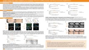


 沪公网安备31010102008431号