Lopez-Izquierdo A et al. (NOV 2014)
American journal of physiology. Heart and circulatory physiology 307 9 H1370--7
A near-infrared fluorescent voltage-sensitive dye allows for moderate-throughput electrophysiological analyses of human induced pluripotent stem cell-derived cardiomyocytes.
Human induced pluripotent stem cell-derived cardiomyocyte (iPSC-CM)-based assays are emerging as a promising tool for the in vitro preclinical screening of QT interval-prolonging side effects of drugs in development. A major impediment to the widespread use of human iPSC-CM assays is the low throughput of the currently available electrophysiological tools. To test the precision and applicability of the near-infrared fluorescent voltage-sensitive dye 1-(4-sulfanatobutyl)-4-β[2-(di-n-butylamino)-6-naphthyl]butadienylquinolinium betaine (di-4-ANBDQBS) for moderate-throughput electrophysiological analyses,we compared simultaneous transmembrane voltage and optical action potential (AP) recordings in human iPSC-CM loaded with di-4-ANBDQBS. Optical AP recordings tracked transmembrane voltage with high precision,generating nearly identical values for AP duration (AP durations at 10%,50%,and 90% repolarization). Human iPSC-CMs tolerated repeated laser exposure,with stable optical AP parameters recorded over a 30-min study period. Optical AP recordings appropriately tracked changes in repolarization induced by pharmacological manipulation. Finally,di-4-ANBDQBS allowed for moderate-throughput analyses,increasing throughput textgreater10-fold over the traditional patch-clamp technique. We conclude that the voltage-sensitive dye di-4-ANBDQBS allows for high-precision optical AP measurements that markedly increase the throughput for electrophysiological characterization of human iPSC-CMs.
View Publication
Pei Y et al. (MAY 2016)
Brain research 1638 Pt A 57--73
Comparative neurotoxicity screening in human iPSC-derived neural stem cells, neurons and astrocytes.
Induced pluripotent stem cells (iPSC) and their differentiated derivatives offer a unique source of human primary cells for toxicity screens. Here,we report on the comparative cytotoxicity of 80 compounds (neurotoxicants,developmental neurotoxicants,and environmental compounds) in iPSC as well as isogenic iPSC-derived neural stem cells (NSC),neurons,and astrocytes. All compounds were tested over a 24-h period at 10 and 100$\$,in duplicate,with cytotoxicity measured using the MTT assay. Of the 80 compounds tested,50 induced significant cytotoxicity in at least one cell type; per cell type,32,38,46,and 41 induced significant cytotoxicity in iPSC,NSC,neurons,and astrocytes,respectively. Four compounds (valinomycin,3,3',5,5'-tetrabromobisphenol,deltamethrin,and triphenyl phosphate) were cytotoxic in all four cell types. Retesting these compounds at 1,10,and 100$\$ using the same exposure protocol yielded consistent results as compared with the primary screen. Using rotenone,we extended the testing to seven additional iPSC lines of both genders; no substantial difference in the extent of cytotoxicity was detected among the cell lines. Finally,the cytotoxicity assay was simplified by measuring luciferase activity using lineage-specific luciferase reporter iPSC lines which were generated from the parental iPSC line. This article is part of a Special Issue entitled SI: PSC and the brain.
View Publication
Xu H et al. (JUL 2016)
Organic & biomolecular chemistry 14 26 6179--83
Cellular thermal shift and clickable chemical probe assays for the determination of drug-target engagement in live cells.
Proof of drug-target engagement in physiologically-relevant contexts is a key pillar of successful therapeutic target validation. We developed two orthogonal technologies,the cellular thermal shift assay (CETSA) and a covalent chemical probe reporter approach (harnessing sulfonyl fluoride tyrosine labeling and subsequent click chemistry) to measure the occupancy of the mRNA-decapping scavenger enzyme DcpS by a small molecule inhibitor in live cells. Enzyme affinity determined using isothermal dose response fingerprinting (ITDRFCETSA) and the concentration required to occupy 50% of the enzyme (OC50) using the chemical probe reporter assay were very similar. In this case,the chemical probe method worked well due to the long offset kinetics of the reversible inhibitor (determined using a fluorescent dye-tagged probe). This work suggests that CETSA could become the first choice assay to determine in-cell target engagement due to its simplicity.
View Publication
High-Dose Fluoride Impairs the Properties of Human Embryonic Stem Cells via JNK Signaling
Fluoride is a ubiquitous natural substance that is often used in dental products to prevent dental caries. The biphasic actions of fluoride imply that excessive systemic exposure to fluoride can cause harmful effects on embryonic development in both animal models and humans. However,insufficient information is available on the effects of fluoride on human embryonic stem cells (hESCs),which is a novel in vitro humanized model for analyzing the embryotoxicities of chemical compounds. Therefore,we investigated the effects of sodium fluoride (NaF) on the proliferation,differentiation and viability of H9 hESCs. For the first time,we showed that 1 mM NaF did not significantly affect the proliferation of hESCs but did disturb the gene expression patterns of hESCs during embryoid body (EB) differentiation. Higher doses of NaF (2 mM and above) markedly decreased the viability and proliferation of hESCs. The mode and underlying mechanism of high-dose NaF-induced cell death were further investigated by assessing the sub-cellular morphology,mitochondrial membrane potential (MMP),caspase activities,cellular reactive oxygen species (ROS) levels and activation of mitogen-activated protein kinases (MAPKs). High-dose NaF caused the death of hESCs via apoptosis in a caspase-mediated but ROS-independent pathway,coupled with an increase in the phospho-c-Jun N-terminal kinase (p-JNK) levels. Pretreatment with a pJNK-specific inhibitor (SP600125) could effectively protect hESCs from NaF-induced cell death in a concentration- and time-dependent manner. These findings suggest that NaF might interfere with early human embryogenesis by disturbing the specification of the three germ layers as well as osteogenic lineage commitment and that high-dose NaF could cause apoptosis through a JNK-dependent pathway in hESCs.
View Publication
Amelioration of murine beta-thalassemia through drug selection of hematopoietic stem cells transduced with a lentiviral vector encoding both gamma-globin and the MGMT drug-resistance gene.
Correction of murine models of beta-thalassemia has been achieved through high-level globin lentiviral vector gene transfer into mouse hematopoietic stem cells (HSCs). However,transduction of human HSCs is less robust and may be inadequate to achieve therapeutic levels of genetically modified erythroid cells. We therefore developed a double gene lentiviral vector encoding both human gamma-globin under the transcriptional control of erythroid regulatory elements and methylguanine methyltransferase (MGMT),driven by a constitutive cellular promoter. MGMT expression provides cellular resistance to alkylator drugs,which can be administered to kill residual untransduced,diseased HSCs,whereas transduced cells are protected. Mice transplanted with beta-thalassemic HSCs transduced with a gamma-globin/MGMT vector initially had subtherapeutic levels of red cells expressing gamma-globin. To enrich gamma-globin-expressing cells,transplanted mice were treated with the alkylator agent 1,3-bis-chloroethyl-1-nitrosourea. This resulted in significant increases in the number of gamma-globin-expressing red cells and the amount of fetal hemoglobin,leading to resolution of anemia. Selection of transduced HSCs was also obtained when cells were drug-treated before transplantation. Mice that received these cells demonstrated reconstitution with therapeutic levels of gamma-globin-expressing cells. These data suggest that MGMT-based drug selection holds promise as a modality to improve gene therapy for beta-thalassemia.
View Publication
产品类型:
产品号#:
09600
09650
产品名:
StemSpan™ SFEM
StemSpan™ SFEM
文献
Khalid O et al. (MAY 2014)
Stem Cell Research 12 3 791--806
Gene expression signatures affected by alcohol-induced DNA methylomic deregulation in human embryonic stem cells
Stem cells,especially human embryonic stem cells (hESCs),are useful models to study molecular mechanisms of human disorders that originate during gestation. Alcohol (ethanol,EtOH) consumption during pregnancy causes a variety of prenatal and postnatal disorders collectively referred to as fetal alcohol spectrum disorders (FASDs). To better understand the molecular events leading to FASDs,we performed a genome-wide analysis of EtOH's effects on the maintenance and differentiation of hESCs in culture. Gene Co-expression Network Analysis showed significant alterations in gene profiles of EtOH-treated differentiated or undifferentiated hESCs,particularly those associated with molecular pathways for metabolic processes,oxidative stress,and neuronal properties of stem cells. A genome-wide DNA methylome analysis revealed widespread EtOH-induced alterations with significant hypermethylation of many regions of chromosomes. Undifferentiated hESCs were more vulnerable to EtOH's effect than their differentiated counterparts,with methylation on the promoter regions of chromosomes 2,16 and 18 in undifferentiated hESCs most affected by EtOH exposure. Combined transcriptomic and DNA methylomic analysis produced a list of differentiation-related genes dysregulated by EtOH-induced DNA methylation changes,which likely play a role in EtOH-induced decreases in hESC pluripotency. DNA sequence motif analysis of genes epigenetically altered by EtOH identified major motifs representing potential binding sites for transcription factors. These findings should help in deciphering the precise mechanisms of alcohol-induced teratogenesis. ?? 2014 Published by Elsevier B.V.
View Publication

 EasySep™小鼠TIL(CD45)正选试剂盒


 文献
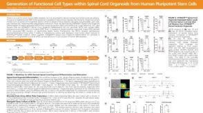
文献 科学海报Generation of Functional 3D Spinal Cord Organoids from Human Pluripotent Stem Cells
 实验方案How to Culture Human Pluripotent Stem Cell (hPSC)-Derived Forebrain Neurons for MEA Analysis Using the Maestro MEA™ System
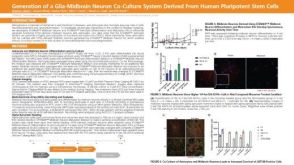 科学海报Generation of a Glia-Midbrain Neuron Co-Culture System Derived From Human Pluripotent Stem Cells

 沪公网安备31010102008431号