Song DH et al. (AUG 2000)
Journal of Biological Chemistry 275 31 23790--97
Endogenous protein kinase CK2 participates in Wnt signaling in mammary epithelial cells
Protein kinase CK2 (formerly casein kinase II) is a serine/threonine kinase overexpressed in many human tumors,transformed cell lines,and rapidly proliferating tissues. Recent data have shown that many cancers involve inappropriate reactivation of Wnt signaling through ectopic expression of Wnts themselves,as has been seen in a number of human breast cancers,or through mutation of intermediates in the Wnt pathway,such as adenomatous polyposis coli or beta-catenin,as described in colon and other cancers. Wnts are secreted factors that are important in embryonic development,but overexpression of certain Wnts,such as Wnt-1,leads to proliferation and transformation of cells. We report that upon stable transfection of Wnt-1 into the mouse mammary epithelial cell line C57MG,morphological changes and increased proliferation are accompanied by increased levels of CK2,as well as of beta-catenin. CK2 and beta-catenin co-precipitate with the Dvl proteins,which are Wnt signaling intermediates. A major phosphoprotein of the size of beta-catenin appears in in vitro kinase reactions performed on the Dvl immunoprecipitates. In vitro translated beta-catenin,Dvl-2,and Dvl-3 are phosphorylated by CK2. The selective CK2 inhibitor apigenin blocks proliferation of Wnt-1-transfected cells,abrogates phosphorylation of beta-catenin,and reduces beta-catenin and Dvl protein levels. These results demonstrate that endogenous CK2 is a positive regulator of Wnt signaling and growth of mammary epithelial cells.
View Publication
产品类型:
产品号#:
03800
03801
03802
03803
03804
03805
03806
产品名:
ClonaCell™-HY 杂交瘤试剂盒
ClonaCell™-HY Medium
ClonaCell™-HY Medium
ClonaCell™-HY Medium
ClonaCell™-HY Medium
ClonaCell™-HY Medium
ClonaCell™-HY PEG (融合)
Kaur R et al. (DEC 2013)
Journal of biomolecular screening 18 10 1223--33
A phenotypic screening approach in cord blood-derived mast cells to identify anti-inflammatory compounds.
Mast cells are unique hematopoietic cells that are richly distributed in the skin and mucosal surfaces of the respiratory and gastrointestinal tract. They play a key role in allergic inflammation by releasing a cocktail of granular constituents,including histamine,serine proteases,and various eicosanoids and cytokines. As such,a number of drugs target either inhibition of mast cell degranulation or the products of degranulation. To identify potential novel drugs and mechanisms in mast cell biology,assays were developed to identify inhibitors of mast cell degranulation and activation in a phenotypic screen. Due to the challenges associated with obtaining primary mast cells,cord blood-derived mononuclear cells were reproducibly differentiated to mast cells and assays developed to monitor tryptase release and prostaglandin D2 generation. The tryptase assay was particularly sensitive,requiring only 500 cells per data point,which permitted a set of approximately 12,000 compounds to be screened robustly and cost-effectively. Active compounds were tested for concomitant inhibition of prostaglandin D2 generation. This study demonstrates the robustness and effectiveness of this approach in the identification of potential novel compounds and mechanisms targeting mast cell-driven inflammation,to enable innovative drug discovery efforts to be prosecuted.
View Publication
Lee Y-KK et al. (JAN 2016)
International journal of cardiology 203 964--971
Efficient attenuation of Friedreich's ataxia (FRDA) cardiomyopathy by modulation of iron homeostasis-human induced pluripotent stem cell (hiPSC) as a drug screening platform for FRDA.
BACKGROUND Friedreich's ataxia (FRDA),a recessive neurodegenerative disorder commonly associated with hypertrophic cardiomyopathy,is caused by silencing of the frataxin (FXN) gene encoding the mitochondrial protein involved in iron-sulfur cluster biosynthesis. METHODS Application of our previously established FRDA human induced pluripotent stem cell (hiPSC) derived cardiomyocytes model as a platform to assess the efficacy of treatment with either the antioxidant coenzyme Q10 analog,idebenone (IDE) or the iron chelator,deferiprone (DFP),which are both under clinical trial. RESULTS DFP was able to more significantly suppress synthesis of reactive oxygen species (ROS) than IDE at the dosages of 25 $\$ and 10nM respectively which agreed with the reduced rate of intracellular accumulation of iron by DFP treatment from 25 to 50 $\$ With regard to cardiac electrical-contraction (EC) coupling function,decay velocity of calcium handling kinetics in FRDA-hiPSC-cardiomyocytes was significantly improved by DFP treatment but not by IDE. Further mechanistic studies revealed that DFP also modulated iron induced mitochondrial stress as reflected by mitochondria network disorganization and decline level of respiratory chain protein,succinate dehydrogenase (CxII) and cytochrome c oxidase (COXIV). In addition,iron-response protein (IRP-1) regulatory loop was overridden by DFP as reflected by resumed level of ferritin (FTH) back to basal level and the attenuated transferrin receptor (TSFR) mRNA level suppression thereby reducing further iron uptake. CONCLUSIONS DFP modulated iron homeostasis in FRDA-hiPSC-cardiomyocytes and effectively relieved stress-stimulation related to cardiomyopathy. The resuming of redox condition led to the significantly improved cardiac prime events,cardiac electrical-coupling during contraction.
View Publication
Chen C et al. (JUL 2014)
Nature communications 5 4430
Role of astroglia in Down's syndrome revealed by patient-derived human-induced pluripotent stem cells.
Down's syndrome (DS),caused by trisomy of human chromosome 21,is the most common genetic cause of intellectual disability. Here we use induced pluripotent stem cells (iPSCs) derived from DS patients to identify a role for astrocytes in DS pathogenesis. DS astroglia exhibit higher levels of reactive oxygen species and lower levels of synaptogenic molecules. Astrocyte-conditioned medium collected from DS astroglia causes toxicity to neurons,and fails to promote neuronal ion channel maturation and synapse formation. Transplantation studies show that DS astroglia do not promote neurogenesis of endogenous neural stem cells in vivo. We also observed abnormal gene expression profiles from DS astroglia. Finally,we show that the FDA-approved antibiotic drug,minocycline,partially corrects the pathological phenotypes of DS astroglia by specifically modulating the expression of S100B,GFAP,inducible nitric oxide synthase,and thrombospondins 1 and 2 in DS astroglia. Our studies shed light on the pathogenesis and possible treatment of DS by targeting astrocytes with a clinically available drug.
View Publication
High-Dose Fluoride Impairs the Properties of Human Embryonic Stem Cells via JNK Signaling
Fluoride is a ubiquitous natural substance that is often used in dental products to prevent dental caries. The biphasic actions of fluoride imply that excessive systemic exposure to fluoride can cause harmful effects on embryonic development in both animal models and humans. However,insufficient information is available on the effects of fluoride on human embryonic stem cells (hESCs),which is a novel in vitro humanized model for analyzing the embryotoxicities of chemical compounds. Therefore,we investigated the effects of sodium fluoride (NaF) on the proliferation,differentiation and viability of H9 hESCs. For the first time,we showed that 1 mM NaF did not significantly affect the proliferation of hESCs but did disturb the gene expression patterns of hESCs during embryoid body (EB) differentiation. Higher doses of NaF (2 mM and above) markedly decreased the viability and proliferation of hESCs. The mode and underlying mechanism of high-dose NaF-induced cell death were further investigated by assessing the sub-cellular morphology,mitochondrial membrane potential (MMP),caspase activities,cellular reactive oxygen species (ROS) levels and activation of mitogen-activated protein kinases (MAPKs). High-dose NaF caused the death of hESCs via apoptosis in a caspase-mediated but ROS-independent pathway,coupled with an increase in the phospho-c-Jun N-terminal kinase (p-JNK) levels. Pretreatment with a pJNK-specific inhibitor (SP600125) could effectively protect hESCs from NaF-induced cell death in a concentration- and time-dependent manner. These findings suggest that NaF might interfere with early human embryogenesis by disturbing the specification of the three germ layers as well as osteogenic lineage commitment and that high-dose NaF could cause apoptosis through a JNK-dependent pathway in hESCs.
View Publication
Khalid O et al. (MAY 2014)
Stem Cell Research 12 3 791--806
Gene expression signatures affected by alcohol-induced DNA methylomic deregulation in human embryonic stem cells
Stem cells,especially human embryonic stem cells (hESCs),are useful models to study molecular mechanisms of human disorders that originate during gestation. Alcohol (ethanol,EtOH) consumption during pregnancy causes a variety of prenatal and postnatal disorders collectively referred to as fetal alcohol spectrum disorders (FASDs). To better understand the molecular events leading to FASDs,we performed a genome-wide analysis of EtOH's effects on the maintenance and differentiation of hESCs in culture. Gene Co-expression Network Analysis showed significant alterations in gene profiles of EtOH-treated differentiated or undifferentiated hESCs,particularly those associated with molecular pathways for metabolic processes,oxidative stress,and neuronal properties of stem cells. A genome-wide DNA methylome analysis revealed widespread EtOH-induced alterations with significant hypermethylation of many regions of chromosomes. Undifferentiated hESCs were more vulnerable to EtOH's effect than their differentiated counterparts,with methylation on the promoter regions of chromosomes 2,16 and 18 in undifferentiated hESCs most affected by EtOH exposure. Combined transcriptomic and DNA methylomic analysis produced a list of differentiation-related genes dysregulated by EtOH-induced DNA methylation changes,which likely play a role in EtOH-induced decreases in hESC pluripotency. DNA sequence motif analysis of genes epigenetically altered by EtOH identified major motifs representing potential binding sites for transcription factors. These findings should help in deciphering the precise mechanisms of alcohol-induced teratogenesis. ?? 2014 Published by Elsevier B.V.
View Publication
产品类型:
产品号#:
05850
05857
05870
05875
07920
85850
85857
85870
85875
产品名:
ACCUTASE™
mTeSR™1
mTeSR™1
Kumagai T et al. (JUN 2003)
Journal of the National Cancer Institute 95 12 896--905
Vitamin D2 analog 19-nor-1,25-dihydroxyvitamin D2: antitumor activity against leukemia, myeloma, and colon cancer cells.
BACKGROUND: 1,25-Dihydroxyvitamin D(3) inhibits growth of several types of human cancer cells in vitro,but its therapeutic use is hampered because it causes hypercalcemia. 19-nor-1,25-Dihydroxyvitamin D(2) (paricalcitol) is a noncalcemic vitamin D analog that is approved by the Food and Drug Administration for the treatment of secondary hyperparathyroidism. We investigated the antitumor activity and mechanism of action of paricalcitol in vitro and in vivo. METHODS: Effects of paricalcitol on proliferation,the cell cycle,differentiation,and apoptosis were examined in cancer cell lines. Effects on tumor growth were examined with colon cancer cell xenografts in nude mice (five in the experimental group and five in the control group). The interaction of paricalcitol with the vitamin D receptor (VDR) in mononuclear spleen cells and myeloid stem cells from wild-type and VDR knockout mice was examined. All statistical tests were two-sided. RESULTS: Paricalcitol inhibited the proliferation of myeloid leukemia cell lines HL-60,NB-4,and THP-1 cells at an effective dose that inhibited growth 50% (ED(50)) of 2.4-5.8 x 10(-9) M by inducing cell cycle arrest and differentiation. Paricalcitol inhibited the proliferation of NCI-H929 myeloma cells at an ED(50) of 2.0 x 10(-10) M by inducing cell cycle arrest and apoptosis. Paricalcitol also inhibited the proliferation of colon cancer cell lines HT-29 (ED(50) = 1.7 x 10(-8) M) and SW837 (ED(50) = 3.2 x 10(-8) M). HT-29 colon cancer xenografts in paricalcitol-treated nude mice were smaller (1044 mm(3) and 1752 mm(3),difference = 708 mm(3),95% confidence interval = 311 to 1104 mm(3); P =.03) and weighed less (1487 mg and 4162 mg,difference = 2675 mg,95% confidence interval = 2103 to 3248 mg; Ptextless.001) than those in vehicle-treated mice. Paricalcitol induced committed myeloid hematopoietic stem cells from wild-type but not from VDR knockout mice to differentiate as macrophages. CONCLUSION: Paricalcitol has anticancer activity against myeloid leukemia,myeloma,and colon cancer cells that may be mediated through the VDR. Because it has been approved by the Food and Drug Administration,clinical trials of this agent in certain cancers are reasonable.
View Publication

 EasySep™小鼠TIL(CD45)正选试剂盒


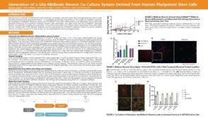 科学海报Generation of a Glia-Midbrain Neuron Co-Culture System Derived From Human Pluripotent Stem Cells


 沪公网安备31010102008431号