Adherent cells generated during long-term culture of human umbilical cord blood CD34+ cells have characteristics of endothelial cells and beneficial effect on cord blood ex vivo expansion.
Hematopoiesis depends on the association of hematopoietic stem cells with stromal cells that constitute the hematopoietic microenvironment. The in vitro development of the endothelial cell from umbilical cord blood (UCB) is not well established and has met very limited success. In this study,UCB CD34(+) cells were cultured for 5 weeks in a stroma-free liquid culture system using thrombopoietin,flt3 ligand,and granulocyte-colony stimulating factor. By week 4-5,we found that firmly adherent fibroblast-like cells were established. These cells showed characteristics of endothelial cells expressing von Willebrand factor,human vascular cell adhesion molecule-1,human intracellular adhesion molecule-1,human CD31,E-selectin,and human macrophage. Furthermore,when comparing an ex vivo system without an established endothelial monolayer to an ex vivo system with an established endothelial monolayer,better expansion of total nucleated cells,CD34(+) cells,and colony-forming units (CFUs)-granulocyte-macrophage and CFUs-granulocyte-erythroid-megakaryocyte-macrophage were found during culture. This phenomenon was in part due to the fact that a significant reduction of apoptotic fractions was found in the CD34(+) cells,which were cultured on the adherent monolayer for up to 5 weeks. To gather quantitative data on the number of endothelial cells derived from a given number of CD34 cells,we performed limiting dilution assay by using Poisson distribution: the number of tested cells (linear scale) producing a 37% negative culture (logarithmic scale) is the number of cells containing one endothelial cell. By this method,one endothelial cell may be found from 314 CD34(+) cells after 5 weeks of culture. These results suggest that the UCB CD34(+) cell fraction contains endothelial cell precursors,establishing the hematopoietic microenvironment and providing the beneficial effects through downregulating apoptosis on UCB expansion protocols. These observations may provide insight for future cellular therapy or graft engineering.
View Publication
产品类型:
产品号#:
04434
04444
产品名:
MethoCult™ H4434 Classic
MethoCult™ H4434 Classic
Liu C et al. (DEC 2010)
Blood 116 25 5518--27
Progenitor cell dose determines the pace and completeness of engraftment in a xenograft model for cord blood transplantation.
Two critical concerns in clinical cord blood transplantation are the initial time to engraftment and the subsequent restoration of immune function. These studies measured the impact of progenitor cell dose on both the pace and strength of hematopoietic reconstitution by transplanting nonobese diabetic/severe combined immunodeficiency/interleukin-2 receptor-gamma-null (NSγ) mice with lineage-depleted aldehyde dehydrogenase-bright CD34(+) human cord blood progenitors. The progress of each transplant was monitored over an extended time course by repeatedly analyzing the peripheral blood for human hematopoietic cells. In vivo human hematopoietic development was complete. After long-term transplantation assays (≥ 19 weeks),human T-cell development was documented within multiple tissues in 16 of 32 NSγ mice. Human T-cell differentiation was active within NSγ thymuses,as documented by the presence of CD4(+) CD8(+) T-cell progenitors as well as T-cell receptor excision circles. It is important to note that although myeloid and B-cell engraftment was detected as early as 4 weeks after transplantation,human T-cell development was exclusively late onset. High progenitor cell doses were associated with a robust human hematopoietic chimerism that accelerated both initial time to engraftment and subsequent T-cell development. At lower progenitor cell doses,the chimerism was weak and the human hematopoietic lineage development was frequently incomplete.
View Publication
Identification of a novel class of human adherent CD34- stem cells that give rise to SCID-repopulating cells.
Here we describe the in vitro generation of a novel adherent cell fraction derived from highly enriched,mobilized CD133(+) peripheral blood cells after their culture with Flt3/Flk2 ligand and interleukin-6 for 3 to 5 weeks. These cells lack markers of hematopoietic stem cells,endothelial cells,mesenchymal cells,dendritic cells,and stromal fibroblasts. However,all adherent cells expressed the adhesion molecules VE-cadherin,CD54,and CD44. They were also positive for CD164 and CD172a (signal regulatory protein-alpha) and for a stem cell antigen defined by the recently described antibody W7C5. Adherent cells can either spontaneously or upon stimulation with stem cell factor give rise to a transplantable,nonadherent CD133(+)CD34(-) stem cell subset. These cells do not generate in vitro hematopoietic colonies. However,their transplantation into nonobese diabetic/severe combined immunodeficiency (NOD/SCID) mice induced substantially higher long-term multilineage engraftment compared with that of freshly isolated CD34(+) cells,suggesting that these cells are highly enriched in SCID-repopulating cells. In addition to cells of the myeloid lineage,nonadherent CD34(-) cells were able to give rise to human cells with B-,T-,and natural killer-cell phenotype. Hence,these cells possess a distinct in vivo differentiation potential compared with that of CD34(+) stem cells and may therefore provide an alternative to CD34(+) progenitor cells for transplantation.
View Publication
Jaatinen T et al. (MAR 2006)
Stem cells (Dayton,Ohio) 24 3 631--41
Global gene expression profile of human cord blood-derived CD133+ cells.
Human cord blood (CB)-derived CD133+ cells carry characteristics of primitive hematopoietic cells and proffer an alternative for CD34+ cells in hematopoietic stem cell (HSC) transplantation. To characterize the CD133+ cell population on a genetic level,a global expression analysis of CD133+ cells was performed using oligonucleotide microarrays. CD133+ cells were purified from four fresh CB units by immunomagnetic selection. All four CD133+ samples showed significant similarity in their gene expression pattern,whereas they differed clearly from the CD133- control samples. In all,690 transcripts were differentially expressed between CD133+ and CD133- cells. Of these,393 were increased and 297 were decreased in CD133+ cells. The highest overexpression was noted in genes associated with metabolism,cellular physiological processes,cell communication,and development. A set of 257 transcripts expressed solely in the CD133+ cell population was identified. Colony-forming unit (CFU) assay was used to detect the clonal progeny of precursors present in the studied cell populations. The results demonstrate that CD133+ cells express primitive markers and possess clonogenic progenitor capacity. This study provides a gene expression profile for human CD133+ cells. It presents a set of genes that may be used to unravel the properties of the CD133+ cell population,assumed to be highly enriched in HSCs.
View Publication
Chen X et al. (SEP 2006)
Stem cells (Dayton,Ohio) 24 9 2052--9
Bioreactor expansion of human adult bone marrow-derived mesenchymal stem cells.
Supplementation of mesenchymal stem cells (MSCs) during hematopoietic stem cell (HSC) transplantation alleviates complications such as graft-versus-host disease,leading to a speedy recovery of hematopoiesis. To meet this clinical demand,a fast MSC expansion method is required. In the present study,we examined the feasibility of using a rotary bioreactor system to expand MSCs from isolated bone marrow mononuclear cells. The cells were cultured in a rotary bioreactor with Myelocult medium containing a combination of supplementary factors,including stem cell factor and interleukin-3 and -6. After 8 days of culture,total cell numbers,Stro-1(+)CD44(+)CD34(-) MSCs,and CD34(+)CD44(+)Stro-1(-) HSCs were increased 9-,29-,and 8-fold,respectively. Colony-forming efficiency-fibroblast per day of the bioreactor-treated cells was 1.44-fold higher than that of the cells without bioreactor treatment. The bioreactor-expanded MSCs showed expression of primitive MSC markers endoglin (SH2) and vimentin,whereas markers associated with lineage differentiation,including osteocalcin (osteogenesis),type II collagen (chondrogenesis),and C/EBP-alpha (CCAAT/enhancer-binding protein-alpha) (adipogenesis),were not detected. Upon induction,the bioreactor-expanded MSCs were able to differentiate into osteoblasts,chondrocytes,and adipocytes. We conclude that the rotary bioreactor with the modified Myelocult medium reported in this study may be used to rapidly expand MSCs.
View Publication
Clonal analysis of deletions on chromosome 20q and JAK2-V617F in MPD suggests that del20q acts independently and is not one of the predisposing mutations for JAK2-V617F.
We developed a real-time copy number polymerase chain reaction assay for deletions on chromosome 20q (del20q),screened peripheral blood granulocytes from 664 patients with myeloproliferative disorders,and identified 19 patients with del20q (2.9%),of which 14 (74%) were also positive for JAK2-V617F. To examine the temporal relationship between the occurrence of del20q and JAK2-V617F,we performed colony assays in methylcellulose,picked individual burst-forming units-erythroid (BFU-E) and colony-forming units-granulocyte (CFU-G) colonies,and genotyped each colony individually for del20q and JAK2-V617F. In 2 of 9 patients,we found that some colonies with del20q carried only wild-type JAK2,whereas other del20q colonies were JAK2-V617F positive,indicating that del20q occurred before the acquisition of JAK2-V617F. However,in colonies from 3 of 9 patients,we observed the opposite order of events. The lack of a strict temporal order of occurrence makes it doubtful that del20q represents a predisposing event for JAK2-V617F. In 2 patients with JAK2-V617F and 1 patient with MPL-W515L,microsatellite analysis revealed that del20q affected chromosomes of different parental origin and/or 9pLOH occurred at least twice. The fact that rare somatic events,such as del20q or 9pLOH,occurred more than once in subclones from the same patients suggests that the myeloproliferative disorder clone carries a predisposition to acquiring such genetic alterations.
View Publication

 EasySep™小鼠TIL(CD45)正选试剂盒


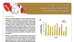 技术公告Depletion of Red Blood Cells from Fresh Blood Samples


 沪公网安备31010102008431号