Distinct roles of integrins alpha6 and alpha4 in homing of fetal liver hematopoietic stem and progenitor cells.
Homing of hematopoietic stem cells (HSCs) into the bone marrow (BM) is a prerequisite for establishment of hematopoiesis during development and following transplantation. However,the molecular interactions that control homing of HSCs,in particular,of fetal HSCs,are not well understood. Herein,we studied the role of the alpha6 and alpha4 integrin receptors for homing and engraftment of fetal liver (FL) HSCs and hematopoietic progenitor cells (HPCs) to adult BM by using integrin alpha6 gene-deleted mice and function-blocking antibodies. Both integrins were ubiquitously expressed in FL Lin(-)Sca-1(+)Kit(+) (LSK) cells. Deletion of integrin alpha6 receptor or inhibition by a function-blocking antibody inhibited FL LSK cell adhesion to its extracellular ligands,laminins-411 and -511 in vitro,and significantly reduced homing of HPCs to BM. In contrast,the anti-integrin alpha6 antibody did not inhibit BM homing of HSCs. In agreement with this,integrin alpha6 gene-deleted FL HSCs did not display any homing or engraftment defect compared with wild-type littermates. In contrast,inhibition of integrin alpha4 receptor by a function-blocking antibody virtually abrogated homing of both FL HSCs and HPCs to BM,indicating distinct functions for integrin alpha6 and alpha4 receptors during homing of fetal HSCs and HPCs.
View Publication
Reconstitution of the functional human hematopoietic microenvironment derived from human mesenchymal stem cells in the murine bone marrow compartment.
Hematopoiesis is maintained by specific interactions between both hematopoietic and nonhematopoietic cells. Whereas hematopoietic stem cells (HSCs) have been extensively studied both in vitro and in vivo,little is known about the in vivo characteristics of stem cells of the nonhematopoietic component,known as mesenchymal stem cells (MSCs). Here we have visualized and characterized human MSCs in vivo following intramedullary transplantation of enhanced green fluorescent protein-marked human MSCs (eGFP-MSCs) into the bone marrow (BM) of nonobese diabetic/severe combined immunodeficiency (NOD/SCID) mice. Between 4 to 10 weeks after transplantation,eGFP-MSCs that engrafted in murine BM integrated into the hematopoietic microenvironment (HME) of the host mouse. They differentiated into pericytes,myofibroblasts,BM stromal cells,osteocytes in bone,bone-lining osteoblasts,and endothelial cells,which constituted the functional components of the BM HME. The presence of human MSCs in murine BM resulted in an increase in functionally and phenotypically primitive human hematopoietic cells. Human MSC-derived cells that reconstituted the HME appeared to contribute to the maintenance of human hematopoiesis by actively interacting with primitive human hematopoietic cells.
View Publication
产品类型:
产品号#:
04034
04044
产品名:
MethoCult™ H4034 Optimum
MethoCult™ H4034 Optimum
Andreani M et al. (JAN 2011)
Haematologica 96 1 128--33
Quantitatively different red cell/nucleated cell chimerism in patients with long-term, persistent hematopoietic mixed chimerism after bone marrow transplantation for thalassemia major or sickle cell disease.
BACKGROUND: Persistent mixed chimerism represents a state in which recipient and donor cells stably co-exist after hematopoietic stem cell transplantation. However,since in most of the studies reported in literature the engraftment state was observed in the nucleated cells,in this study we determined the donor origin of the mature erythrocytes of patients with persistent mixed chimerism after transplantation for hemoglobinopathies. Results were compared with the engraftment state observed in singly picked out burst-forming unit - erythroid colonies and in the nucleated cells collected from the peripheral blood and from the bone marrow. DESIGN AND METHODS: The donor origin of the erythrocytes was determined analyzing differences on the surface antigens of the erythrocyte suspension after incubation with anti-ABO and/or anti-C,-c,-D,-E and -e monoclonal antibodies by a flow cytometer. Analysis of short tandem repeats was used to determine the donor origin of nucleated cells and burst-forming unit - erythroid colonies singly picked out after 14 days of incubation. RESULTS: The proportions of donor-derived nucleated cells in four transplanted patients affected by hemoglobinopathies were 71%,46%,15% and 25% at day 1364,1385,1314 and 932,respectively. Similar results were obtained for the erythroid precursors,analyzing the donor/recipient origin of the burst-forming unit - erythroid colonies. In contrast,on the same days of observation,the proportions of donor-derived erythrocytes in the four patients with persistent mixed chimerism were 100%,100%,73% and 90%. Conclusions Our results showed that most of the erythrocytes present in four long-term transplanted patients affected by hemoglobinopathies and characterized by the presence of few donor engrafted nucleated cells were of donor origin. The indication that small proportions of donor engrafted cells might be sufficient for clinical control of the disease in patients affected by hemoglobinopathies is relevant,although the biological mechanisms underlying these observations need further investigation.
View Publication
产品类型:
产品号#:
04434
04444
84434
84444
产品名:
MethoCult™ H4434 Classic
MethoCult™ H4434 Classic
Zhu F et al. (SEP 2014)
Stem cells and development 23 17 2119--2125
A modified method for implantation of pluripotent stem cells under the rodent kidney capsule.
Teratoma formation,the standard in vivo pluripotency assay,is also frequently used as a tumorigenicity assay. A common concern in therapeutic stem cell applications is the tumorigenicity potential of a small number of cell impurities in the final product. Estimation of this small number is hampered by the inaccurate methodology of the tumorigenicity assay. Hence,a protocol for tumorigenicity assay that can deliver a defined number of cells,without error introduced by leakage or migration of cells is needed. In this study,we tested our modified transplantation method that allows for transplant of small numbers of pluripotent stem cells (PSCs) under the kidney capsule with minimal cell leakage. A glass capillary with a finely shaped tip and an attached mouth pipette was used to inject PSCs into the rodent kidney capsule. H9 embryonic and induced PSCs were tagged with Fluc and green fluorescence protein reporter genes and divided in different cell doses for transplantation. Bioluminescence imaging (BLI) on the day of surgery showed that the cell signal was confined to the kidney and signal intensity correlated with increasing transplant cell numbers. The overall cell leakage rate was 17% and the rodent survival rate was 96%. Teratoma formation was observed in rodents transplanted with cell numbers between 1 × 10(5)-2 × 10(6). We conclude that this modified procedure for transplanting PSCs under the kidney capsule allows for transplantation of a defined number of PSCs with significant reduction of error associated with cell leakage from the transplant site.
View Publication
产品类型:
产品号#:
05850
05857
05870
05875
85850
85857
85870
85875
产品名:
mTeSR™1
mTeSR™1
Jeong J et al. (OCT 2014)
Experimental and Molecular Pathology 97 2 253--258
Patient-tailored application for Duchene muscular dystrophy on mdx mice based induced mesenchymal stem cells
Mesenchymal stem cells (MSCs) may be used as powerful tools for the repair and regeneration of damaged tissues. However,isolating tissue specific-derived MSCs may cause pain and increased infection rates in patients,and repetitive isolations may be required. To overcome these difficulties,we have examined alternative methods for MSC production. Here,we show that induced pluripotent stem cells (iPSCs) may be differentiated into mesenchymal stem cells (iMSCs) following exposure to SB431542. Purified iMSCs were administered to mdx mice to study skeletal muscle regeneration in a murine model of muscular dystrophy. Purified iMSCs displayed fibroblast-like morphology,formed three-dimensional spheroid structures,and expressed characteristic mesenchymal stem cell surface markers such as CD29,CD33,CD73,CD90,and CD105. Moreover,iMSCs were capable of differentiating into adipogenic,osteogenic,and chondrogenic lineages. Transplanting iMSC cells to tibialis anterior skeletal muscle tissue in mdx mice lowered oxidative damage as evidenced by a reduction in nitrotyrosine levels,and normal dystrophin expression levels were restored. This study demonstrates the therapeutic potential of purified iMSCs in skeletal muscle regeneration in mdx mice,and suggests that iPSCs are a viable alternate source for deriving MSCs as needed. textcopyright 2014 Elsevier Inc.
View Publication
产品类型:
产品号#:
05850
05857
05870
05875
85850
85857
85870
85875
产品名:
mTeSR™1
mTeSR™1
Zimmerman Z et al. (AUG 2005)
Biology of Blood and Marrow Transplantation 11 8 576--86
Effector cells derived from host CD8 memory T cells mediate rapid resistance against minor histocompatibility antigen-mismatched allogeneic marrow grafts without participation of perforin, Fas ligand, and the simultaneous inhibition of 3 tumor necrosis Fa
Reduced-intensity conditioning regimens for transplant recipients have heightened awareness of immunologic resistance to allogeneic bone marrow transplants (BMT). Although T cell-mediated cytotoxicity has been assumed to play a role in the resistance against donor allogeneic hematopoietic stem and progenitor cell grafts,several studies have reported relatively unimpaired resistance by recipients who lack perforin,Fas ligand (FasL),and other cytotoxic mediators. This study compared the early kinetics of T cell-mediated resistance in B6 (H2b) cytotoxically normal versus deficient recipients after transplantation with major histocompatibility complex-matched,minor histocompatibility antigen (MiHA)-mismatched allogeneic marrow grafts. Wild-type B6 or cytotoxic double-deficient perforin-/-/ gld+/+ (B6-cdd) mice were sensitized against major histocompatibility complex-matched BALB.B or C3H.SW (H2b) MiHA and transplanted with a high dose (1 ?? 107) of T cell-depleted bone marrow. CD8 T memory cells were shown to be present in recipients before BMT,and anti-CD8 monoclonal antibody infusion abolished resistance,thus demonstrating that CD8 T cells are the host effector population. Donor-committed and high proliferative potential progenitor numbers were markedly diminished by 48 hours after transplantation in both wild-type B6 and B6-cdd anti-donor MiHA-sensitized recipients. These observations indicate that the resistance pathway used in the cytotoxic deficient mice was both potent and rapidly induced - consistent with a CD8 memory T-cell response. To examine the role of Tumor necrosis factor-like weak inducer of apoptosis (TWEAK)- and TL1A-mediated cytotoxicity in this strong resistance,newly generated monoclonal antibodies specific for these ligands were administered to B6-cdd recipients sensitized to donor antigens. Recipients of syngeneic B6-gfp bone marrow exhibited significant donor colony-forming unit numbers after BMT. In contrast,low or absent colony-forming unit levels were detected in allogeneic recipients,including those that lacked perforin and FasL and that received anti-TWEAK,anti-tumor necrosis factor-related apoptosis-inducing ligand,and anti-TL1A monoclonal antibodies. These findings extend previous observations by demonstrating the existence of a rapidly effected resistance pathway mediated by memory CD8 effector T cells independent of the 2 major pathways of cytotoxicity. Together with previous findings,these results support the notion that effector cells derived from memory CD8 T-cell populations can mediate strong resistance against donor allogeneic MiHA-disparate hematopoietic engraftment by using a mechanism that is independent of the contribution of perforin,FasL,and the known death ligand receptor pathways. ?? 2005 American Society for Blood and Marrow Transplantation.
View Publication
产品类型:
产品号#:
03800
03801
03802
03803
03804
03805
03806
产品名:
ClonaCell™-HY杂交瘤试剂盒
ClonaCell™-HY培养基A
ClonaCell™-HY 培养基 B
ClonaCell™-HY 培养基 C
ClonaCell™-HY 培养基 D
ClonaCell™-HY 培养基 E
ClonaCell™-HY PEG
Shimakura Y et al. (JAN 2000)
Stem cells (Dayton,Ohio) 18 3 183--9
Murine stromal cell line HESS-5 maintains reconstituting ability of Ex vivo-generated hematopoietic stem cells from human bone marrow and cytokine-mobilized peripheral blood.
Human bone marrow (BM) or mobilized peripheral blood (mPB) CD34(+) cells have been shown to loose their stem cell quality during culture period more easily than those from cord blood (CB). We previously reported that human umbilical CB stem cells could effectively be expanded in the presence of human recombinant cytokines and a newly established murine bone marrow stromal cell line HESS-5. In this study we assessed the efficacy of this xenogeneic coculture system using human BM and mPB CD34(+) cells as materials. We measured the generation of CD34(+)CD38(-) cells and colony-forming units,and assessed severe-combined immunodeficient mouse-repopulating cell (SRC) activity using cells five days after serum-free cytokine-containing culture in the presence or the absence of a direct contact with HESS-5 cells. As compared with the stroma-free culture,the xenogeneic coculture was significantly superior on expansion of CD34(+)CD38(-) cells and colony-forming cells and on maintenance of SRC activity. The PKH26 study demonstrated that cell division was promoted faster in cells cocultured with HESS-5 cells than in cells cultured without HESS-5 cells. These results indicate that HESS-5 supports rapid generation of primitive progenitor cells (PPC) and maintains reconstituting ability of newly generated stem cells during ex vivo culture irrespective of the source of samples. This xenogeneic coculture system will be useful for ex vivo manipulation such as gene transduction to promote cell division and the generation of PPC and to prevent loss of stem cell quality.
View Publication
产品类型:
产品号#:
04064
04034
04044
产品名:
MethoCult™ H4034 Optimum 入门试剂盒
MethoCult™ H4034 Optimum
MethoCult™ H4034 Optimum
Kang L et al. ( 2013)
Frontiers in immunology 4 MAY 101
Characterization and ex vivo Expansion of Human Placenta-Derived Natural Killer Cells for Cancer Immunotherapy.
Recent clinical studies suggest that adoptive transfer of donor-derived natural killer (NK) cells may improve clinical outcome in hematological malignancies and some solid tumors by direct anti-tumor effects as well as by reduction of graft versus host disease (GVHD). NK cells have also been shown to enhance transplant engraftment during allogeneic hematopoietic stem cell transplantation (HSCT) for hematological malignancies. The limited ex vivo expansion potential of NK cells from peripheral blood (PB) or umbilical cord blood (UCB) has however restricted their therapeutic potential. Here we define methods to efficiently generate NK cells from donor-matched,full-term human placenta perfusate (termed Human Placenta-Derived Stem Cell,HPDSC) and UCB. Following isolation from cryopreserved donor-matched HPDSC and UCB units,CD56+CD3- placenta-derived NK cells,termed pNK cells,were expanded in culture for up to 3 weeks to yield an average of 1.2 billion cells per donor that were textgreater80% CD56+CD3-,comparable to doses previously utilized in clinical applications. Ex vivo-expanded pNK cells exhibited a marked increase in anti-tumor cytolytic activity coinciding with the significantly increased expression of NKG2D,NKp46,and NKp44 (p textless 0.001,p textless 0.001,and p textless 0.05,respectively). Strong cytolytic activity was observed against a wide range of tumor cell lines in vitro. pNK cells display a distinct microRNA (miRNA) expression profile,immunophenotype,and greater anti-tumor capacity in vitro compared to PB NK cells used in recent clinical trials. With further development,pNK may represent a novel and effective cellular immunotherapy for patients with high clinical needs and few other therapeutic options.
View Publication
Pirson L et al. (JUL 2006)
Stem cells (Dayton,Ohio) 24 7 1814--21
Despite inhibition of hematopoietic progenitor cell growth in vitro, the tyrosine kinase inhibitor imatinib does not impair engraftment of human CD133+ cells into NOD/SCIDbeta2mNull mice.
There is potential interest for combining allogeneic hematopoietic cell transplantation (HCT),and particularly allogeneic HCT with a nonmyeloablative regimen,to the tyrosine kinase inhibitor imatinib (Glivec; Novartis,Basel,Switzerland,http://www.novartis.com) in order to maximize anti-leukemic activity against Philadelphia chromosome-positive leukemias. However,because imatinib inhibits c-kit,the stem cell factor receptor,it could interfere with bone marrow engraftment. In this study,we examined the impact of imatinib on normal progenitor cell function. Imatinib decreased the colony-forming capacity of mobilized peripheral blood human CD133(+) cells but not that of long-term culture-initiating cells. Imatinib also decreased the proliferation of cytokine-stimulated CD133(+) cells but did not induce apoptosis of these cells. Expression of very late antigen (VLA)-4,VLA-5,and CXCR4 of CD133(+) cells was not modified by imatinib,but imatinib decreased the ability of CD133(+) cells to migrate. Finally,imatinib did not decrease engraftment of CD133(+) cells into irradiated nonobese diabetic/severe combined immunodeficient/beta2m(null) mice conditioned with 3 or 1 Gy total body irradiation. In summary,our results suggest that,despite inhibition of hematopoietic progenitor cell growth in vitro,imatinib does not interfere with hematopoietic stem cell engraftment.
View Publication
产品类型:
产品号#:
05150
04435
04445
04960
04902
04900
04961
04901
04963
04962
04970
04971
产品名:
MyeloCult™ H5100
MethoCult™ H4435 Enriched
MethoCult™ H4435 Enriched
MegaCult™-C胶原和无细胞因子培养基
胶原蛋白溶液
MegaCult™-C无细胞因子培养基
MegaCult™-C胶原和含细胞因子培养基
MegaCult™-C含细胞因子培养基
双室载玻片套件
MegaCult™-C CFU-Mk染色试剂盒
MegaCult™-C无细胞因子全套试剂盒
MegaCult™-C含细胞因子全套试剂盒
Seeger FH et al. (MAR 2007)
European heart journal 28 6 766--72
Cell isolation procedures matter: a comparison of different isolation protocols of bone marrow mononuclear cells used for cell therapy in patients with acute myocardial infarction.
AIM: The recently published REPAIR-AMI and ASTAMI trial showed differences in contractile recovery of left ventricular function after infusion of bone marrow-derived cells in acute myocardial infarction. Since the trials used different protocols for cell isolation and storage (REPAIR-AMI: Ficoll,storage in X-vivo 10 medium plus serum; ASTAMI: Lymphoprep,storage in NaCl plus plasma),we compared the functional activity of BMC isolated by the two different protocols. METHODS AND RESULTS: The recovery of total cell number,colony-forming units (CFU),and the number of mesenchymal stem cells were significantly reduced to 77 +/- 4%,83 +/- 16%,and 65 +/- 15%,respectively,when using the ASTAMI protocol compared with the REPAIR protocol. The capacity of the isolated BMC to migrate in response to stromal cell-derived factor 1 (SDF-1) was profoundly reduced when using the ASTAMI cell isolation procedure (42 +/- 8% and 78 +/- 3% reduction in healthy and CAD-patient cells,respectively). Finally,infusion of BMC into a hindlimb ischaemia model demonstrated a significantly blunted blood-flow-recovery by BMC isolated with the ASTAMI protocol (54 +/- 6% of the effect obtained by REPAIR cells). Comparison of the individual steps identified the use of NaCl and plasma for cell storage as major factors for functional impairment of the BMC. CONCLUSION: Cell isolation protocols have a major impact on the functional activity of bone marrow-derived progenitor cells. The assessment of cell number and viability may not entirely reflect the functional capacity of cells in vivo. Additional functional testing appears to be mandatory to assure proper cell function before embarking on clinical cell therapy trials.
View Publication

 EasySep™小鼠TIL(CD45)正选试剂盒


 产品手册NeuroCult™-XF: Xeno-Free Culture Medium for the Proliferation of Human Neural Stem Cells
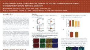 科学海报A Fully Defined Animal Component Free Medium for Efficient Differentiation of Human Pluripotent Stem Cells to Definitive Endoderm
 沪公网安备31010102008431号