Fortin G et al. (AUG 2009)
The Journal of experimental medicine 206 9 1995--2011
A role for CD47 in the development of experimental colitis mediated by SIRPalpha+CD103- dendritic cells.
Mesenteric lymph node (mLN) CD103 (alphaE integrin)(+) dendritic cells (DCs) induce regulatory T cells and gut tolerance. However,the function of intestinal CD103(-) DCs remains to be clarified. CD47 is the ligand of signal regulatory protein alpha (SIRPalpha) and promotes SIRPalpha(+) myeloid cell migration. We first show that mucosal CD103(-) DCs selectively express SIRPalpha and that their frequency was augmented in the lamina propria and mLNs of mice that developed Th17-biased colitis in response to trinitrobenzene sulfonic acid. In contrast,the percentage of SIRPalpha(+)CD103(-) DCs and Th17 responses were decreased in CD47-deficient (CD47 knockout [KO]) mice,which remained protected from colitis. We next demonstrate that transferring wild-type (WT),but not CD47 KO,SIRPalpha(+)CD103(-) DCs in CD47 KO mice elicited severe Th17-associated wasting disease. CD47 expression was required on the SIRPalpha(+)CD103(-) DCs for efficient trafficking to mLNs in vivo,whereas it was dispensable on both DCs and T cells for Th17 polarization in vitro. Finally,administration of a CD47-Fc molecule resulted in reduced SIRPalpha(+)CD103(-) DC-mediated Th17 responses and the protection of WT mice from colitis. We thus propose SIRPalpha(+)CD103(-) DCs as a pathogenic DC subset that drives Th17-biased responses and colitis,and the CD47-SIRPalpha axis as a potential therapeutic target for inflammatory bowel disease.
View Publication
产品类型:
产品号#:
18556
18556RF
产品名:
Houtenbos I et al. (JUL 2003)
Cancer immunology,immunotherapy : CII 52 7 455--62
Serum-free generation of antigen presenting cells from acute myeloid leukaemic blasts for active specific immunisation.
PURPOSE: Immunotherapy holds promise as a new strategy for the eradication of residual cells in acute myeloid leukaemia (AML). Leukaemic antigen presenting cells (APCs) combining optimal antigen presentation and tumour antigenicity could be used as potent T cell activators. For clinical purposes it is desirable to culture APCs under serum-free conditions. Therefore,we compared morphological,immunophenotypical and functional outcome of the serum-free culture of AML-APCs to their serum-enriched culture. METHODS: AML blasts (n=19) were cultured in the presence of either a cytokine mix or calcium ionophore (CI) for 14 and 2 days,respectively,in FCS-containing medium (FCS),StemSpan serum-free medium (SP) and CellGro serum-free medium (CG). After culture relative yields were calculated and immunophenotypic analysis of APC markers was performed. The mixed leukocyte reaction (MLR) was used to determine T cell stimulating capacity. RESULTS: Serum-free culture of AML-APCs resulted in comparable morphology,relative yields and immunophenotype to serum-enriched culture. By comparing both serum-free media we observed a trend towards a more mature phenotype of CI-cultured AML-APCs in SP. MLR showed that serum-free cultured cells have equal T cell stimulatory capacity in comparison with serum-enriched culture. CONCLUSION: These data show that the serum-free culture of AML-APCs is feasible and that these APCs are comparable to serum-enriched cultured AML-APCs with regard to morphological,immunophenotypical and functional characteristics. These AML-APCs are suitable for the development of active specific immunisation protocols which meet the criteria for good clinical practise (GCP).
View Publication
Hoxa3 promotes the differentiation of hematopoietic progenitor cells into proangiogenic Gr-1+CD11b+ myeloid cells.
Injury induces the recruitment of bone marrow-derived cells (BMDCs) that contribute to the repair and regeneration process. The behavior of BMDCs in injured tissue has a profound effect on repair,but the regulation of BMDC behavior is poorly understood. Aberrant recruitment/retention of these cells in wounds of diabetic patients and animal models is associated with chronic inflammation and impaired healing. BMD Gr-1(+)CD11b(+) cells function as immune suppressor cells and contribute significantly to tumor-induced neovascularization. Here we report that Gr-1(+)CD11b(+) cells also contribute to injury-induced neovascularization,but show altered recruitment/retention kinetics in the diabetic environment. Moreover,diabetic-derived Gr-1(+)CD11b(+) cells fail to stimulate neovascularization in vivo and have aberrant proliferative,chemotaxis,adhesion,and differentiation potential. Previously we demonstrated that gene transfer of HOXA3 to wounds of diabetic mice is taken up by and expressed by recruited BMDCs. This is associated with a suppressed inflammatory response,enhanced neovascularization,and accelerated wound healing. Here we show that sustained expression of Hoxa3 in diabetic-derived BMD Gr-1(+)CD11b(+) cells reverses their diabetic phenotype. These findings demonstrate that manipulation of adult stem/progenitor cells ex vivo could be used as a potential therapy in patients with impaired wound healing.
View Publication
产品类型:
产品号#:
03434
03444
产品名:
MethoCult™ GF M3434
MethoCult™ GF M3434
Goldman FD et al. (MAY 2008)
Blood 111 9 4523--31
Characterization of primitive hematopoietic cells from patients with dyskeratosis congenita.
Dyskeratosis congenita (DC) is an inherited bone marrow (BM) failure syndrome associated with mutations in telomerase genes and the acquisition of shortened telomeres in blood cells. To investigate the basis of the compromised hematopoiesis seen in DC,we analyzed cells from granulocyte colony-stimulating factor mobilized peripheral blood (mPB) collections from 5 members of a family with autosomal dominant DC with a hTERC mutation. Premobilization BM samples were hypocellular,and percentages of CD34(+) cells in marrow and mPB collections were significantly below values for age-matched controls in 4 DC subjects. Directly clonogenic cells,although present at normal frequencies within the CD34(+) subset,were therefore absolutely decreased. In contrast,even the frequency of long-term culture-initiating cells within the CD34(+) DC mPB cells was decreased,and the telomere lengths of these cells were also markedly reduced. Nevertheless,the different lineages of mature cells were produced in normal numbers in vitro. These results suggest that marrow failure in DC is caused by a reduction in the ability of hematopoietic stem cells to sustain their numbers due to telomere impairment rather than a qualitative defect in their commitment to specific lineages or in the ability of their lineage-restricted progeny to execute normal differentiation programs.
View Publication
Liu Z et al. (JUN 2011)
The Journal of biological chemistry 286 23 20606--14
Multiple apoptotic defects in hematopoietic cells from mice lacking lipocalin 24p3.
The lipocalin mouse 24p3 has been implicated in diverse physiological processes,including apoptosis,iron trafficking,development and innate immunity. Studies from our laboratory as well as others demonstrated the proapoptotic activity of 24p3 in a variety of cultured models. However,a general role for the lipocalin 24p3 in the hematopoietic system has not been tested in vivo. To study the role of 24p3,we derived 24p3 null mice and back-crossed them onto C57BL/6 and 129/SVE backgrounds. Homozygous 24p3(-/-) mice developed a progressive accumulation of lymphoid,myeloid,and erythroid cells,which was not due to enhanced hematopoiesis because competitive repopulation and recovery from myelosuppression were the same as for wild type. Instead,apoptotic defects were unique to many mature hematopoietic cell types,including neutrophils,cytokine-dependent mast cells,thymocytes,and erythroid cells. Thymocytes isolated from 24p3 null mice also displayed resistance to apoptosis-induced by dexamethasone. Bim response to various apoptotic stimuli was attenuated in 24p3(-/-) cells,thus explaining their resistance to the ensuing cell death. The results of these studies,in conjunction with those of previous studies,reveal 24p3 as a regulator of the hematopoietic compartment with important roles in normal physiology and disease progression. Interestingly,these functions are limited to relatively mature blood cell compartments.
View Publication
产品类型:
产品号#:
03234
产品名:
MethoCult™ M3234
L. Starck et al. ( 2014)
The Journal of Immunology 192 206-213
Immunotherapy with TCR-Redirected T Cells: Comparison of TCR-Transduced and TCR-Engineered Hematopoietic Stem Cell-Derived T Cells
Redirecting Ag specificity by transfer of TCR genes into PBLs is an attractive method to generate large numbers of cytotoxic T cells for immunotherapy of cancer and viral diseases. However,transferred TCR chains can pair with endogenous TCR chains,resulting in the formation of mispaired TCR dimers and decreased or unspecific reactivity. TCR gene transfer into hematopoietic stem cells (HSCs) is an alternative to create T cells with desired Ag specificity,because in this case expression of endogenous TCR chains is then less likely owing to allelic exclusion. We generated TCR-transduced T cells from peripheral T cells using the lymphocytic choriomeningitis virus-specific P14 TCR. After transfer of the P14 TCR genes into HSCs and subsequent reconstitution of irradiated mice,TCR-engineered HSC-derived T cells were produced. We then compared the Ag-specific T cell populations with P14 TCR-transgenic T cells for their therapeutic efficiency in three in vivo models. In this study,we demonstrate that TCR-transduced T cells and TCR-engineered HSC-derived T cells are comparable in controlling lymphocytic choriomeningitis virus infection in mice and suppress growth of B16 tumor cells expressing the cognate Ag in a comparable manner.
View Publication
产品类型:
产品号#:
18756
18756RF
产品名:
EasySep™小鼠SCA1正选试剂盒
RoboSep™ 小鼠SCA1正选试剂盒含滤芯吸头
Jensen H et al. ( 2017)
Journal of immunology (Baltimore,Md. : 1950) 199 6 1967--1972
Cutting Edge: IL-2-Induced Expression of the Amino Acid Transporters SLC1A5 and CD98 Is a Prerequisite for NKG2D-Mediated Activation of Human NK Cells.
Priming of human NK cells with IL-2 is necessary to render them functionally competent upon NKG2D engagement. We examined the underlying mechanisms that control NKG2D responsiveness in NK cells and found that IL-2 upregulates expression of the amino acid transporters SLC1A5 and CD98. Using specific inhibitors to block SLC1A5 and CD98 function,we found that production of IFN-γ and degranulation by CD56bright and CD56dim NK cells following NKG2D stimulation were dependent on both transporters. IL-2 priming increased the activity of mTORC1,and inhibition of mTORC1 abrogated the ability of the IL-2-primed NK cells to produce IFN-γ in response to NKG2D-mediated stimulation. This study identifies a series of IL-2-induced cellular changes that regulates the NKG2D responsiveness in human NK cells.
View Publication
产品类型:
产品号#:
19055
19055RF
产品名:
EasySep™人NK细胞富集试剂盒
RoboSep™ 人NK细胞富集试剂盒含滤芯吸头
Akoto C et al. (MAR 2017)
Clinical and experimental allergy : journal of the British Society for Allergy and Clinical Immunology 47 3 351--360
Mast cells are permissive for rhinovirus replication: potential implications for asthma exacerbations.
BACKGROUND Human rhinoviruses (HRVs) are a major trigger of asthma exacerbations,with the bronchial epithelium being the major site of HRV infection and replication. Mast cells (MCs) play a key role in asthma where their numbers are increased in the bronchial epithelium with increasing disease severity. OBJECTIVE In view of the emerging role of MCs in innate immunity and increased localization to the asthmatic bronchial epithelium,we investigated whether HRV infection of MCs generated innate immune responses which were protective against infection. METHODS The LAD2 MC line or primary human cord blood-derived MCs (CBMCs) were infected with HRV or UV-irradiated HRV at increasing multiplicities of infection (MOI) without or with IFN-β or IFN-λ. After 24 h,innate immune responses were assessed by RT-qPCR and IFN protein release by ELISA. Viral replication was determined by RT-qPCR and virion release by TCID50 assay. RESULTS HRV infection of LAD2 MCs induced expression of IFN-β,IFN-λ and IFN-stimulated genes. However,LAD2 MCs were permissive for HRV replication and release of infectious HRV particles. Similar findings were observed with CBMCs. Neutralization of the type I IFN receptor had minimal effects on viral shedding,suggesting that endogenous type I IFN signalling offered limited protection against HRV. However,augmentation of these responses by exogenous IFN-β,but not IFN-λ,protected MCs against HRV infection. CONCLUSION AND CLINICAL RELEVANCE MCs are permissive for the replication and release of HRV,which is prevented by exogenous IFN-β treatment. Taken together,these findings suggest a novel mechanism whereby MCs may contribute to HRV-induced asthma exacerbations.
View Publication

 EasySep™小鼠TIL(CD45)正选试剂盒


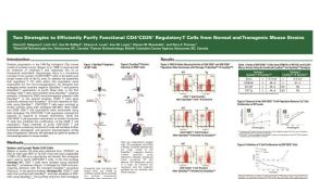 科学海报Cell Isolation of Functional CD4+CD25+ Regulatory T Cells from Mouse Strains
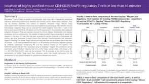 科学海报Isolation of Highly Purified Mouse CD4+CD25+Foxp3+ Regulatory T Cells in Less
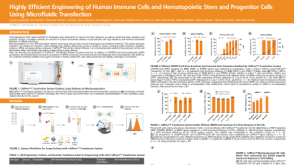 科学海报Highly Efficient Engineering of Human Immune Cells and Hematopoietic Stem and Progenitor Cells Using Microfluidic Transfection
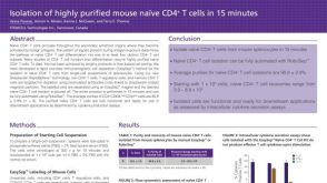 科学海报Immunomagnetic Cell Isolation of Highly Purified Mouse Naïve CD4+ T Cells in 15 Minutes
 沪公网安备31010102008431号