Crane CA et al. (JAN 2010)
Neuro-oncology 12 1 7--13
TGF-beta downregulates the activating receptor NKG2D on NK cells and CD8+ T cells in glioma patients.
The activating receptor NKG2D,expressed by natural killer (NK) cells and CD8(+) T cells,has a role in the specific killing of transformed cells. We examined NKG2D expression in patients with glioblastoma multiforme and found that NKG2D was downregulated on NK cells and CD8(+) T cells. Expression of NKG2D on lymphocytes significantly increased following tumor resection and correlated with an increased ability to kill NKG2D ligand-positive tumor targets. Despite the presence of soluble NKG2D ligands in the sera of glioblastoma patients,NKG2D downregulation was primarily caused by tumor-derived tumor growth factor-beta,suggesting that blocking of this cytokine may have therapeutic benefit.
View Publication
产品类型:
产品号#:
19055
19055RF
产品名:
EasySep™人NK细胞富集试剂盒
RoboSep™ 人NK细胞富集试剂盒含滤芯吸头
McIntyre BAS et al. (JUL 2015)
Innate immunity 21 5 504--511
Innate immune response of human pluripotent stem cell-derived airway epithelium.
The acquisition of innate immune response is requisite to having bona fide differentiation of airway epithelium. Procedures developed to differentiate lung airway from human pluripotent stem cells (hPSCs) have demonstrated anecdotal evidence for innate immune response,but an in-depth exploration of response levels is lacking. Herein,using an established method of airway epithelial generation from hPSCs,we show that hPSC-derived epithelial cells are able to up-regulate expression of TNF$\$,IL8 and IL1$\$ response to challenge with bacterial endotoxin LPS,but lack response from genes associated with innate immune response in other cell types. Further,stimulation of cells with TNF-$\$ in auto-induction of TNF$\$,as well as cytokine responses of IL8 and IL1$\$ The demonstration of innate immune induction in hPSC-derived airway epithelia gives further strength to the functionality of in vitro protocols aimed at generating differentiated airway cells that can potentially be used in a translational setting. Finally,we propose that innate immune challenge of airway epithelium from human pluripotent stem cell sources be used as a robust validation of functional in vitro differentiation.
View Publication
Bentley C et al. (NOV 2011)
Nutrition,metabolism,and cardiovascular diseases : NMCD 21 11 871--8
Influence of chylomicron remnants on human monocyte activation in vitro.
BACKGROUND AND AIMS: Atherosclerosis is known to be an inflammatory disease and there is increasing evidence that chylomicron remnants (CMR),the lipoproteins which carry dietary fats in the blood,cause macrophage foam cell formation and inflammation. In early atherosclerosis the frequency of activated monocytes in the peripheral circulation is increased,and clearance of CMR from blood may be delayed,however,whether CMR contribute directly to monocyte activation and subsequent egress into the arterial wall has not been established. Here,the contribution of CMR to activation of monocyte pro-inflammatory pathways was assessed using an in vitro model. METHODS AND RESULTS: Primary human monocytes and CMR-like particles (CRLP) were used to measure several endpoints of monocyte activation. Treatment with CRLP caused rapid and prolonged generation of reactive oxygen species by monocytes. The pro-inflammatory chemokines MCP-1 and IL-8 were secreted in nanogram quantities by the cells in the absence of CRLP. IL-8 secretion was transiently increased after CRLP treatment,and CRLP maintained secretion in the presence of pharmacological inhibitors of IL-8 production. In contrast,exposure to CRLP significantly reduced MCP-1 secretion. Chemotaxis towards MCP-1 was increased in monocytes pre-exposed to CRLP and was reversed by addition of exogenous MCP-1. CONCLUSION: Our findings indicate that CRLP activate human monocytes and augment their migration in vitro by reducing cellular MCP-1 expression. Our data support the current hypothesis that CMR contribute to the inflammatory milieu of the arterial wall in early atherosclerosis,and suggest that this may reflect direct interaction with circulating blood monocytes.
View Publication
产品类型:
产品号#:
15028
15068
产品名:
RosetteSep™人单核细胞富集抗体混合物
RosetteSep™人单核细胞富集抗体混合物
Chen S et al. (AUG 2007)
Journal of immunology (Baltimore,Md. : 1950) 179 3 1634--47
Modulatory effects of 1,25-dihydroxyvitamin D3 on human B cell differentiation.
1,25-Dihydroxyvitamin D(3) (1,25(OH)(2)D(3)) can modulate immune responses,but whether it directly affects B cell function is unknown. Patients with systemic lupus erythematosus,especially those with antinuclear Abs and increased disease activity,had decreased 1,25(OH)(2)D(3) levels,suggesting that vitamin D might play a role in regulating autoantibody production. To address this,we examined the effects of 1,25(OH)(2)D(3) on B cell responses and found that it inhibited the ongoing proliferation of activated B cells and induced their apoptosis,whereas initial cell division was unimpeded. The generation of plasma cells and postswitch memory B cells was significantly inhibited by 1,25(OH)(2)D(3),although the up-regulation of genetic programs involved in B cell differentiation was only modestly affected. B cells expressed mRNAs for proteins involved in vitamin D activity,including 1 alpha-hydroxylase,24-hydroxylase,and the vitamin D receptor,each of which was regulated by 1,25(OH)(2)D(3) and/or activation. Importantly,1,25(OH)(2)D(3) up-regulated the expression of p27,but not of p18 and p21,which may be important in regulating the proliferation of activated B cells and their subsequent differentiation. These results indicate that 1,25(OH)(2)D(3) may play an important role in the maintenance of B cell homeostasis and that the correction of vitamin D deficiency may be useful in the treatment of B cell-mediated autoimmune disorders.
View Publication
产品类型:
产品号#:
15024
15064
产品名:
RosetteSep™人B细胞富集抗体混合物
RosetteSep™人B细胞富集抗体混合物
Ichikawa S et al. (MAY 2011)
Journal of immunology (Baltimore,Md. : 1950) 186 10 5549--55
Hepatic stellate cells function as regulatory bystanders.
Regulatory T cells (Tregs) contribute significantly to the tolerogenic nature of the liver. The mechanisms,however,underlying liver-associated Treg induction are still elusive. We recently identified the vitamin A metabolite,retinoic acid (RA),as a key controller that promotes TGF-β-dependent Foxp3(+) Treg induction but inhibits TGF-β-driven Th17 differentiation. To investigate whether the RA producing hepatic stellate cells (HSC) are part of the liver tolerance mechanism,we investigated the ability of HSC to function as regulatory APC. Different from previous reports,we found that highly purified HSC did not express costimulatory molecules and only upregulated MHC class II after in vitro culture in the presence of exogenous IFN-γ. Consistent with an insufficient APC function,HSC failed to stimulate naive OT-II TCR transgenic CD4(+) T cells and only moderately stimulated α-galactosylceramide-primed invariant NKT cells. In contrast,HSC functioned as regulatory bystanders and promoted enhanced Foxp3 induction by OT-II TCR transgenic T cells primed by spleen dendritic cells,whereas they greatly inhibited the Th17 differentiation. Furthermore,the regulatory bystander capacity of the HSC was completely dependent on their ability to produce RA. Our data thus suggest that HSC can function as regulatory bystanders,and therefore,by promoting Tregs and suppressing Th17 differentiation,they might represent key players in the mechanism that drives liver-induced tolerance.
View Publication
产品类型:
产品号#:
01700
01705
01701
01702
19755
产品名:
ALDEFLUOR™ 试剂盒
ALDEFLUOR™ DEAB试剂, 1.5 mM, 1 mL
ALDEFLUOR™检测缓冲液
Cobb JP et al. (MAR 2005)
Proceedings of the National Academy of Sciences of the United States of America 102 13 4801--6
Application of genome-wide expression analysis to human health and disease.
The application of genome-wide expression analysis to a large-scale,multicentered program in critically ill patients poses a number of theoretical and technical challenges. We describe here an analytical and organizational approach to a systematic evaluation of the variance associated with genome-wide expression analysis specifically tailored to study human disease. We analyzed sources of variance in genome-wide expression analyses performed with commercial oligonucleotide arrays. In addition,variance in gene expression in human blood leukocytes caused by repeated sampling in the same subject,among different healthy subjects,among different leukocyte subpopulations,and the effect of traumatic injury,were also explored. We report that analytical variance caused by sample processing was acceptably small. Blood leukocyte gene expression in the same individual over a 24-h period was remarkably constant. In contrast,genome-wide expression varied significantly among different subjects and leukocyte subpopulations. Expectedly,traumatic injury induced dramatic changes in apparent gene expression that were greater in magnitude than the analytical noise and interindividual variance. We demonstrate that the development of a nation-wide program for gene expression analysis with careful attention to analytical details can reduce the variance in the clinical setting to a level where patterns of gene expression are informative among different healthy human subjects,and can be studied with confidence in human disease.
View Publication
产品类型:
产品号#:
15021
15061
15028
15068
产品名:
RosetteSep™人T细胞富集抗体混合物
RosetteSep™人T细胞富集抗体混合物
RosetteSep™人单核细胞富集抗体混合物
RosetteSep™人单核细胞富集抗体混合物
Darce JR et al. (DEC 2007)
Journal of immunology (Baltimore,Md. : 1950) 179 11 7276--86
Regulated expression of BAFF-binding receptors during human B cell differentiation.
BAFF plays a central role in B-lineage cell biology; however,the regulation of BAFF-binding receptor (BBR) expression during B cell activation and differentiation is not completely understood. In this study,we provide a comprehensive ex vivo analysis of BBRs in human B-lineage cells at various stages of maturation,as well as describe the events that drive and regulate receptor expression. Our data reveal that B-lineage cells ranging from naive to plasma cells (PCs),excluding bone marrow PCs,express BAFF-R uniformly. In contrast,only tonsillar memory B cells (MB) and PCs,from both tonsil and bone marrow tissues,express BCMA. Furthermore,we show that TACI is expressed by MB cells and PCs,as well as a subpopulation of activated CD27(neg) B cells. In this regard,we demonstrate that TACI is inducible early upon B cell activation and this is independent of B cell turnover. In addition,we found that TACI expression requires activation of the ERK1/2 pathway,since its expression was blocked by ERK1/2-specific inhibitors. Expression of BAFF-R and B cell maturation Ag (BCMA) is also highly regulated and we demonstrate that BCMA expression is only acquired in MB cells and in a manner accompanied by loss of BAFF-R expression. This inverse expression coincides with MB cell differentiation into Ig-secreting cells (ISC),since blocking differentiation inhibited both induction of BCMA expression and loss of BAFF-R. Collectively,our data suggest that the BBR profile may serve as a footprint of the activation history and stage of differentiation of normal human B cells.
View Publication
Norman JM et al. (OCT 2011)
Nature immunology 12 10 975--83
The antiviral factor APOBEC3G enhances the recognition of HIV-infected primary T cells by natural killer cells.
APOBEC3G (A3G) is an intrinsic antiviral factor that inhibits the replication of human immunodeficiency virus (HIV) by deaminating cytidine residues to uridine. This causes guanosine-to-adenosine hypermutation in the opposite strand and results in inactivation of the virus. HIV counteracts A3G through the activity of viral infectivity factor (Vif),which promotes degradation of A3G. We report that viral protein R (Vpr),which interacts with a uracil glycosylase,also counteracted A3G by diminishing the incorporation of uridine. However,this process resulted in activation of the DNA-damage–response pathway and the expression of natural killer (NK) cell–activating ligands. Our results show that pathogen-induced deamination of cytidine and the DNA-damage response to virus-mediated repair of the incorporation of uridine enhance the recognition of HIV-infected cells by NK cells.
View Publication

 EasySep™小鼠TIL(CD45)正选试剂盒


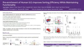 科学海报Pre-enrichment of Human ILCs Improves Sorting Efficiency While Maintaining Functionality


 沪公网安备31010102008431号