Vasir B et al. (FEB 2005)
Journal of immunology (Baltimore,Md. : 1950) 174 4 2376--86
Dendritic cells induce MUC1 expression and polarization on human T cells by an IL-7-dependent mechanism.
The MUC1 transmembrane mucin is expressed on the surface of activated human T cells; however,the physiologic signals responsible for the regulation of MUC1 in T cells are not known. The present studies demonstrate that IL-7,but not IL-2 or IL-4,markedly induces MUC1 expression on CD3+ T cells. MUC1 was also up-regulated by IL-15,but to a lesser extent than that found with IL-7. The results show that IL-7 up-regulates MUC1 on CD4+,CD8+,CD25+,CD69+,naive CD45RA+,and memory CD45RO+ T cells. In concert with induction of MUC1 expression by IL-7,activated dendritic cells (DC) that produce IL-7 up-regulate MUC1 on allogeneic CD3+ T cells. DC also induce MUC1 expression on autologous CD3+ T cells in the presence of recall Ag. Moreover,DC-induced MUC1 expression on T cells is blocked by a neutralizing anti-IL-7 Ab. The results also demonstrate that DC induce polarization of MUC1 on T cells at sites opposing the DC-T cell synapse. These findings indicate that DC-mediated activation of Ag-specific T cells is associated with induction and polarization of MUC1 expression by an IL-7-dependent mechanism.
View Publication
Muthuswamy R et al. (JUL 2008)
Cancer research 68 14 5972--8
Ability of mature dendritic cells to interact with regulatory T cells is imprinted during maturation.
Preferential activation of regulatory T (Treg) cells limits autoimmune tissue damage during chronic immune responses but can also facilitate tumor growth. Here,we show that tissue-produced inflammatory mediators prime maturing dendritic cells (DC) for the differential ability of attracting anti-inflammatory Treg cells. Our data show that prostaglandin E(2) (PGE(2)),a factor overproduced in chronic inflammation and cancer,induces stable Treg-attracting properties in maturing DC,mediated by CCL22. The elevated production of CCL22 by PGE(2)-matured DC persists after the removal of PGE(2) and is further elevated after secondary stimulation of DC in a neutral environment. This PGE(2)-induced overproduction of CCL22 and the resulting attraction of FOXP3(+) Tregs are counteracted by IFN alpha,a mediator of acute inflammation,which also restores the ability of the PGE(2)-exposed DC to secrete the Th1-attracting chemokines: CXCL9,CXCL10,CXCL11,and CCL5. In accordance with these observations,different DCs clinically used as cancer vaccines show different Treg-recruiting abilities,with PGE(2)-matured DC,but not type 1-polarized DC,generated in the presence of type I and type II IFNs,showing high Treg-attracting activity. The current data,showing that the ability of mature DC to interact with Treg cells is predetermined at the stage of DC maturation,pave the way to preferentially target the regulatory versus proinflammatory T cells in autoimmunity and transplantation,as opposed to intracellular infections and cancer.
View Publication
产品类型:
产品号#:
19052
19052RF
产品名:
EasySep™人CD4+ T细胞富集试剂盒
RoboSep™ 人CD4+ T细胞富集试剂盒含滤芯吸头
S. Fan et al. ( 2019)
NPJ vaccines 4 14
Role of innate lymphoid cells and dendritic cells in intradermal immunization of the enterovirus antigen.
Enterovirus type 71 (EV71) and coxsackievirus A 16 (CA16) are the major pathogens of human hand,foot,and mouth disease (HFMD). In our previous study,intramuscular immunization with the inactivated EV71 vaccine elicited effective immunity,while immunization with the inactivated CA16 vaccine did not. In this report,we focused on innate immune responses elicited by inactivated EV71 and CA16 antigens administered intradermally or intramuscularly. The distributions of the EV71 and CA16 antigens administered intradermally or intramuscularly were not obviously different,but the antigens were detected for a shorter period of time when administered intradermally. The expression levels of NF-kappaB pathway signaling molecules,which were identified as being capable of activating DCs,ILCs,and T cells,were higher in the intradermal group than in the intramuscular group. Antibodies for the EV71 and CA16 antigens colocalized with ILCs and DCs in skin and muscle tissues under fluorescence microscopy. Interestingly,ILC colocalization decreased over time,while DC colocalization increased over time. ELISpot analysis showed that coordination between DCs and ILCs contributed to successful adaptive immunity against vaccine antigens in the skin. EV71 and/or CA16 antigen immunization via the intradermal route was more capable of significantly increasing neutralizing antibody titers and activating specific T cell responses than immunization via the intramuscular route. Furthermore,neonatal mice born to mothers immunized with the EV71 and CA16 antigens were 100{\%} protected against wild-type EV71 or CA16 viral challenge. Together,our results provide new insights into the development of vaccines for HFMD.
View Publication
产品类型:
产品号#:
19851
19851RF
产品名:
EasySep™小鼠T细胞分选试剂盒
RoboSep™ 小鼠T细胞分选试剂盒
Kovats S et al. (NOV 2016)
Clinical and experimental immunology 186 2 214--226
West Nile virus-infected human dendritic cells fail to fully activate invariant natural killer T cells.
West Nile virus (WNV) infection is a mosquito-borne zoonosis with increasing prevalence in the United States. WNV infection begins in the skin,and the virus replicates initially in keratinocytes and dendritic cells (DCs). In the skin and cutaneous lymph nodes,infected DCs are likely to interact with invariant natural killer T cells (iNKTs). Bidirectional interactions between DCs and iNKTs amplify the innate immune response to viral infections,thus controlling viral load and regulating adaptive immunity. iNKTs are stimulated by CD1d-bound lipid antigens or activated indirectly by inflammatory cytokines. We exposed human monocyte-derived DCs to WNV Kunjin and determined their ability to activate isolated blood iNKTs. DCs became infected as judged by synthesis of viral mRNA and Envelope and NS-1 proteins,but did not undergo significant apoptosis. Infected DCs up-regulated the co-stimulatory molecules CD86 and CD40,but showed decreased expression of CD1d. WNV infection induced DC secretion of type I interferon (IFN),but no or minimal interleukin (IL)-12,IL-23,IL-18 or IL-10. Unexpectedly,we found that the WNV-infected DCs stimulated human iNKTs to up-regulate CD69 and produce low amounts of IL-10,but not proinflammatory cytokines such as IFN-γ or tumour necrosis factor (TNF)-α. Both CD1d and IFNAR blockade partially abrogated this iNKT response,suggesting involvement of a T cell receptor (TCR)-CD1d interaction and type I interferon receptor (IFNAR) signalling. Thus,WNV infection interferes with DC-iNKT interactions by preventing the production of proinflammatory cytokines. iNKTs may be a source of IL-10 observed in human flavivirus infections and initiate an anti-inflammatory innate response that limits adaptive immunity and immune pathology upon WNV infection.
View Publication
产品类型:
产品号#:
19059
19059RF
产品名:
EasySep™人单核细胞富集试剂盒
RoboSep™ 人单核细胞富集试剂盒含滤芯吸头
Bemark M et al. ( 2016)
Nature communications 7 12698
Limited clonal relatedness between gut IgA plasma cells and memory B cells after oral immunization.
Understanding how memory B cells are induced and relate to long-lived plasma cells is important for vaccine development. Immunity to oral vaccines has been considered short-lived because of a poor ability to develop IgA B-cell memory. Here we demonstrate that long-lived mucosal IgA memory is readily achieved by oral but not systemic immunization in mouse models with NP hapten conjugated with cholera toxin and transfer of B1-8(high)/GFP(+) NP-specific B cells. Unexpectedly,memory B cells are poorly related to long-lived plasma cells and less affinity-matured. They are α4β7-integrin(+)CD73(+)PD-L2(+)CD80(+) and at systemic sites mostly IgM(+),while 80% are IgA(+) in Peyer's patches. On reactivation,most memory B cells in Peyer's patches are GL7(-),but expand in germinal centres and acquire higher affinity and more mutations,demonstrating strong clonal selection. CCR9 expression is found only in Peyer's patches and appears critical for gut homing. Thus,gut mucosal memory possesses unique features not seen after systemic immunization.
View Publication
产品类型:
产品号#:
19854
19854RF
产品名:
EasySep™小鼠B细胞分选试剂盒
RoboSep™ 小鼠B细胞分选试剂盒
D. Park et al. (may 2019)
Scientific reports 9 1 7094
Differences in the molecular signatures of mucosal-associated invariant T cells and conventional T cells.
Mucosal-associated invariant T (MAIT) cells exhibit different characteristics from those of TCRalpha7.2- conventional T cells. They play important roles in various inflammatory diseases,including rheumatoid arthritis and inflammatory bowel disease. MAIT cells express a single T cell receptor alpha chain,TCRalpha7.2 segment associated with Jalpha33 and CDR3 with fixed length,which recognizes bacteria-derived vitamin B metabolites. However,the characteristics of MAIT cells and TCRalpha7.2+ CD161- T cells have never been compared. Here,we performed RNA sequencing to compare the properties of MAIT cells,TCRalpha7.2- conventional T cells and TCRalpha7.2+ CD161- T cells. Genome-wide transcriptomes of MAIT cells,TCRalpha7.2- conventional T cells,and TCRalpha7.2+ CD161- T cells were compared and analyzed using causal network analysis. This is the first report comparing the transcriptomes of MAIT cells,TCRalpha7.2- conventional T cells and TCRalpha7.2+ CD161- T cells. We also identified the predominant signaling pathways of MAIT cells,which differed from those of TCRalpha7.2- conventional T cells and TCRalpha7.2+ CD161- T cells,through a gene set enrichment test and upstream regulator analysis and identified the genes responsible for the characteristic MAIT cell phenotypes. Our study advances the complete understanding of MAIT biology.
View Publication
产品类型:
产品号#:
15021
15061
产品名:
RosetteSep™人T细胞富集抗体混合物
RosetteSep™人T细胞富集抗体混合物
Li Y et al. (FEB 2007)
Journal of immunology (Baltimore,Md. : 1950) 178 3 1938--47
Phosphorylated ERM is responsible for increased T cell polarization, adhesion, and migration in patients with systemic lupus erythematosus.
Systemic lupus erythematosus (SLE) is an autoimmune/inflammatory disease characterized by autoantibody production and abnormal T cells that infiltrate tissues through not well-known mechanisms. We report that SLE T lymphocytes display increased levels of CD44,ezrin,radixin,and moesin (ERM) phosphorylation,stronger actin polymerization,higher polar cap formation,and enhanced adhesion and chemotactic migration compared with T cells from patients with rheumatoid arthritis and normal individuals. Silencing of CD44 by CD44 small interfering RNA in SLE T cells inhibited significantly their ability to adhere and migrate as did treatment with Rho kinase and actin polymerization inhibitors. Forced expression of T567D-ezrin,a phosphorylation-mimic form,enhanced remarkably the adhesion and migration rate of normal T cells. Anti-CD3/TCR autoantibodies present in SLE sera caused increased ERM phosphorylation,adhesion,and migration in normal T cells. pERM and CD44 are highly expressed in T cells infiltrating in the kidneys of patients with lupus nephritis. These data prove that increased ERM phosphorylation represents a key molecular abnormality that guides T cell adhesion and migration in SLE patients.
View Publication
Costantini C et al. (JAN 2009)
Immunobiology 214 9-10 828--34
On the co-purification of 6-sulfo LacNAc(+) dendritic cells (slanDC) with NK cells enriched from human blood.
The ability of NK cells to directly recognize pathogens and be activated via Toll-like receptors (TLR) is increasingly recognized. Nevertheless,controversial results on the NK cell ability to be directly activated by lipopolysaccharide (LPS),the ligand of TLR4,have been recently reported. To start elucidating the reasons explaining the contrasting observations of the literature,we focused on the potential role of currently used NK cell purification procedures to condition putative NK cell responsiveness to LPS. To do so,human NK cells were isolated by negative selection,using three different commercial kits,to be comparatively evaluated for the production of IFNgamma in response to ultra-pure LPS and/or IL-2. Despite the lack of surface TLR4,we found that two out of the three NK cell-enriched populations released IFNgamma (and one of the two,IL-12p70 as well) in response to the LPS plus IL-2 combination,whereas the last one did not. However,the two LPS plus IL-2-responsive NK cell populations were found variably contaminated with 6-sulfo LacNAc(+) dendritic cells (slanDC),demonstrated responsible for triggering,via the production of IL-12p70 in response to LPS,the release of IFNgamma by IL-2-stimulated NK cells. Accordingly,slanDC depletion completely abrogated the capacity to produce both IL-12p70 and IFNgamma in response to LPS plus IL-2 by slanDC-containing NK cells. Taken together,our data uncover that two commercially available kits,specifically designed to isolate NK cells by negative selection,also co-purify variable amounts of slanDC. The latter cells may dramatically affect the outcome of experiments carried on to evaluate NK cell responsiveness to TLR agonists such as LPS.
View Publication

 EasySep™小鼠TIL(CD45)正选试剂盒


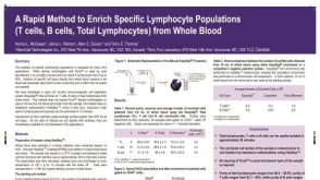 科学海报Cell Enrichment of Specific Lymphocyte Populations (T Cells, B Cells, Total Lymphocytes) from Whole Blood


 沪公网安备31010102008431号