R. Gupta et al. (may 2019)
Journal of immunology (Baltimore,Md. : 1950) 202 10 2924--2944
Mechanism for IL-15-Driven B Cell Chronic Lymphocytic Leukemia Cycling: Roles for AKT and STAT5 in Modulating Cyclin D2 and DNA Damage Response Proteins.
Clonal expansion of B cell chronic lymphocytic leukemia (B-CLL) occurs within lymphoid tissue pseudofollicles. IL-15,a stromal cell-associated cytokine found within spleens and lymph nodes of B-CLL patients,significantly boosts in vitro cycling of blood-derived B-CLL cells following CpG DNA priming. Both IL-15 and CpG DNA are elevated in microbe-draining lymphatic tissues,and unraveling the basis for IL-15-driven B-CLL growth could illuminate new therapeutic targets. Using CpG DNA-primed human B-CLL clones and approaches involving both immunofluorescent staining and pharmacologic inhibitors,we show that both PI3K/AKT and JAK/STAT5 pathways are activated and functionally important for IL-15→CD122/ɣc signaling in ODN-primed cells expressing activated pSTAT3. Furthermore,STAT5 activity must be sustained for continued cycling of CFSE-labeled B-CLL cells. Quantitative RT-PCR experiments with inhibitors of PI3K and STAT5 show that both contribute to IL-15-driven upregulation of mRNA for cyclin D2 and suppression of mRNA for DNA damage response mediators ATM,53BP1,and MDC1. Furthermore,protein levels of these DNA damage response molecules are reduced by IL-15,as indicated by Western blotting and immunofluorescent staining. Bioinformatics analysis of ENCODE chromatin immunoprecipitation sequencing data from cell lines provides insight into possible mechanisms for STAT5-mediated repression. Finally,pharmacologic inhibitors of JAKs and STAT5 significantly curtailed B-CLL cycling when added either early or late in a growth response. We discuss how the IL-15-induced changes in gene expression lead to rapid cycling and possibly enhanced mutagenesis. STAT5 inhibitors might be an effective modality for blocking B-CLL growth in patients.
View Publication
产品类型:
产品号#:
15024
15064
产品名:
RosetteSep™人B细胞富集抗体混合物
RosetteSep™人B细胞富集抗体混合物
Pua HH et al. (APR 2009)
Journal of immunology (Baltimore,Md. : 1950) 182 7 4046--55
Autophagy is essential for mitochondrial clearance in mature T lymphocytes.
Macroautophagy plays an important role in the regulation of cell survival,metabolism,and the lysosomal degradation of cytoplasmic material. In the immune system,autophagy contributes to the clearance of intracellular pathogens,MHCII cross-presentation of endogenous Ags,as well as cell survival. We and others have demonstrated that autophagy occurs in T lymphocytes and contributes to the regulation of their cellular function,including survival and proliferation. Here we show that the essential autophagy gene Atg7 is required in a cell-intrinsic manner for the survival of mature primary T lymphocytes. We also find that mitochondrial content is developmentally regulated in T but not in B cells,with exit from the thymus marking a transition from high mitochondrial content in thymocytes to lower mitochondrial content in mature T cells. Macroautophagy has been proposed to play an important role in the clearance of intracellular organelles,and autophagy-deficient mature T cells fail to reduce their mitochondrial content in vivo. Consistent with alterations in mitochondrial content,autophagy-deficient T cells have increased reactive oxygen species production as well as an imbalance in pro- and antiapoptotic protein expression. With much recent interest in the possibility of autophagy-dependent developmentally programmed clearance of organelles in lens epithelial cells and erythrocytes,our data demonstrate that autophagy may have a physiologically significant role in the clearance of superfluous mitochondria in T lymphocytes as part of normal T cell homeostasis.
View Publication
产品类型:
产品号#:
19751
19751RF
产品名:
Wang X et al. (MAR 2009)
Journal of immunology (Baltimore,Md. : 1950) 182 6 3597--608
MEKK3 is essential for lymphopenia-induced T cell proliferation and survival.
T cell homeostasis is crucial for maintaining an efficient and balanced T cell immunity. The interaction between TCR and self peptide (sp) MHC ligands is known to be the key driving force in this process,and it is believed to be functionally and mechanistically different from that initiated by the antigenic TCR stimulation. Yet,very little is known about the downstream signaling events triggered by this TCR-spMHC interaction and how they differ from those triggered by antigenic TCR stimulation. In this study,we show that T cell conditional ablation of MEKK3,a Ser/Thr kinase in the MAPK cascade,causes a significant reduction in peripheral T cell numbers in the conditional knockout mice,but does not perturb thymic T cell development and maturation. Using an adoptive mixed transfer method,we show that MEKK3-deficient T cells are severely impaired in lymphopenia-induced cell proliferation and survival. Interestingly,the Ag-induced T cell proliferation proceeds normally in the absence of MEKK3. Finally,we found that the activity of ERK1/2,but not p38 MAPK,was attenuated during the lymphopenia-driven response in MEKK3-deficient T cells. Together,these data suggest that MEKK3 may play a crucial selective role for spMHC-mediated T cell homeostasis.
View Publication
Kawakami Y et al. (JUN 2009)
The Journal of experimental medicine 206 6 1219--25
Inhibition of NK cell activity by IL-17 allows vaccinia virus to induce severe skin lesions in a mouse model of eczema vaccinatum.
Threats of bioterrorism have renewed efforts to better understand poxvirus pathogenesis and to develop a safer vaccine against smallpox. Individuals with atopic dermatitis are excluded from smallpox vaccination because of their propensity to develop eczema vaccinatum,a disseminated vaccinia virus (VACV) infection. To study the underlying mechanism of the vulnerability of atopic dermatitis patients to VACV infection,we developed a mouse model of eczema vaccinatum. Virus infection of eczematous skin induced severe primary erosive skin lesions,but not in the skin of healthy mice. Eczematous mice exhibited lower natural killer (NK) cell activity but similar cytotoxic T lymphocyte activity and humoral immune responses. The role of NK cells in controlling VACV-induced skin lesions was demonstrated by experiments depleting or transferring NK cells. The proinflammatory cytokine interleukin (IL)-17 reduced NK cell activity in mice with preexisting dermatitis. Given low NK cell activities and increased IL-17 expression in atopic dermatitis patients,these results can explain the increased susceptibility of atopic dermatitis patients to eczema vaccinatum.
View Publication
产品类型:
产品号#:
19755
产品名:
Ng Y-S et al. (OCT 2004)
The Journal of experimental medicine 200 7 927--34
Bruton's tyrosine kinase is essential for human B cell tolerance.
Most polyreactive and antinuclear antibodies are removed from the human antibody repertoire during B cell development. To elucidate how B cell receptor (BCR) signaling may regulate human B cell tolerance,we tested the specificity of recombinant antibodies from single peripheral B cells isolated from patients suffering from X-linked agammaglobulinemia (XLA). These patients carry mutations in the Bruton's tyrosine kinase (BTK) gene that encode an essential BCR signaling component. We find that in the absence of Btk,peripheral B cells show a distinct antibody repertoire consistent with extensive secondary V(D)J recombination. Nevertheless,XLA B cells are enriched in autoreactive clones. Our results demonstrate that Btk is essential in regulating thresholds for human B cell tolerance.
View Publication
Vessillier S et al. (SEP 2015)
Journal of immunological methods 424 43--52
Cytokine release assays for the prediction of therapeutic mAb safety in first-in man trials--Whole blood cytokine release assays are poorly predictive for TGN1412 cytokine storm.
The therapeutic monoclonal antibody (mAb) TGN1412 (anti-CD28 superagonist) caused near-fatal cytokine release syndrome (CRS) in all six volunteers during a phase-I clinical trial. Several cytokine release assays (CRAs) with reported predictivity for TGN1412-induced CRS have since been developed for the preclinical safety testing of new therapeutic mAbs. The whole blood (WB) CRA is the most widely used,but its sensitivity for TGN1412-like cytokine release was recently criticized. In a comparative study,using group size required for 90% power with 5% significance as a measure of sensitivity,we found that WB and 10% (v/v) WB CRAs were the least sensitive for TGN1412 as these required the largest group sizes (n = 52 and 79,respectively). In contrast,the peripheral blood mononuclear cell (PBMC) solid phase (SP) CRA was the most sensitive for TGN1412 as it required the smallest group size (n = 4). Similarly,the PBMC SP CRA was more sensitive than the WB CRA for muromonab-CD3 (anti-CD3) which stimulates TGN1412-like cytokine release (n = 4 and 4519,respectively). Conversely,the WB CRA was far more sensitive than the PBMC SP CRA for alemtuzumab (anti-CD52) which stimulates FcγRI-mediated cytokine release (n = 8 and 180,respectively). Investigation of potential factors contributing to the different sensitivities revealed that removal of red blood cells (RBCs) from WB permitted PBMC-like TGN1412 responses in a SP CRA,which in turn could be inhibited by the addition of the RBC membrane protein glycophorin A (GYPA); this observation likely underlies,at least in part,the poor sensitivity of WB CRA for TGN1412. The use of PBMC SP CRA for the detection of TGN1412-like cytokine release is recommended in conjunction with adequately powered group sizes for dependable preclinical safety testing of new therapeutic mAbs.
View Publication
产品类型:
产品号#:
18352
18352RF
产品名:
Schneider E et al. (SEP 2009)
Journal of immunology (Baltimore,Md. : 1950) 183 6 3591--7
IL-33 activates unprimed murine basophils directly in vitro and induces their in vivo expansion indirectly by promoting hematopoietic growth factor production.
IL-33,a new member of the IL-1 family,has been described as an important inducer of Th2 cytokines and mediator of inflammatory responses. In this study,we demonstrate that murine basophils sorted directly from the bone marrow,without prior exposure to IL-3 or Fc(epsilon)R cross-linking,respond to IL-33 alone by producing substantial amounts of histamine,IL-4,and IL-6. These cells express ST2 constitutively and generate a cytokine profile that differs from their IL-3-induced counterpart by a preferential production of IL-6. In vivo,IL-33 promotes basophil expansion in the bone marrow (BM) through an indirect mechanism of action depending on signaling through the beta(c) chain shared by receptors for IL-3,GM-CSF,and IL-5. IL-3 can still signal through its specific beta(IL-3) chain in these mutant mice,which implies that it is not the unique growth-promoting mediator in this setup,but requires IL-5 and/or GMCSF. Our results support a major role of the latter growth factor,which is readily generated by total BM cells as well as sorted basophils in response to IL-33 along with low amounts of IL-3. Furthermore,GM-CSF amplifies IL-3-induced differentiation of basophils from BM cells,whereas IL-5 that is also generated in vivo,affects neither their functions nor their growth in vitro or in vivo. In conclusion,our data provide the first evidence that IL-33 not only activates unprimed basophils directly,but also promotes their expansion in vivo through induction of GM-CSF and IL-3.
View Publication
产品类型:
产品号#:
18755
18755RF
产品名:
EasySep™小鼠CD49b正选试剂盒
RoboSep™ 小鼠CD49b正选试剂盒含滤芯吸头
Menon MP et al. (MAR 2006)
The Journal of clinical investigation 116 3 683--94
Signals for stress erythropoiesis are integrated via an erythropoietin receptor-phosphotyrosine-343-Stat5 axis.
Anemia due to chronic disease or chemotherapy often is ameliorated by erythropoietin (Epo). Present studies reveal that,unlike steady-state erythropoiesis,erythropoiesis during anemia depends sharply on an Epo receptor-phosphotyrosine-343-Stat5 signaling axis. In mice expressing a phosphotyrosine-null (PY-null) Epo receptor allele (EpoR-HM),severe and persistent anemia was induced by hemolysis or 5-fluorouracil. In short-term transplantation experiments,donor EpoR-HM bone marrow cells also failed to efficiently repopulate the erythroid compartment. In each context,stress erythropoiesis was rescued to WT levels upon the selective restoration of an EpoR PY343 Stat5-binding site (EpoR-H allele). As studied using a unique primary culture system,EpoR-HM erythroblasts exhibited marked stage-specific losses in Epo-dependent growth and survival. EpoR-H PY343 signals restored efficient erythroblast expansion,and the selective Epo induction of the Stat5 target genes proviral integration site-1 (Pim-1) and oncostatin-M. Bcl2-like 1 (Bcl-x),in contrast,was not significantly induced via WT-EpoR,EpoR-HM,or EpoR-H alleles. In Kit+ CD71+ erythroblasts,EpoR-PY343 signals furthermore enhanced SCF growth effects,and SCF modulation of Pim-1 kinase and oncostatin-M expression. In maturing Kit- CD71+ erythroblasts,oncostatin-M exerted antiapoptotic effects that likewise depended on EpoR PY343-mediated events. Stress erythropoiesis,therefore,requires stage-specific EpoR-PY343-Stat5 signals,some of which selectively bolster SCF and oncostatin-M action.
View Publication

 EasySep™小鼠TIL(CD45)正选试剂盒


 产品手册Highway1™: Fast, Gentle, and Automated Cell Sorting for Every Lab
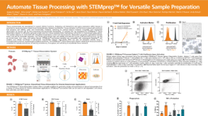 科学海报Automate Tissue Processing with STEMprep™ for Versatile Sample Preparation
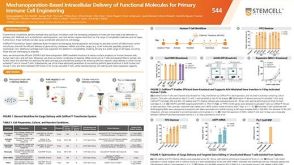 科学海报Mechanoporation-Based Intracellular Delivery of Functional Molecules for Primary Immune Cell Engineering

 沪公网安备31010102008431号