Rhee C et al. (FEB 2017)
Developmental biology 422 2 83--91
ARID3A is required for mammalian placenta development.
Previous studies in the mouse indicated that ARID3A plays a critical role in the first cell fate decision required for generation of trophectoderm (TE). Here,we demonstrate that ARID3A is widely expressed during mouse and human placentation and essential for early embryonic viability. ARID3A localizes to trophoblast giant cells and other trophoblast-derived cell subtypes in the junctional and labyrinth zones of the placenta. Conventional Arid3a knockout embryos suffer restricted intrauterine growth with severe defects in placental structural organization. Arid3a null placentas show aberrant expression of subtype-specific markers as well as significant alteration in cytokines,chemokines and inflammatory response-related genes,including previously established markers of human placentation disorders. BMP4-mediated induction of trophoblast stem (TS)-like cells from human induced pluripotent stem cells results in ARID3A up-regulation and cytoplasmic to nuclear translocation. Overexpression of ARID3A in BMP4-mediated TS-like cells up-regulates TE markers,whereas pluripotency markers are down-regulated. Our results reveal an essential,conserved function for ARID3A in mammalian placental development through regulation of both intrinsic and extrinsic developmental programs.
View Publication
产品类型:
产品号#:
05850
05857
05870
05875
85850
85857
85870
85875
产品名:
mTeSR™1
mTeSR™1
Storms RW et al. (JUL 2005)
Blood 106 1 95--102
Distinct hematopoietic progenitor compartments are delineated by the expression of aldehyde dehydrogenase and CD34.
A broad range of hematopoietic stem cells and progenitors reside within a fraction of umbilical cord blood (UCB) that exhibits low light scatter properties (SSC(lo)) and high expression of aldehyde dehydrogenase (ALDH(br)). Many SSC(lo) ALDH(br) cells coexpress CD34; however,other cells express either ALDH or CD34. To investigate the developmental potential of these cell subsets,purified ALDH(br) CD34+,ALDH(neg) CD34+,and ALDH(br) CD34(neg) UCB cells were characterized within a variety of in vivo and in vitro assays. Primitive progenitors capable of multilineage development were monitored in long- and short-term repopulation assays performed on nonobese diabetic/severe combined immunodeficiency (NOD/SCID) mice,and in primary and secondary long-term culture assays. These progenitors were highly enriched within the ALDH(br) CD34+ fraction. This cell fraction also enriched short-term myeloid progenitors that were detected in vitro. By comparison,ALDH(neg) CD34+ cells contained few primitive progenitors and had diminished short-term myeloid potential but exhibited enhanced short-term natural killer (NK) cell development in vitro. The ALDH(br) CD34(neg) cells were not efficiently supported by any of the assays used. These studies suggested that in particular the expression of ALDH delineated distinct CD34+ stem cell and progenitor compartments. The differential expression of ALDH may provide a means to explore normal and malignant processes associated with myeloid and lymphoid development.
View Publication
Loss of the Rho GTPase activating protein p190-B enhances hematopoietic stem cell engraftment potential.
Hematopoietic stem cell (HSC) engraftment is a multistep process involving HSC homing to bone marrow,self-renewal,proliferation,and differentiation to mature blood cells. Here,we show that loss of p190-B RhoGTPase activating protein,a negative regulator of Rho GTPases,results in enhanced long-term engraftment during serial transplantation. This effect is associated with maintenance of functional HSC-enriched cells. Furthermore,loss of p190-B led to marked improvement of HSC in vivo repopulation capacity during ex vivo culture without altering proliferation and multilineage differentiation of HSC and progeny. Transcriptional analysis revealed that p190-B deficiency represses the up-regulation of p16(Ink4a) in HSCs in primary and secondary transplantation recipients,providing a possible mechanism of p190-B-mediated HSC functions. Our study defines p190-B as a critical transducer element of HSC self-renewal activity and long-term engraftment,thus suggesting that p190-B is a target for HSC-based therapies requiring maintenance of engraftment phenotype.
View Publication
产品类型:
产品号#:
09600
09650
产品名:
StemSpan™ SFEM
StemSpan™ SFEM
Punzel M et al. (APR 2003)
Experimental hematology 31 4 339--47
The symmetry of initial divisions of human hematopoietic progenitors is altered only by the cellular microenvironment.
OBJECTIVE: We examined if cellular elements or adhesive ligands were able to alter asymmetric divisions of CD34(+)/CD38(-) cells in contrast to soluble factors at a single cell level. MATERIALS AND METHODS: After single cell deposition onto 96-well plates,cells were cocultured for 10 days with the stem cell supporting cell line AFT024,fibronectin (FN),or bovine serum albumin (BSA). The divisional history was monitored with time-lapse microscopy. Subsequent function for the most primitive cells was assessed using the myeloid-lymphoid-initiating cell (ML-IC) assay. Committed progenitors were measured using colony-forming cells (CFC). RESULTS: Only contact with AFT024 recruited significant numbers of CD34(+)/CD38(-) cells into cell cycle and increased asymmetric divisions. Although most ML-IC were still identified among cells that have divided fewer than 3 times,a significant number of ML-IC shifted into the fast-dividing fraction after exposure to AFT024. The increase in ML-IC frequency was predominantly due to recruitment of quiescent and slow-dividing cells from the starting population. Increase in CFC activity induced by AFT024 was found only among rapidly dividing cells. CONCLUSIONS: For the first time,we have demonstrated that asymmetric divisions can be altered upon exposure with a stem cell-supporting microenvironment. For the primitive subset of cells (ML-IC),this was predominantly due to recruitment into cell cycle and increased rounds of cycling without loss of function. Exposure to AFT024 cells also increased proliferation and asymmetric divisions of committed CFC. Hence direct communication between hematopoietic progenitors with stroma cells is required for maintaining self-renewal potential.
View Publication
产品类型:
产品号#:
05150
产品名:
MyeloCult™ H5100
Yu C et al. (DEC 2010)
Blood 116 23 4786--94
Retinoic acid enhances the generation of hematopoietic progenitors from human embryonic stem cell-derived hemato-vascular precursors.
Current induction schemes directing hematopoietic differentiation of human embryonic stem cells (hESCs) are not well defined to mimic the sequential stages of hematopoietic development in vivo. Here,we report a 3-stage method to direct differentiation of hESCs toward hematopoietic progenitors in chemically defined mediums. In the first 2 stages,we efficiently generated T-positive primitive streak/mesendoderm cells and kinase domain receptor-positive (KDR(+)) platelet-derived growth factor receptor α-negative (PDGFRα(-)) hemato-vascular precursors sequentially. In the third stage,we found that cells in a spontaneous differentiation condition mainly formed erythroid colonies. Addition of all-trans retinoic acid (RA) greatly enhanced generation of hematopoietic progenitors in this stage while suppressing erythroid development. The RA-treated cells highly expressed definitive hematopoietic genes,formed large numbers of multilineage and myeloid colonies,and gave rise to greater than 45% CD45(+) hematopoietic cells. When hematopoietic progenitors were selected with CD34 and C-Kit,greater than 95% CD45(+) hematopoietic cells could be generated. In addition,we found that endogenous RA signaling at the second stage was required for vascular endothelial growth factor/basic fibroblast growth factor-induced hemato-vascular specification,whereas exogenously applied RA efficiently induced KDR(-)PDGFRα(+) paraxial mesoderm cells. Our study suggests that RA signaling plays diverse roles in human mesoderm and hematopoietic development.
View Publication
产品类型:
产品号#:
04436
09600
09650
84435
84445
产品名:
MethoCult™ SF H4436
StemSpan™ SFEM
StemSpan™ SFEM
Fiedler K et al. (JAN 2011)
Blood 117 4 1329--39
Neutrophil development and function critically depend on Bruton tyrosine kinase in a mouse model of X-linked agammaglobulinemia.
Bruton tyrosine kinase (Btk) is essential for B cell development and function and also appears to be important for myeloid cells. The bone marrow of Btk-deficient mice shows enhanced granulopoiesis compared with that of wild-type mice. In purified granulocyte-monocyte-progenitors (GMP) from Btk-deficient mice,the development of granulocytes is favored at the expense of monocytes. However,Btk-deficient neutrophils are impaired in maturation and function. Using bone marrow chimeras,we show that this defect is cell-intrinsic to neutrophils. In GMP and neutrophils,Btk plays a role in GM-CSF- and Toll-like receptor-induced differentiation. Molecular analyses revealed that expression of the lineage-determining transcription factors C/EBPα,C/EBPβ,and PU.1,depends on Btk. In addition,expression of several granule proteins,including myeloperoxidase,neutrophilic granule protein,gelatinase and neutrophil elastase,is Btk-dependent. In the Arthus reaction,an acute inflammatory response,neutrophil migration into tissues,edema formation,and hemorrhage are significantly reduced in Btk-deficient animals. Together,our findings implicate Btk as an important regulator of neutrophilic granulocyte maturation and function in vivo.
View Publication
O'Connor MD et al. (JAN 2011)
Methods in molecular biology (Clifton,N.J.) 690 67--80
Functional assays for human embryonic stem cell pluripotency.
Realizing the potential that human embryonic stem cells (hESCs) hold,both for the advancement of biomedical science and the development of new treatments for many human disorders,will be greatly facilitated by the introduction of standardized methods for assessing and altering the biological properties of these cells. The 7-day in vitro alkaline phosphatase colony-forming cell (AP(+)-CFC) assay currently offers the most sensitive and specific method to quantify the frequency of undifferentiated cells present in a culture. In this regard,it is superior to any phenotypic assessment protocol. The AP(+)-CFC assay,thus,provides a valuable tool for monitoring the quality of hESC cultures,and also for evaluating quantitative changes in pluripotent cell numbers following manipulations that may affect the self-renewal and differentiation properties of the treated cells. Two other methods routinely used to evaluate hESC pluripotency involve either culturing the cells under conditions that promote the formation of nonadherent differentiating cell aggregates (termed embryoid bodies),or transplanting the cells into immunodeficient mice to obtain teratomas containing differentiated cells representative of endoderm,mesoderm,and ectoderm lineages.
View Publication
McKinney-Freeman SL et al. (MAY 2008)
Blood 111 10 4944--53
Modulation of murine embryonic stem cell-derived CD41+c-kit+ hematopoietic progenitors by ectopic expression of Cdx genes.
Cdx1,Cdx2,and Cdx4 comprise the caudal-like Cdx gene family in mammals,whose homologues regulate hematopoietic development in zebrafish. Previously,we reported that overexpression of Cdx4 enhances hematopoietic potential from murine embryonic stem cells (ESCs). Here we compare the effect of ectopic Cdx1,Cdx2,and Cdx4 on the differentiation of murine ESC-derived hematopoietic progenitors. The 3 Cdx genes differentially influence the formation and differentiation of hematopoietic progenitors within a CD41(+)c-kit(+) population of embryoid body (EB)-derived cells. Cdx1 and Cdx4 enhance,whereas Cdx2 strongly inhibits,the hematopoietic potential of CD41(+)ckit(+) EB-derived cells,changes that are reflected by effects on hematopoietic lineage-specific and Hox gene expression. When we subject stromal cell and colony assay cultures of EB-derived hematopoietic progenitors to ectopic expression of Cdx genes,Cdx4 dramatically enhances,whereas Cdx1 and Cdx2 both inhibit hematopoietic activity,probably by blocking progenitor differentiation. These data demonstrate distinct effects of Cdx genes on hematopoietic progenitor formation and differentiation,insights that we are using to facilitate efforts at in vitro culture of hematopoietic progenitors from ESC. The behavior of Cdx genes in vitro suggests how derangement of these developmental regulators might contribute to leukemogenesis.
View Publication


 EasySep™小鼠TIL(CD45)正选试剂盒





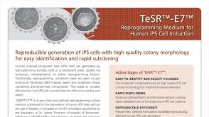 产品手册TeSR™-E7™ Reprogramming Medium for Human iPS Cell Induction


 沪公网安备31010102008431号