Trowbridge JJ et al. (SEP 2006)
Proceedings of the National Academy of Sciences of the United States of America 103 38 14134--9
Hedgehog modulates cell cycle regulators in stem cells to control hematopoietic regeneration.
The signals that control the regenerative ability of hematopoietic stem cells (HSCs) in response to damage are unknown. Here,we demonstrate that downstream activation of the Hedgehog (Hh) signaling pathway induces cycling and expansion of primitive bone marrow hematopoietic cells under homeostatic conditions and during acute regeneration. However,this effect is at the expense of HSC function,because continued Hh activation during regeneration represses expression of specific cell cycle regulators,leading to HSC exhaustion. In vivo treatment with an inhibitor of the Hh pathway rescues these transcriptional and functional defects in HSCs. Our study establishes Hh signaling as a regulator of the HSC cell cycle machinery that balances hematopoietic homeostasis and regeneration in vivo.
View Publication
产品类型:
产品号#:
03434
03444
产品名:
MethoCult™ GF M3434
MethoCult™ GF M3434
Van Meter MEM et al. (MAY 2007)
Blood 109 9 3945--52
K-RasG12D expression induces hyperproliferation and aberrant signaling in primary hematopoietic stem/progenitor cells.
Defining how cancer-associated mutations perturb signaling networks in stem/progenitor populations that are integral to tumor formation and maintenance is a fundamental problem with biologic and clinical implications. Point mutations in RAS genes contribute to many cancers,including myeloid malignancies. We investigated the effects of an oncogenic Kras(G12D) allele on phosphorylated signaling molecules in primary c-kit(+) lin(-/low) hematopoietic stem/progenitor cells. Comparison of wild-type and Kras(G12D) c-kit(+) lin(-/low) cells shows that K-Ras(G12D) expression causes hyperproliferation in vivo and results in abnormal levels of phosphorylated STAT5,ERK,and S6 under basal and stimulated conditions. Whereas Kras(G12D) cells demonstrate hyperactive signaling after exposure to granulocyte-macrophage colony-stimulating factor,we unexpectedly observe a paradoxical attenuation of ERK and S6 phosphorylation in response to stem cell factor. These studies provide direct biochemical evidence that cancer stem/progenitor cells remodel signaling networks in response to oncogenic stress and demonstrate that multi-parameter flow cytometry can be used to monitor the effects of targeted therapeutics in vivo. This strategy has broad implications for defining the architecture of signaling networks in primary cancer cells and for implementing stem cell-targeted interventions.
View Publication
产品类型:
产品号#:
03231
03434
03444
产品名:
MethoCult™ M3231
MethoCult™ GF M3434
MethoCult™ GF M3434
Miyake N et al. (MAR 2006)
Stem cells (Dayton,Ohio) 24 3 653--61
HOXB4-induced self-renewal of hematopoietic stem cells is significantly enhanced by p21 deficiency.
Enforced expression of the HOXB4 transcription factor and downregulation of p21(Cip1/Waf) (p21) can each independently increase proliferation of murine hematopoietic stem cells (HSCs). We asked whether the increase in HSC self-renewal generated by overexpression of HOXB4 is enhanced in p21-deficient HSCs. HOXB4 was overexpressed in hematopoietic cells from wild-type (wt) and p21-/- mice. Bone marrow (BM) cells were transduced with a retroviral vector expressing HOXB4 together with GFP (MIGB4),or a control vector containing GFP alone (MIG) and maintained in liquid culture for up to 11 days. At day 11 of the expansion culture,the number of primary CFU-GM (colony-forming unit granulocyte-macrophage) colonies and the repopulating ability were significantly increased in MIGB4 p21-/- BM (p21B4) cells compared with MIGB4-transduced wt BM (wtB4) cells. To test proliferation of HSCs in vivo,we performed competitive repopulation experiments and obtained significantly higher long-term engraftment of expanded p21B4 cells compared with wtB4 cells. The 5-day expansion of p21B4 HSCs generated 100-fold higher numbers of competitive repopulating units compared with wtMIG and threefold higher numbers compared with wtB4. The findings demonstrate that increased expression of HOXB4,in combination with suppression of p21 expression,could be a useful strategy for effective and robust expansion of HSCs.
View Publication
产品类型:
产品号#:
03534
产品名:
MethoCult™ GF M3534
Ramadan A et al. (SEP 2010)
Genes to cells : devoted to molecular & cellular mechanisms 15 9 983--94
Cells with hematopoietic activity in the mouse placenta reside in side population.
The discovery of a major hematopoietic stem cell pool in midgestation mouse embryo has defined the placenta as an important hematopoietic anatomical site. In this study,we examined the flow cytometric pattern of mouse placenta cells on embryonic days (E) 10.5 to E15.5,in view of CD45 and c-Kit expression. We also determined which population of these cells shows differentiation potential toward multiple hematopoietic lineages by performing coculture with OP9 stromal cells and colony-forming assay in methylcellulose. Only CD45(+)c-Kit(+) population showed the ability to form hematopoietic colonies including multiple lineages. To distinguish which fraction of placenta cells have the hematopoietic activity,we used GFP transgenic mice in which the fetal part of the placenta is GFP positive and the maternal part is GFP negative. E11.5 and E13.5 CD45(+)c-Kit(+) placental cells that have ability to form hematopoietic colonies are the fetal GFP positive placental cells. E11.5 and E13.5 CD45(+)c-Kit(+) placental cells that have an ability to form hematopoietic colonies mainly reside in Hoechst dye-effluxing side population area (SP). Taken together,in the placenta of mouse embryo,we conclude that SP cells in the CD45(+)c-Kit(+) fetal placental cells have the ability to form hematopoietic colonies.
View Publication
产品类型:
产品号#:
03434
03444
产品名:
MethoCult™ GF M3434
MethoCult™ GF M3434
Tchernychev B et al. (DEC 2010)
Proceedings of the National Academy of Sciences of the United States of America 107 51 22255--9
Discovery of a CXCR4 agonist pepducin that mobilizes bone marrow hematopoietic cells.
The G protein-coupled receptor (GPCR),chemokine CXC-type receptor 4 (CXCR4),and its ligand,CXCL12,mediate the retention of polymorphonuclear neutrophils (PMNs) and hematopoietic stem and progenitor cells (HSPCs) in the bone marrow. Agents that disrupt CXCL12-mediated chemoattraction of CXCR4-expressing cells mobilize PMNs and HSPCs into the peripheral circulation and are therapeutically useful for HSPC collection before autologous bone marrow transplantation (ABMT). Our aim was to develop unique CXCR4-targeted therapeutics using lipopeptide GPCR modulators called pepducins. A pepducin is a synthetic molecule composed of a peptide derived from the amino acid sequence of one of the intracellular (IC) loops of a target GPCR coupled to a lipid tether. We prepared and screened a small CXCR4-targeted pepducin library and identified several pepducins with in vitro agonist activity,including ATI-2341,whose peptide sequence derives from the first IC loop. ATI-2341 induced CXCR4- and G protein-dependent signaling,receptor internalization,and chemotaxis in CXCR4-expressing cells. It also induced dose-dependent peritoneal recruitment of PMNs when administered i.p. to mice. However,when administered systemically by i.v. bolus,ATI-2341 acted as a functional antagonist and dose-dependently mediated release of PMNs from the bone marrow of both mice and cynomolgus monkeys. ATI-2341-mediated release of granulocyte/macrophage progenitor cells from the bone marrow was confirmed by colony-forming assays. We conclude that ATI-2341 is a potent and efficacious mobilizer of bone marrow PMNs and HSPCs and could represent a previously undescribed therapeutic approach for the recruitment of HSPCs before ABMT.
View Publication
产品类型:
产品号#:
03534
产品名:
MethoCult™ GF M3534
Zhang CC and Lodish HF (JUN 2005)
Blood 105 11 4314--20
Murine hematopoietic stem cells change their surface phenotype during ex vivo expansion.
Ex vivo expansion of hematopoietic stem cells (HSCs) is important for many clinical applications,and knowledge of the surface phenotype of ex vivo-expanded HSCs will be critical to their purification and analysis. Here,we developed a simple culture system for bone marrow (BM) HSCs using low levels of stem cell factor (SCF),thrombopoietin (TPO),insulin-like growth factor 2 (IGF-2),and fibroblast growth factor-1 (FGF-1) in serum-free medium. As measured by competitive repopulation analyses,there was a more than 20-fold increase in numbers of long-term (LT)-HSCs after a 10-day culture of total BM cells. Culture of BM side population" (SP) cells�
View Publication
产品类型:
产品号#:
09600
09650
28600
产品名:
StemSpan™ SFEM
StemSpan™ SFEM
L-Calc™有限稀释软件
Ling K-W et al. (OCT 2004)
The Journal of experimental medicine 200 7 871--82
GATA-2 plays two functionally distinct roles during the ontogeny of hematopoietic stem cells.
GATA-2 is an essential transcription factor in the hematopoietic system that is expressed in hematopoietic stem cells (HSCs) and progenitors. Complete deficiency of GATA-2 in the mouse leads to severe anemia and embryonic lethality. The role of GATA-2 and dosage effects of this transcription factor in HSC development within the embryo and adult are largely unexplored. Here we examined the effects of GATA-2 gene dosage on the generation and expansion of HSCs in several hematopoietic sites throughout mouse development. We show that a haploid dose of GATA-2 severely reduces production and expansion of HSCs specifically in the aorta-gonad-mesonephros region (which autonomously generates the first HSCs),whereas quantitative reduction of HSCs is minimal or unchanged in yolk sac,fetal liver,and adult bone marrow. However,HSCs in all these ontogenically distinct anatomical sites are qualitatively defective in serial or competitive transplantation assays. Also,cytotoxic drug-induced regeneration studies show a clear GATA-2 dose-related proliferation defect in adult bone marrow. Thus,GATA-2 plays at least two functionally distinct roles during ontogeny of HSCs: the production and expansion of HSCs in the aorta-gonad-mesonephros and the proliferation of HSCs in the adult bone marrow.
View Publication
产品类型:
产品号#:
03434
03444
产品名:
MethoCult™ GF M3434
MethoCult™ GF M3434
Zhuge Y et al. (AUG 2014)
2014 6171--6174
Human pluripotent stem cell tools for cardiac optogenetics
It is likely that arrhythmias should be avoided for therapies based on human pluripotent stem cell (hPSC)-derived cardiomyocytes (CM) to be effective. Towards achieving this goal,we introduced light-activated channelrhodopsin-2 (ChR2),a cation channel activated with 480 nm light,into human embryonic stem cells (hESC). By using in vitro approaches,hESC-CM are able to be activated with light. ChR2 is stably transduced into undifferentiated hESC via a lentiviral vector. Via directed differentiation,hESCChR2-CM are produced and subjected to optical stimulation. hESCChR2-CM respond to traditional electrical stimulation and produce similar contractility features as their wild-type counterparts but only hESCChR2-CM can be activated by optical stimulation. Here it is shown that a light sensitive protein can enable in vitro optical control of hESC-CM and that this activation occurs optimally above specific light stimulation intensity and pulse width thresholds. For future therapy,in vivo optical stimulation along with optical inhibition could allow for acute synchronization of implanted hPSC-CM with patient cardiac rhythms.
View Publication
产品类型:
产品号#:
05850
05857
05870
05875
85850
85857
85870
85875
产品名:
mTeSR™1
mTeSR™1
Lagarkova MA et al. (NOV 2008)
Cell Cycle 7 22 3610--3612
CD 30 is a marker of undifferentiated human embryonic stem cells rather than a biomarker of transformed hESCs
Recently it has been demonstrated that CD30 expression was rather specific for transformed than for normal human ES cells and therefore CD30 maybe suggested as a potential marker for human ES cells bearing chromosomal abnormalities. Using immunohistochemistry and RT-PCR analysis we examined �?¡D30 expression in 10 hESCs lines with normal and abberant karyotypes. All hESC lines expressed CD30 antigen and RNA in undifferentiated state whether cell line beared chromosomal abnormalities or not. In contrast to previous notions our data demonstrate that CD30 could be considered as marker of undifferentiated hESCs without respect to karyotype changes.
View Publication
产品类型:
产品号#:
05850
05857
05870
05875
85850
85857
85870
85875
产品名:
mTeSR™1
mTeSR™1
Orelio C et al. (APR 2009)
Haematologica 94 4 462--9
Interleukin-1 regulates hematopoietic progenitor and stem cells in the midgestation mouse fetal liver.
BACKGROUND: Hematopoietic progenitors are generated in the yolk sac and aorta-gonad-mesonephros region during early mouse development. At embryonic day 10.5 the first hematopoietic stem cells emerge in the aorta-gonad-mesonephros. Subsequently,hematopoietic stem cells and progenitors are found in the fetal liver. The fetal liver is a potent hematopoietic site,playing an important role in the expansion and differentiation of hematopoietic progenitors and hematopoietic stem cells. However,little is known concerning the regulation of fetal liver hematopoietic stem cells. In particular,the role of cytokines such as interleukin-1 in the regulation of hematopoietic stem cells in the embryo has been largely unexplored. Recently,we observed that the adult pro-inflammatory cytokine interleukin-1 is involved in regulating aorta-gonad-mesonephros hematopoietic progenitor and hematopoietic stem cell activity. Therefore,we set out to investigate whether interleukin-1 also plays a role in regulating fetal liver progenitor cells and hematopoietic stem cells. DESIGN AND METHODS: We examined the interleukin-1 ligand and receptor expression pattern in the fetal liver. The effects of interleukin-1 on hematopoietic progenitor cells and hematopoietic stem cells were studied by FACS and transplantation analyses of fetal liver explants,and in vivo effects on hematopoietic stem cell and progenitors were studied in Il1r1(-/-) embryos. RESULTS: We show that fetal liver hematopoietic progenitor cells express the IL-1RI and that interleukin-1 increases fetal liver hematopoiesis,progenitor cell activity and promotes hematopoietic cell survival. Moreover,we show that in Il1r1(-/-) embryos,hematopoietic stem cell activity is impaired and myeloid progenitor activity is increased. CONCLUSIONS: The IL-1 ligand and receptor are expressed in the midgestation liver and act in the physiological regulation of fetal liver hematopoietic progenitor cells and hematopoietic stem cells.
View Publication
产品类型:
产品号#:
03434
03444
产品名:
MethoCult™ GF M3434
MethoCult™ GF M3434
Storms RW et al. (AUG 1999)
Proceedings of the National Academy of Sciences of the United States of America 96 16 9118--23
Isolation of primitive human hematopoietic progenitors on the basis of aldehyde dehydrogenase activity.
Because hematopoietic stem cells are rich in aldehyde dehydrogenase (ALDH) activity,we developed a fluorescent substrate for ALDH,termed BODIPY aminoacetaldehyde (BAAA),and tested its potential for isolating primitive human hematopoietic cells. A population of cells with low orthogonal light scattering and bright fluorescence intensity (SSC(lo)ALDH(br) cells) could be readily fractionated from human umbilical cord blood cells costained with BAAA and the multidrug-resistance inhibitor verapamil. The SSC(lo)ALDH(br) population was depleted of lineage-committed cells,40-90% pure for CD34(+)CD38(lo/-) cells,and enriched 50- to 100-fold for primitive hematopoietic progenitors detected in short- and long-term culture analyses. Together,these observations indicate that fractionating human hematopoietic stem cells on the basis of ALDH activity using BAAA is an effective method for isolating primitive human hematopoietic progenitors. This technique may be useful for isolating stem cells from other tissues as well.
View Publication

 EasySep™小鼠TIL(CD45)正选试剂盒


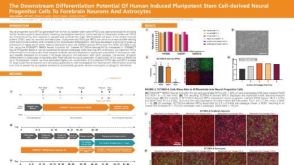 科学海报The Downstream Differentiation Potential of Human Induced Pluripotent Stem Cell-Derived Neural Progenitor Cells to Forebrain Neurons and Astrocytes

 沪公网安备31010102008431号