Vodyanik MA et al. (SEP 2006)
Blood 108 6 2095--105
Leukosialin (CD43) defines hematopoietic progenitors in human embryonic stem cell differentiation cultures.
During hematopoietic differentiation of human embryonic stem cells (hESCs),early hematopoietic progenitors arise along with endothelial cells within the CD34(+) population. Although hESC-derived hematopoietic progenitors have been previously identified by functional assays,their phenotype has not been defined. Here,using hESC differentiation in coculture with OP9 stromal cells,we demonstrate that early progenitors committed to hematopoietic development could be identified by surface expression of leukosialin (CD43). CD43 was detected on all types of emerging clonogenic progenitors before expression of CD45,persisted on differentiating hematopoietic cells,and reliably separated the hematopoietic CD34(+) population from CD34(+)CD43(-)CD31(+)KDR(+) endothelial and CD34(+)CD43(-)CD31(-)KDR(-) mesenchymal cells. Furthermore,we demonstrated that the first-appearing CD34(+)CD43(+)CD235a(+)CD41a(+/-)CD45(-) cells represent precommitted erythro-megakaryocytic progenitors. Multipotent lymphohematopoietic progenitors were generated later as CD34(+)CD43(+)CD41a(-)CD235a(-)CD45(-) cells. These cells were negative for lineage-specific markers (Lin(-)),expressed KDR,VE-cadherin,and CD105 endothelial proteins,and expressed GATA-2,GATA-3,RUNX1,C-MYB transcription factors that typify initial stages of definitive hematopoiesis originating from endothelial-like precursors. Acquisition of CD45 expression by CD34(+)CD43(+)CD45(-)Lin(-) cells was associated with progressive myeloid commitment and a decrease of B-lymphoid potential. CD34(+)CD43(+)CD45(+)Lin(-) cells were largely devoid of VE-cadherin and KDR expression and had a distinct FLT3(high)GATA3(low)RUNX1(low)PU1(high)MPO(high)IL7RA(high) gene expression profile.
View Publication
High-efficiency induction of neural conversion in human ESCs and human induced pluripotent stem cells with a single chemical inhibitor of transforming growth factor beta superfamily receptors.
Chemical compounds have emerged as powerful tools for modulating ESC functions and deriving induced pluripotent stem cells (iPSCs),but documentation of compound-induced efficient directed differentiation in human ESCs (hESCs) and human iPSC (hiPSCs) is limited. By screening a collection of chemical compounds,we identified compound C (also denoted as dorsomorphin),a protein kinase inhibitor,as a potent regulator of hESC and hiPSC fate decisions. Compound C suppresses mesoderm,endoderm,and trophoectoderm differentiation and induces rapid and high-efficiency neural conversion in both hESCs and hiPSCs,88.7% and 70.4%,respectively. Interestingly,compound C is ineffective in inducing neural conversion in mouse ESCs (mESCs). Large-scale kinase assay revealed that compound C targets at least seven transforming growth factor beta (TGF-β) superfamily receptors,including both type I and type II receptors,and thereby blocks both the Activin and bone morphogenesis protein (BMP) signaling pathways in hESCs. Dual inhibition of Activin and BMP signaling accounts for the effects of compound C on hESC differentiation and neural conversion. We also identified muscle segment homeobox gene 2 (MSX2) as a downstream target gene of compound C and a key signaling intermediate of the BMP pathway in hESCs. Our findings provide a single-step cost-effective method for efficient derivation of neural progenitor cells in adherent culture from human pluripotent stem cells. Therefore,it will be uniquely suitable for the production of neural progenitor cells in large scale and should facilitate the use of stem cells in drug screening and regenerative medicine and study of early human neural development.
View Publication
产品类型:
产品号#:
05850
05857
05870
05875
72102
85850
85857
85870
85875
100-0246
产品名:
Dorsomorphin
mTeSR™1
mTeSR™1
白消安(Busulfan)
Liu H and Roy K ( )
Tissue engineering 11 1-2 319--30
Stem cell-based tissue engineering is a promising technology in the effort to create functional tissues of choice. To establish an efficient approach for generating hematopoietic cell lineages directly from embryonic stem (ES) cells and to study the effects of three-dimensional (3D) biomaterials on ES cell differentiation,we cultured mouse ES cells on 3D,highly porous,biomimetic scaffolds. Cell differentiation was evaluated by microscopy and flow cytometry analysis with a variety of hematopoiesis- specific markers. Our data indicate that ES cells differentiated on porous 3D scaffold structures developed embryoid bodies (EBs) similar to those in traditional two-dimensional (2D) cultures; however,unlike 2D differentiation,these EBs integrated with the scaffold and appeared embedded in a network of extracellular matrix. Most significantly,the efficiency of hematopoietic precursor cell (HPC) generation on 3D,as indicated by the expression of various HPC-specific surface markers (CD34,Sca-1,Flk-1,and c-Kit) and colony-forming cell (CFC) assays,was reproducibly increased (about 2-fold) over their 2D counterparts. Comparison of static and dynamic 3D cultures demonstrated that spinner flask technology also contributed to the higher hematopoietic differentiation efficiency of ES cells seeded on scaffolds. Continued differentiation of 3D-derived HPCs into the myeloid lineage demonstrated increased efficiency (2-fold) of generating myeloid compared with differentiation from 2D-derived HPCs.
View Publication
Inhibition of aldehyde dehydrogenase expands hematopoietic stem cells with radioprotective capacity.
Hematopoietic stem cells (HSCs) are enriched for aldehyde dehydrogenase (ALDH) activity and ALDH is a selectable marker for human HSCs. However,the function of ALDH in HSC biology is not well understood. We sought to determine the function of ALDH in regulating HSC fate. Pharmacologic inhibition of ALDH with diethylaminobenzaldehyde (DEAB) impeded the differentiation of murine CD34(-)c-kit(+)Sca-1(+)lineage(-) (34(-)KSL) HSCs in culture and facilitated a ninefold expansion of cells capable of radioprotecting lethally irradiated mice compared to input 34(-)KSL cells. Treatment of bone marrow (BM) 34(-)KSL cells with DEAB caused a fourfold increase in 4-week competitive repopulating units,verifying the amplification of short-term HSCs (ST-HSCs) in response to ALDH inhibition. Targeted siRNA of ALDH1a1 in BM HSCs caused a comparable expansion of radioprotective progenitor cells in culture compared to DEAB treatment,confirming that ALDH1a1 was the target of DEAB inhibition. The addition of all trans retinoic acid blocked DEAB-mediated expansion of ST-HSCs in culture,suggesting that ALDH1a1 regulates HSC differentiation via augmentation of retinoid signaling. Pharmacologic inhibition of ALDH has therapeutic potential as a means to amplify ST-HSCs for transplantation purposes.
View Publication
产品类型:
产品号#:
01700
01705
01701
01702
产品名:
ALDEFLUOR™ 试剂盒
ALDEFLUOR™ DEAB试剂, 1.5 mM, 1 mL
ALDEFLUOR™检测缓冲液
Maldonado M et al. (MAY 2015)
Biomaterials 50 1 10--19
The effects of electrospun substrate-mediated cell colony morphology on the self-renewal of human induced pluripotent stem cells
The development of xeno-free,chemically defined stem cell culture systems has been a primary focus in the field of regenerative medicine to enhance the clinical application of pluripotent stem cells (PSCs). In this regard,various electrospun substrates with diverse physiochemical properties were synthesized utilizing various polymer precursors and surface treatments. Human induced pluripotent stem cells (IPSCs) cultured on these substrates were characterized by their gene and protein expression to determine the effects of the substrate physiochemical properties on the cells' self-renewal,i.e.,proliferation and the maintenance of pluripotency. The results showed that surface chemistry significantly affected cell colony formation via governing the colony edge propagation. More importantly,when surface chemistry of the substrates was uniformly controlled by collagen conjugation,the stiffness of substrate was inversely related to the sphericity,a degree of three dimensionality in colony morphology. The differences in sphericity subsequently affected spontaneous differentiation of IPSCs during a long-term culture,implicating that the colony morphology is a deciding factor in the lineage commitment of PSCs. Overall,we show that the capability of controlling IPSC colony morphology by electrospun substrates provides a means to modulate IPSC self-renewal.
View Publication
产品类型:
产品号#:
05850
05857
05870
05875
85850
85857
85870
85875
产品名:
mTeSR™1
mTeSR™1
Kolhar P et al. (APR 2010)
Journal of biotechnology 146 3 143--6
Synthetic surfaces for human embryonic stem cell culture.
Human embryonic stem cells (hESCs) have numerous potential biomedical applications owing to their unique abilities for self-renewal and pluripotency. Successful clinical application of hESCs and derivatives necessitates the culture of these cells in a fully defined environment. We have developed a novel peptide-based surface that uses a high-affinity cyclic RGD peptide for culture of hESCs under chemically defined conditions.
View Publication
Alkaline phosphatase-positive colony formation is a sensitive, specific, and quantitative indicator of undifferentiated human embryonic stem cells.
Human embryonic stem cells (hESCs) can be maintained in vitro as immortal pluripotent cells but remain responsive to many differentiation-inducing signals. Investigation of the initial critical events involved in differentiation induction would be greatly facilitated if a specific,robust,and quantitative assay for pluripotent hESCs with self-renewal potential were available. Here we describe the results of a series of experiments to determine whether the formation of adherent alkaline phosphatase-positive (AP(+)) colonies under conditions optimized for propagating undifferentiated hESCs would meet this need. The findings can be summarized as follows. (a) Most colonies obtained under these conditions consist of textgreateror=30 AP(+) cells that coexpress OCT4,NANOG,SSEA3,SSEA4,TRA-1-60,and TRA-1-81. (b) Most such colonies are derived from SSEA3(+) cells. (c) Primary colonies contain cells that produce secondary colonies of the same composition,including cells that initiate multilineage differentiation in embryoid bodies (EBs). (d) Colony formation is independent of plating density or the colony-forming cell (CFC) content of the test population over a wide range of cell concentrations. (e) CFC frequencies decrease when differentiation is induced by exposure either to retinoic acid or to conditions that stimulate EB formation. Interestingly,this loss of AP(+) clonogenic potential also occurs more rapidly than the loss of SSEA3 or OCT4 expression. The CFC assay thus provides a simple,reliable,broadly applicable,and highly specific functional assay for quantifying undifferentiated hESCs with self-renewal potential. Its use under standardized assay conditions should enhance future elucidation of the mechanisms that regulate hESC propagation and their early differentiation.
View Publication
产品类型:
产品号#:
05850
05857
05870
05875
07923
36254
85850
85857
85870
85875
产品名:
Dispase (1 U/mL)
DMEM/F-12 with 15 mM HEPES
mTeSR™1
mTeSR™1
Chua SJ et al. (FEB 2009)
Biochemical and biophysical research communications 379 2 217--21
Neural progenitors, neurons and oligodendrocytes from human umbilical cord blood cells in a serum-free, feeder-free cell culture.
We have previously demonstrated that lineage negative cells (Lin(neg)) from umbilical cord blood (UCB) develop into multipotent cells capable of differentiation into bone,muscle,endothelial and neural cells. The objective of this study was to determine the optimal conditions required for Lin(neg) UCB cells to differentiate into neuronal cells and oligodendrocytes. We demonstrate that early neural stage markers (nestin,neurofilament,A2B5 and Sox2) are expressed in Lin(neg) cells cultured in FGF4,SCF,Flt3-ligand reprogramming culture media followed by the early macroglial cell marker O4. Early stage oligodendrocyte markers CNPase,GalC,Olig2 and the late-stage marker MOSP are observed,as is the Schwann cell marker PMP22. In summary,Lin(neg) UCB cells,when appropriately cultured,are able to exhibit characteristics of neuronal and macroglial cells that can specifically differentiate into oligodendrocytes and Schwann cells and express proteins associated with myelin production after in vitro differentiation.
View Publication
产品类型:
产品号#:
09600
09650
产品名:
StemSpan™ SFEM
StemSpan™ SFEM
Pettinato G et al. (DEC 2014)
Scientific reports 4 7402
Formation of well-defined embryoid bodies from dissociated human induced pluripotent stem cells using microfabricated cell-repellent microwell arrays.
A simple,scalable,and reproducible technology that allows direct formation of large numbers of homogeneous and synchronized embryoid bodies (EBs) of defined sizes from dissociated human induced pluripotent stem cells (hiPSCs) was developed. Non-cell-adhesive hydrogels were used to create round-bottom microwells to host dissociated hiPSCs. No Rho-associated kinase inhibitor (ROCK-i),or centrifugation was needed and the side effects of ROCK-i can be avoided. The key requirement for the successful EB formation in addition to the non-cell-adhesive round-bottom microwells is the input cell density per microwell. Too few or too many cells loaded into the microwells will compromise the EB formation process. In parallel,we have tested our microwell-based system for homogeneous hEB formation from dissociated human embryonic stem cells (hESCs). Successful production of homogeneous hEBs from dissociated hESCs in the absence of ROCK-i and centrifugation was achieved within an optimal range of input cell density per microwell. Both the hiPSC- and hESC-derived hEBs expressed key proteins characteristic of all the three developmental germ layers,confirming their EB identity. This novel EB production technology may represent a versatile platform for the production of homogeneous EBs from dissociated human pluripotent stem cells (hPSCs).
View Publication
产品类型:
产品号#:
05850
05857
05870
05875
85850
85857
85870
85875
产品名:
mTeSR™1
mTeSR™1
Nemeth MJ et al. (SEP 2007)
Proceedings of the National Academy of Sciences of the United States of America 104 39 15436--41
Wnt5a inhibits canonical Wnt signaling in hematopoietic stem cells and enhances repopulation.
The mechanisms that regulate hematopoietic stem cell (HSC) fate decisions between proliferation and multilineage differentiation are unclear. Members of the Wnt family of ligands that activate the canonical Wnt signaling pathway,which utilizes beta-catenin to relay the signal,have been demonstrated to regulate HSC function. In this study,we examined the role of noncanonical Wnt signaling in regulating HSC fate. We observed that noncanonical Wnt5a inhibited Wnt3a-mediated canonical Wnt signaling in HSCs and suppressed Wnt3a-mediated alterations in gene expression associated with HSC differentiation,such as increased expression of myc. Wnt5a increased short- and long-term HSC repopulation by maintaining HSCs in a quiescent G(0) state. From these data,we propose that Wnt5a regulates hematopoiesis by the antagonism of the canonical Wnt pathway,resulting in a pool of quiescent HSCs.
View Publication

 EasySep™小鼠TIL(CD45)正选试剂盒


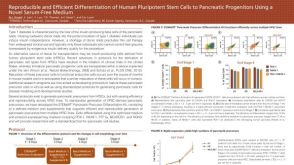 科学海报Reproducible and Efficient Differentiation of Human Pluripotent Stem Cells to Pancreatic Progenitors Using a Novel Serum-Free Medium
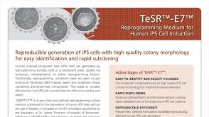 产品手册TeSR™-E7™ Reprogramming Medium for Human iPS Cell Induction
 沪公网安备31010102008431号