Lawrence HJ et al. (DEC 2005)
Blood 106 12 3988--94
Loss of expression of the Hoxa-9 homeobox gene impairs the proliferation and repopulating ability of hematopoietic stem cells.
The homeobox gene Hoxa-9 is normally expressed in primitive bone marrow cells,and overexpression of Hoxa-9 markedly expands hematopoietic stem cells,suggesting a function in early hematopoiesis. We present evidence for major functional defects in Hoxa-9-/- hematopoietic stem cells. Hoxa-9-/- marrow cells have normal numbers of immunophenotypic stem cells (Lin(-)c-kit(+)flk-2(-)Sca-1+ [KLFS] cells). However,sublethally irradiated Hoxa-9-/- mice develop persistent pancytopenia,indicating unusual sensitivity to ionizing irradiation. In competitive transplantation assays,Hoxa-9-/- cells showed an 8-fold reduction in multilineage long-term repopulating ability,a defect not seen in marrow cells deficient for the adjacent Hoxa-10 gene. Single-cell cultures of KLFS cells showed a 4-fold reduction in large high-proliferation potential colonies. In liquid cultures,Hoxa-9-deficient Lin(-)Sca-1(+) cells showed slowed proliferation (a 5-fold reduction in cell numbers at day 8) and delayed emergence of committed progenitors (a 5-fold decrease in colony-forming cells). Slowing of proliferation was accompanied by a delay in myeloid maturation,with a decrease in Gr-1hiMac-1hi cells at the end of the culture. Retroviral transduction with a Hoxa-9 expression vector dramatically enhanced the cytokine-driven proliferation and in vivo engraftment of Hoxa-9-/- marrow cells. Hoxa-9 appears to be specifically required for normal hematopoietic stem cell function both in vitro and in vivo.
View Publication
产品类型:
产品号#:
03231
09600
09650
产品名:
MethoCult™ M3231
StemSpan™ SFEM
StemSpan™ SFEM
Ma ACH et al. (DEC 2010)
Leukemia 24 12 2090--9
A DEAB-sensitive aldehyde dehydrogenase regulates hematopoietic stem and progenitor cells development during primitive hematopoiesis in zebrafish embryos.
Although aldehyde dehydrogenase (ALDH) activity has become a surrogate of hematopoietic stem and progenitor cells (HSPCs),its function during hematopoiesis was unclear. Here,we examined its role in zebrafish hematopoiesis based on pharmacological inhibition and morpholino (MO) knockdown. Zebrafish embryos were treated with diethylaminobenzaldehyde (DEAB,1 μmol/l) between 0- and 48 hour-post-fertilization (hpf). MOs targeting aldhs were injected between 1 and 4-cell stage. The effects on hematopoiesis were evaluated at different stages. DEAB treatment between 0 and 18 hpf increased gene expression associated with HSPC (scl,lmo2),erythropoiesis (gata1,α- and β-eHb) and myelopoiesis (spi1) as well as gfp(+) cells in dissociated Tg(gata1:gfp) embryos. The effects were ameliorated by all-trans retinoic acid (1 nmol/l). Definitive hematopoiesis and the erythromyeloid precursors were unaffected. In all,14 out of 15 zebrafish aldhs were detectable by reverse transcription PCR in 18 hpf embryos,of which only aldh1a2 and aldh16a1 were expressed in sites pertinent to hematopoiesis. Molecular targeting by MOs was demonstrated for 15 aldhs,but none of them,even in combined aldh1a2 and aldh1a3 knockdown,recapitulated the hematopoietic expansion in DEAB-treated embryos. In conclusion,DEAB expands HSPC population during primitive hematopoiesis through inhibition of aldh and retinoic acid synthesis. The specific aldh isoform(s) remains to be determined.
View Publication
产品类型:
产品号#:
01700
01705
01702
产品名:
ALDEFLUOR™ 试剂盒
ALDEFLUOR™ DEAB试剂, 1.5 mM, 1 mL
ALDEFLUOR™检测缓冲液
Ikebe C and Suzuki K ( 2014)
BioMed research international 2014 951512
Mesenchymal stem cells for regenerative therapy: optimization of cell preparation protocols.
Administration of bone marrow-derived mesenchymal stem cells (MSCs) is an innovative approach for the treatment of a range of diseases that are not curable by current therapies including heart failure. A number of clinical trials have been completed and many others are ongoing; more than 2,000 patients worldwide have been administered with culture-expanded allogeneic or autologous MSCs for the treatment of various diseases,showing feasibility and safety (and some efficacy) of this approach. However,protocols for isolation and expansion of donor MSCs vary widely between these trials,which could affect the efficacy of the therapy. It is therefore important to develop international standards of MSC production,which should be evidence-based,regulatory authority-compliant,of good medical practice grade,cost-effective,and clinically practical,so that this innovative approach becomes an established widely adopted treatment. This review article summarizes protocols to isolate and expand bone marrow-derived MSCs in 47 recent clinical trials of MSC-based therapy,which were published after 2007 onwards and provided sufficient methodological information. Identified issues and possible solutions associated with the MSC production methods,including materials and protocols for isolation and expansion,are discussed with reference to relevant experimental evidence with aim of future clinical success of MSC-based therapy.
View Publication
产品类型:
产品号#:
07930
07931
07940
07955
07956
07959
07954
100-1061
07952
产品名:
CryoStor® CS10
CryoStor® CS10
CryoStor® CS10
CryoStor® CS10
CryoStor® CS10
CryoStor® CS10
CryoStor® CS10
Mashimo Y and Kamei K-II ( 2015)
1346 85--98
Microfluidic Image Cytometry for Single-Cell Phenotyping of Human Pluripotent Stem Cells
A microfluidic human pluripotent stem cell (hPSC) array has been developed for robust and reproducible hPSC culture methods to assess chemically defined serum- and feeder-free culture conditions. This microfluidic platform,combined with image cytometry,enables the systematic analysis of multiple simultaneously detected marker expression in individual cells,for screening of various chemically defined media across hPSC lines,and the study of phenotypic responses.
View Publication
产品类型:
产品号#:
05850
05857
05870
05875
85850
85857
85870
85875
产品名:
mTeSR™1
mTeSR™1
Trzonkowski P et al. (MAR 2009)
Cytometry. Part A : the journal of the International Society for Analytical Cytology 75 3 175--88
Ex vivo expansion of CD4(+)CD25(+) T regulatory cells for immunosuppressive therapy.
Immunosuppressants are powerful drugs,capable of triggering severe adverse effects. Hence,there is tremendous interest in replacing them with less-toxic agents. Adoptive therapy with CD25(+)CD4(+) T regulatory cells (Tregs) holds promise as an alternative to immunosuppressants. Tregs have been described as the most potent immunosuppressive cells in the human body. In a number of experimental models,they have been found to quench autoimmune diseases,maintain allogeneic transplants,and prevent allergic diseases. A major stumbling block in their clinical application is related to Treg phenotype and the very limited number of these cells in the periphery,not exceeding 1-5% of total CD4(+) T cells. Recent progress in multicolor flow cytometry and cell sorting as well as cellular immunology has found ways of overcoming these obstacles,and has opened the doors to the clinical application of Tregs. In the review,we describe Treg sorting and expansion techniques that have been developed in recent years. In the experience of our laboratory,as well as some published reports,Treg adoptive therapy is a promising tool in immunosuppressive therapy,and should be considered for clinical trials.
View Publication
Duportet X et al. (DEC 2014)
Nucleic Acids Research 42 21 13440--13451
A platform for rapid prototyping of synthetic gene networks in mammalian cells
Mammalian synthetic biology may provide novel therapeutic strategies,help decipher new paths for drug discovery and facilitate synthesis of valuable molecules. Yet,our capacity to genetically program cells is currently hampered by the lack of efficient approaches to streamline the design,construction and screening of synthetic gene networks. To address this problem,here we present a framework for modular and combinatorial assembly of functional (multi)gene expression vectors and their efficient and specific targeted integration into a well-defined chromosomal context in mammalian cells. We demonstrate the potential of this framework by assembling and integrating different functional mammalian regulatory networks including the largest gene circuit built and chromosomally integrated to date (6 transcription units,27kb) encoding an inducible memory device. Using a library of 18 different circuits as a proof of concept,we also demonstrate that our method enables one-pot/single-flask chromosomal integration and screening of circuit libraries. This rapid and powerful prototyping platform is well suited for comparative studies of genetic regulatory elements,genes and multi-gene circuits as well as facile development of libraries of isogenic engineered cell lines.
View Publication
产品类型:
产品号#:
05850
05857
05870
05875
85850
85857
85870
85875
产品名:
mTeSR™1
mTeSR™1
Imai T et al. ( 2017)
Anticancer research 37 1 47--55
KIF11 Is Required for Spheroid Formation by Oesophageal and Colorectal Cancer Cells.
BACKGROUND Oesophageal squamous cell carcinoma (ESCC) and colorectal cancer (CRC) are common types of human cancer. Spheroid colony formation is used to characterize cancer stem cell (CSCs). In the present study,we analyzed the significance of kinesin family 11 (KIF11 in human ESCC and CRC. MATERIALS AND METHODS Expression of KIF11 in 105 ESCC and 100 CRC cases was determined using immunohistochemistry. RNA interference was used to inhibit KIF11 expression in ESCC and CRC cell lines. RESULTS In total,61 out of 105 (58%) ESCC and 62 out of 100 (62%) CRC cases were positive for KIF11. Expression of KIF11 was not associated with any clinicopathological characteristics. Both the number and size of spheres produced by from TE-5 ESCC cells and DLD-1 CRC cells were significantly reduced upon KIF11 siRNA transfection compared to negative control siRNA transfection. CONCLUSION These results indicate that KIF11 plays an important role in CSCs of ESCC and CRC.
View Publication
产品类型:
产品号#:
05850
05857
05870
05875
85850
85857
85870
85875
产品名:
mTeSR™1
mTeSR™1
Sessarego N et al. (MAR 2008)
Haematologica 93 3 339--46
Multipotent mesenchymal stromal cells from amniotic fluid: solid perspectives for clinical application.
BACKGROUND: Mesenchymal stromal cells are multipotent cells considered to be of great promise for use in regenerative medicine. However,the cell dose may be a critical factor in many clinical conditions and the yield resulting from the ex vivo expansion of mesenchymal stromal cells derived from bone marrow may be insufficient. Thus,alternative sources of mesenchymal stromal cells need to be explored. In this study,mesenchymal stromal cells were successfully isolated from second trimester amniotic fluid and analyzed for chromosomal stability to validate their safety for potential utilization as a cell therapy product. DESIGN AND METHODS: Mesenchymal stromal cells were expanded up to the sixth passage starting from amniotic fluid using different culture conditions to optimize large-scale production. RESULTS: The highest number of mesenchymal stromal cells derived from amniotic fluid was reached at a low plating density; in these conditions the expansion of mesenchymal stromal cells from amniotic fluid was significantly greater than that of adult bone marrow-derived mesenchymal stromal cells. Mesenchymal stromal cells from amniotic fluid represent a relatively homogeneous population of immature cells with immunosuppressive properties and extensive proliferative potential. Despite their high proliferative capacity in culture,we did not observe any karyotypic abnormalities or transformation potential in vitro nor any tumorigenic effect in vivo. CONCLUSIONS: Fetal mesenchymal stromal cells can be extensively expanded from amniotic fluid,showing no karyotypic abnormalities or transformation potential in vitro and no tumorigenic effect in vivo. They represent a relatively homogeneous population of immature mesenchymal stromal cells with long telomeres,immunosuppressive properties and extensive proliferative potential. Our results indicate that amniotic fluid represents a rich source of mesenchymal stromal cells suitable for banking to be used when large amounts of cells are required.
View Publication
Human reconstituting hematopoietic stem cells up-regulate Fas expression upon active cell cycling but remain resistant to Fas-induced suppression.
The Fas receptor and its ligand have been implicated in mediating the bone marrow (BM) suppression observed in graft-versus-host disease and a number of other BM-failure syndromes. However,previous studies have suggested that Fas is probably not expressed on human hematopoietic stem cells (HSCs),but up-regulated as a consequence of their commitment and differentiation,suggesting that progenitors or differentiated blood cells,rather than HSCs,are the targets of Fas-mediated suppression. The present studies confirm that candidate HSCs in human cord blood and BM lack constitutive expression of Fas,but demonstrate that Fas expression on CD34+ progenitor and stem cells is correlated to their cell cycle and activation status. With the use of recently developed in vitro conditions promoting HSC self-renewing divisions,Fas was up-regulated on virtually all HSCs capable of multilineage reconstituting nonobese diabetic/severe combined immunodeficiency (NOD-SCID) mice in vivo,as well as on long-term culture-initiating cells (LTC-ICs). Similarly,in vivo cycling of NOD-SCID repopulating cells upon transplantation,resulted in up-regulation of Fas expression. However,repopulating HSCs expressing high levels of Fas remained highly resistant to Fas-mediated suppression,and HSC function was compromised only upon coactivation with tumor necrosis factor. Thus,reconstituting human HSCs up-regulate Fas expression upon active cycling,demonstrating that HSCs could be targets for Fas-mediated BM suppression.
View Publication
产品类型:
产品号#:
04100
05150
05350
09500
09600
09650
产品名:
MethoCult™ H4100
MyeloCult™ H5100
BIT 9500血清替代物
StemSpan™ SFEM
StemSpan™ SFEM
Woods EJ et al. (OCT 2009)
Cryobiology 59 2 150--7
Optimized cryopreservation method for human dental pulp-derived stem cells and their tissues of origin for banking and clinical use.
Dental pulp is a promising source of mesenchymal stem cells with the potential for cell-mediated therapies and tissue engineering applications. We recently reported that isolation of dental pulp-derived stem cells (DPSC) is feasible for at least 120h after tooth extraction,and that cryopreservation of early passage cultured DPSC leads to high-efficiency recovery post-thaw. This study investigated additional processing and cryobiological characteristics of DPSC,ending with development of procedures for banking. First,we aimed to optimize cryopreservation of established DPSC cultures,with regards to optimizing the cryoprotective agent (CPA),the CPA concentration,the concentration of cells frozen,and storage temperatures. Secondly,we focused on determining cryopreservation characteristics of enzymatically digested tissue as a cell suspension. Lastly,we evaluated the growth,surface markers and differentiation properties of DPSC obtained from intact teeth and undigested,whole dental tissue frozen and thawed using the optimized procedures. In these experiments it was determined that Me(2)SO at a concentration between 1 and 1.5M was the ideal cryopreservative of the three studied. It was also determined that DPSC viability after cryopreservation is not limited by the concentration of cells frozen,at least up to 2x10(6) cells/mL. It was further established that DPSC can be stored at -85 degrees C or -196 degrees C for at least six months without loss of functionality. The optimal results with the least manipulation were achieved by isolating and cryopreserving the tooth pulp tissues,with digestion and culture performed post-thaw. A recovery of cells from textgreater85% of the tissues frozen was achieved and cells isolated post-thaw from tissue processed and frozen with a serum free,defined cryopreservation medium maintained morphological and developmental competence and demonstrated MSC-hallmark trilineage differentiation under the appropriate culture conditions.
View Publication

 EasySep™小鼠TIL(CD45)正选试剂盒


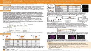 科学海报Generation of Microglia From Human Pluripotent Stem Cells for Neurodegenerative Disease Modeling

 沪公网安备31010102008431号