Neutrophil survival and c-kit(+)-progenitor proliferation in Staphylococcus aureus-infected skin wounds promote resolution.
Polymorphonuclear neutrophils (PMNs) are critical for the formation,maintenance,and resolution of bacterial abscesses. However,the mechanisms that regulate PMN survival and proliferation during the evolution of an abscess are not well defined. Using a mouse model of Staphylococcus aureus abscess formation within a cutaneous wound,combined with real-time imaging of genetically tagged PMNs,we observed that a high bacterial burden elicited a sustained mobilization of PMNs from the bone marrow to the infected wound,where their lifespan was markedly extended. A continuous rise in wound PMN number,which was not accounted for by trafficking from the bone marrow or by prolonged survival,was correlated with the homing of c-kit(+)-progenitor cells from the blood to the wound,where they proliferated and formed mature PMNs. Furthermore,by blocking their recruitment with an antibody to c-kit,which severely limited the proliferation of mature PMNs in the wound and shortened mouse survival,we confirmed that progenitor cells are not only important contributors to PMN expansion in the wound,but are also functionally important for immune protection. We conclude that the abscess environment provides a niche capable of regulating PMN survival and local proliferation of bone marrow-derived c-kit(+)-progenitor cells.
View Publication
产品类型:
产品号#:
03434
03444
产品名:
MethoCult™ GF M3434
MethoCult™ GF M3434
Drayer AL et al. (JAN 2006)
Stem cells (Dayton,Ohio) 24 1 105--14
Mammalian target of rapamycin is required for thrombopoietin-induced proliferation of megakaryocyte progenitors.
Thrombopoietin (TPO) is a potent regulator of megakaryopoiesis and stimulates megakaryocyte (MK) progenitor expansion and MK differentiation. In this study,we show that TPO induces activation of the mammalian target of rapamycin (mTOR) signaling pathway,which plays a central role in translational regulation and is required for proliferation of MO7e cells and primary human MK progenitors. Treatment of MO7e cells,human CD34+,and primary MK cells with the mTOR inhibitor rapamycin inhibits TPO-induced cell cycling by reducing cells in S phase and blocking cells in G0/G1. Rapamycin markedly inhibits the clonogenic growth of MK progenitors with high proliferative capacity but does not reduce the formation of small MK colonies. Addition of rapamycin to MK suspension cultures reduces the number of MK cells,but inhibition of mTOR does not significantly affect expression of glycoproteins IIb/IIIa (CD41) and glycoprotein Ib (CD42),nuclear polyploidization levels,cell size,or cell survival. The downstream effectors of mTOR,p70 S6 kinase (S6K) and 4E-binding protein 1 (4E-BP1),are phosphorylated by TPO in a rapamycin- and LY294002-sensitive manner. Part of the effect of the phosphatidyl inositol 3-kinase pathway in regulating megakaryopoiesis may be mediated by the mTOR/S6K/4E-BP1 pathway. In conclusion,these data demonstrate that the mTOR pathway is activated by TPO and plays a critical role in regulating proliferation of MK progenitors,without affecting differentiation or cell survival.
View Publication
产品类型:
产品号#:
04961
04902
04901
04971
04963
04962
产品名:
MegaCult™-C胶原和含细胞因子培养基
胶原蛋白溶液
MegaCult™-C含细胞因子培养基
MegaCult™-C含细胞因子全套试剂盒
双室载玻片套件
MegaCult™-C CFU-Mk染色试剂盒
Gkountela S et al. (APR 2014)
Stem Cell Reviews and Reports 10 2 230--239
PRMT5 is required for human embryonic stem cell proliferation but not pluripotency.
Human pluripotent stem cells (PSCs) are critical in vitro tools forbackslashnunderstanding mechanisms that regulate lineage differentiation inbackslashnthe human embryo as well as a potentially unlimited supply of stembackslashncells for regenerative medicine. Pluripotent human and mouse embryonicbackslashnstem cells (ESCs) derived from the inner cell mass of blastocystsbackslashnshare a similar transcription factor network to maintain pluripotencybackslashnand self-renewal,yet there are considerable molecular differencesbackslashnreflecting the diverse environments in which mouse and human ESCsbackslashnare derived. In the current study we evaluated the role of Proteinbackslashnarginine methyltransferase 5 (PRMT5) in human ESC (hESC) self-renewalbackslashnand pluripotency given its critical role in safeguarding mouse ESCbackslashnpluripotency. Unlike the mouse,we discovered that PRMT5 has no rolebackslashnin hESC pluripotency. Using microarray analysis we discovered thatbackslashna significant depletion in PRMT5 RNA and protein from hESCs changedbackslashnthe expression of only 78 genes,with the majority being repressed.backslashnFunctionally,we discovered that depletion of PRMT5 had no effectbackslashnon expression of OCT4,NANOG or SOX2,and did not prevent teratomabackslashnformation. Instead,we show that PRMT5 functions in hESCs to regulatebackslashnproliferation in the self-renewing state by regulating the fractionbackslashnof cells in Gap 1 (G1) of the cell cycle and increasing expressionbackslashnof the G1 cell cycle inhibitor P57. Taken together our data unveilsbackslashna distinct role for PRMT5 in hESCs and identifies P57 as new target.
View Publication
产品类型:
产品号#:
05850
05857
05870
05875
85850
85857
85870
85875
产品名:
mTeSR™1
mTeSR™1
Felfly H and Klein OD (JUL 2013)
Scientific Reports 3 2277
Sprouty genes regulate proliferation and survival of human embryonic stem cells.
Sprouty (Spry) genes encode negative regulators of receptor tyrosine kinase (RTK) signaling,which plays important roles in human embryonic stem cells (hESCs). SPRY2 and SPRY4 are the two most highly expressed Sprouty family members in hESCs,suggesting that they may influence self-renewal. To test this hypothesis,we performed siRNA-mediated knock down (KD) studies. SPRY2 KD resulted in increased cell death and decreased proliferation,whereas SPRY4 KD enhanced survival. In both cases,after KD the cells were able to differentiate into cells of the three germ layers,although after SPRY2 KD there was a tendency toward increased ectodermal differentiation. SPRY2 KD cells displayed impaired mitochondrial fusion and cell membrane damage,explaining in part the increased cell death. These data indicate that Sprouty genes regulate pathways involved in proliferation and cell death in hESCs.
View Publication
产品类型:
产品号#:
05850
05857
05870
05875
85850
85857
85870
85875
产品名:
mTeSR™1
mTeSR™1
Derda R et al. (FEB 2010)
Journal of the American Chemical Society 132 4 1289--1295
High-throughput discovery of synthetic surfaces that support proliferation of pluripotent cells.
Synthetic materials that promote the growth or differentiation of cells have advanced the fields of tissue engineering and regenerative medicine. Most functional biomaterials are based on a handful of peptide sequences derived from protein ligands for cell surface receptors. Because few proteins possess short peptide sequences that alone can engage cell surface receptors,the repertoire of receptors that can be targeted with this approach is limited. Materials that bind diverse classes of receptors,however,may be needed to guide cell growth and differentiation. To provide access to such new materials,we utilized phage display to identify novel peptides that bind to the surface of pluripotent cells. Using human embryonal carcinoma (EC) cells as bait,approximately 3 x 10(4) potential cell-binding phage clones were isolated. The pool was narrowed using an enzyme-linked immunoassay: 370 clones were tested,and seven cell-binding peptides were identified. Of these,six sequences possess EC cell-binding ability. Specifically,when displayed by self-assembled monolayers (SAMs) of alkanethiols on gold,they mediate cell adhesion. The corresponding soluble peptides block this adhesion,indicating that the identified peptide sequences are specific. They also are functional. Synthetic surfaces displaying phage-derived peptides support growth of undifferentiated human embryonic stem (ES) cells. When these cells were cultured on SAMs presenting the sequence TVKHRPDALHPQ or LTTAPKLPKVTR in a chemically defined medium (mTeSR),they expressed markers of pluripotency at levels similar to those of cells cultured on Matrigel. Our results indicate that this screening strategy is a productive avenue for the generation of materials that control the growth and differentiation of cells.
View Publication
产品类型:
产品号#:
05850
05857
05870
05875
85850
85857
85870
85875
产品名:
mTeSR™1
mTeSR™1
Matsumoto K et al. (JAN 2000)
Stem cells (Dayton,Ohio) 18 3 196--203
In vitro proliferation potential of AC133 positive cells in peripheral blood.
AC133 antigen is a novel marker for human hematopoietic stem/progenitor cells. In this study,we examined the expression and proliferation potential of AC133(+) cells obtained from steady-state peripheral blood (PB). The proportion of AC133(+) cells in the CD34(+) subpopulation of steady-state PB was significantly lower than that of cord blood (CB),although that of cytokine-mobilized PB was higher than that of CB. The proliferation potential of AC133(+)CD34(+) and AC133(-)CD34(+) cells was examined by colony-forming analysis and analysis of long-term culture-initiating cells (LTC-IC). Although the total number of colony-forming cells was essentially the same in the AC133(+)CD34(+) fraction as in the AC133(-)CD34(+) fraction,the proportion of LTC-IC was much higher in the AC133(+)CD34(+) fraction. Virtually no LTC-IC were detected in the AC133(-)CD34(+) fraction. In addition,the features of the colonies grown from these two fractions were quite different. Approximately 70% of the colonies derived from the AC133(+)CD34(+) fraction were granulocyte-macrophage colonies,whereas more than 90% of the colonies derived from the AC133(-)CD34(+) fraction were erythroid colonies. Furthermore,an ex vivo expansion study observed expansion of colony-forming cells only in the AC133(+)CD34(+) population,and not in the AC133(-)CD34(+) population. These findings suggest that to isolate primitive hematopoietic cells from steady-state PB,selection by AC133 expression is better than selection by CD34 expression.
View Publication
产品类型:
产品号#:
04034
04044
产品名:
MethoCult™ H4034 Optimum
MethoCult™ H4034 Optimum
Cutler AJ et al. (DEC 2010)
Journal of immunology (Baltimore,Md. : 1950) 185 11 6617--23
Umbilical cord-derived mesenchymal stromal cells modulate monocyte function to suppress T cell proliferation.
Mesenchymal stromal cells (MSCs) may be derived from a variety of tissues,with human umbilical cord (UC) providing an abundant and noninvasive source. Human UC-MSCs share similar in vitro immunosuppressive properties as MSCs obtained from bone marrow and cord blood. However,the mechanisms and cellular interactions used by MSCs to control immune responses remain to be fully elucidated. In this paper,we report that suppression of mitogen-induced T cell proliferation by human UC-,bone marrow-,and cord blood-MSCs required monocytes. Removal of monocytes but not B cells from human adult PBMCs (PBMNCs) reduced the immunosuppressive effects of MSCs on T cell proliferation. There was rapid modulation of a number of cell surface molecules on monocytes when PBMCs or alloantigen-activated PBMNCs were cultured with UC-MSCs. Indomethacin treatment significantly inhibited the ability of UC-MSCs to suppress T cell proliferation,indicating an important role for PGE(2). Monocytes purified from UC-MSC coculture had significantly reduced accessory cell and allostimulatory function when tested in subsequent T cell proliferation assays,an effect mediated in part by UC-MSC PGE(2) production and enhanced by PBMNC alloactivation. Therefore,we identify monocytes as an essential intermediary through which UC-MSCs mediate their suppressive effects on T cell proliferation.
View Publication
产品类型:
产品号#:
05401
05402
05411
产品名:
MesenCult™ MSC 基础培养基(人)
MesenCult™ MSC 刺激补充剂(人)
MesenCult™ 增殖试剂盒(人)
Bouscary D et al. (MAY 2003)
Blood 101 9 3436--43
Critical role for PI 3-kinase in the control of erythropoietin-induced erythroid progenitor proliferation.
The production of red blood cells is tightly regulated by erythropoietin (Epo). The phosphoinositide 3-kinase (PI 3-kinase) pathway was previously shown to be activated in response to Epo. We studied the role of this pathway in the control of Epo-induced survival and proliferation of primary human erythroid progenitors. We show that phosphoinositide 3 (PI 3)-kinase associates with 4 tyrosine-phosphorylated proteins in primary human erythroid progenitors,namely insulin receptor substrate-2 (IRS2),Src homology 2 domain-containing inositol 5'-phosphatase (SHIP),Grb2-associated binder-1 (Gab1),and the Epo receptor (EpoR). Using different in vitro systems,we demonstrate that 3 alternative pathways independently lead to Epo-induced activation of PI 3-kinase and phosphorylation of its downstream effectors,Akt,FKHRL1,and P70S6 kinase: through direct association of PI 3-kinase with the last tyrosine residue (Tyr479) of the Epo receptor (EpoR),through recruitment and phosphorylation of Gab proteins via either Tyr343 or Tyr401 of the EpoR,or through phosphorylation of IRS2 adaptor protein. The mitogen-activated protein (MAP) kinase pathway was also activated by Epo in erythroid progenitors,but we found that this process is independent of PI 3-kinase activation. In erythroid progenitors,the functional role of PI 3-kinase was both to prevent apoptosis and to stimulate cell proliferation in response to Epo stimulation. Finally,our results show that PI 3-kinase-mediated proliferation of erythroid progenitors in response to Epo occurs mainly through modulation of the E3 ligase SCF(SKP2),which,in turn,down-regulates p27(Kip1) cyclin-dependent kinase (CDK) inhibitor via proteasome degradation.
View Publication

 EasySep™小鼠TIL(CD45)正选试剂盒


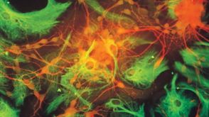
 产品手册NeuroCult™: Reagents for Brain Tumor Stem Cell Research
 产品手册NeuroCult™-XF: Xeno-Free Culture Medium for the Proliferation of Human Neural Stem Cells
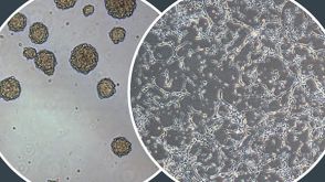

 沪公网安备31010102008431号