Detection, isolation, and stimulation of quiescent primitive leukemic progenitor cells from patients with acute myeloid leukemia (AML).
Although many acute myeloid leukemia (AML) colony-forming cells (CFCs) and long-term culture-initiating cells (LTC-ICs) directly isolated from patients are actively cycling,quiescent progenitors are present in most samples. In the current study,(3)H-thymidine ((3)H-Tdr) suicide assays demonstrated that most NOD/SCID mouse leukemia-initiating cells (NOD/SL-ICs) are quiescent in 6 of 7 AML samples. AML cells in G(0),G(1),and S/G(2)+M were isolated from 4 of these samples using Hoechst 33342/pyroninY staining and cell sorting. The progenitor content of each subpopulation was consistent with the (3)H-Tdr suicide results,with NOD/SL-ICs found almost exclusively among G(0) cells while the cycling status of AML CFCs and LTC-ICs was more heterogeneous. Interestingly,after 72 hours in serum-free culture with or without Steel factor (SF),Flt-3 ligand (FL),and interleukin-3 (IL-3),most G(0) AML cells entered active cell cycle (percentage of AML cells remaining in G(0) at 72 hours,1.2% to 37%,and 0% to 7.6% in cultures without and with growth factors [GFs],respectively) while G(0) cells from normal lineage-depleted bone marrow remained quiescent in the absence of GF. All 4 AML samples showed evidence of autocrine production of 2 or more of SF,FL,IL-3,and granulocyte-macrophage colony-stimulating factor (GM-CSF). In addition,3 of 4 samples contained an internal tandem duplication of the FLT3 gene. In summary,quiescent leukemic cells,including NOD/SL-ICs,are present in most AML patients. Their spontaneous entry into active cell cycle in short-term culture might be explained by the deregulated GF signaling present in many AMLs.
View Publication
Mujtaba T et al. (OCT 1999)
Developmental biology 214 1 113--27
Lineage-restricted neural precursors can be isolated from both the mouse neural tube and cultured ES cells.
We have previously identified multipotent neuroepithelial (NEP) stem cells and lineage-restricted,self-renewing precursor cells termed NRPs (neuron-restricted precursors) and GRPs (glial-restricted precursors) present in the developing rat spinal cord (A. Kalyani,K. Hobson,and M. S. Rao,1997,Dev. Biol. 186,202-223; M. S. Rao and M. Mayer-Proschel,1997,Dev. Biol. 188,48-63; M. Mayer-Proschel,A. J. Kalyani,T. Mujtaba,and M. S. Rao,1997,Neuron 19,773-785). We now show that cells identical to rat NEPs,NRPs,and GRPs are present in mouse neural tubes and that immunoselection against cell surface markers E-NCAM and A2B5 can be used to isolate NRPs and GRPs,respectively. Restricted precursors similar to NRPs and GRPs can also be isolated from mouse embryonic stem cells (ES cells). ES cell-derived NRPs are E-NCAM immunoreactive,undergo self-renewal in defined medium,and differentiate into multiple neuronal phenotypes in mass culture. ES cells also generate A2B5-immunoreactive cells that are similar to E9 NEP-cell-derived GRPs and can differentiate into oligodendrocytes and astrocytes. Thus,lineage restricted precursors can be generated in vitro from cultured ES cells and these restricted precursors resemble those derived from mouse neural tubes. These results demonstrate the utility of using ES cells as a source of late embryonic precursor cells.
View Publication
产品类型:
产品号#:
06902
06952
00321
00322
00323
00324
00325
产品名:
Anderson SA et al. (JAN 2005)
Blood 105 1 420--5
Noninvasive MR imaging of magnetically labeled stem cells to directly identify neovasculature in a glioma model.
Bone marrow-derived endothelial precursor cells incorporate into neovasculature and have been successfully used as vehicles for gene delivery to brain tumors. To determine whether systemically administered Sca1+ bone marrow cells labeled with superparamagnetic iron oxide nanoparticles can be detected by in vivo magnetic resonance imaging in a mouse brain tumor model,mouse Sca1+ cells were labeled in vitro with ferumoxides-poly-L-lysine complexes. Labeled or control cells were administered intravenously to glioma-bearing severe combined immunodeficient (SCID) mice. Magnetic resonance imaging (MRI) was performed during tumor growth. Mice that received labeled cells demonstrated hypointense regions within the tumor that evolved over time and developed a continuous dark hypointense ring at a consistent time point. This effect was not cleared by administration of a gadolinium contrast agent. Histology showed iron-labeled cells around the tumor rim in labeled mice,which expressed CD31 and von Willebrand factor,indicating the transplanted cells detected in the tumor have differentiated into endothelial-like cells. These results demonstrate that MRI can detect the incorporation of magnetically labeled bone marrow-derived precursor cells into tumor vasculature as part of ongoing angiogenesis and neovascularization. This technique can be used to directly identify neovasculature in vivo and to facilitate gene therapy by noninvasively monitoring these cells as gene delivery vectors.
View Publication
产品类型:
产品号#:
09600
09650
09850
产品名:
StemSpan™ SFEM
StemSpan™ SFEM
Ginis I et al. (JUN 2012)
Tissue engineering. Part C,Methods 18 6 453--63
Evaluation of bone marrow-derived mesenchymal stem cells after cryopreservation and hypothermic storage in clinically safe medium.
Achievements in tissue engineering using mesenchymal stem cells (MSC) demand a clinically acceptable off-the-shelf" cell therapy product. Efficacy of cryopreservation of human bone marrow-derived MSC in clinically safe animal product-free medium containing 2% 5% and 10% dimethyl sulfoxide (DMSO) was evaluated by measuring cell recovery viability apoptosis proliferation rate expression of a broad panel of MSC markers and osteogenic differentiation. Rate-controlled freezing in CryoStor media was performed in a programmable cell freezer. About 95% of frozen cells were recovered as live cells after freezing in CryoStor solutions with 5% and 10% DMSO followed by storage in liquid nitrogen for 1 month. Cell recovery after 5 months storage was 72% and 80% for 5% and 10% DMSO respectively. Measurements of caspase 3 activity demonstrated that 15.5% and 12.8% of cells after 1 month and 18.3% and 12.9% of cells after 5 months storage in 5% and 10% DMSO respectively were apoptotic. Proliferation of MSC recovered after cryopreservation was measured during 2 weeks post-plating. Proliferation rate was not compromised and was even enhanced. Cryopreservation did not alter expression of MSC markers. Quantitative analysis of alkaline phosphatase (ALP) activity ALP surface expression and Ca deposition in previously cryopreserved MSC and then differentiated for 3 weeks in osteogenic medium demonstrated the same degree of osteogenic differentiation as in unfrozen parallel cultures. Cell viability and functional parameters were analyzed in MSC after short-term storage at 4°C in HypoThermosol-FRS solution also free of animal products. Hypothermic storage for 2 and 4 days resulted in about 100% and 85% cell recovery respectively less than 10% of apoptotic cells and normal proliferation marker expression and osteogenic potential. Overall our results demonstrate that human MSC could be successfully cryopreserved for banking and clinical applications and delivered to the bedside in clinically safe protective reagents.
View Publication
产品类型:
产品号#:
07930
07931
07940
07955
07956
07959
07954
100-1061
07952
产品名:
CryoStor® CS10
CryoStor® CS10
CryoStor® CS10
CryoStor® CS10
CryoStor® CS10
CryoStor® CS10
CryoStor® CS10
Varga E et al. (OCT 2016)
Stem cell research 17 3 482--484
Generation of Mucopolysaccharidosis type II (MPS II) human induced pluripotent stem cell (iPSC) line from a 1-year-old male with pathogenic IDS mutation.
Peripheral blood was collected from a 1-year-old male patient with an X-linked recessive mutation of Iduronate 2-sulfatase (IDS) gene (NM000202.7(IDS):c.85CtextgreaterT) causing MPS II (OMIM 309900). Peripheral blood mononuclear cells (PBMCs) were reprogrammed by lentiviral delivery of a self-silencing hOKSM polycistronic vector. The pluripotency of the iPSC line was confirmed by the expression of pluripotency-associated markers and in vitro spontaneous differentiation towards the 3 germ layers. The iPSC line showed normal karyotype. The cell line offers a good platform to study MPS II pathophysiology,for drug testing,early biomarker discovery and gene therapy studies.
View Publication
产品类型:
产品号#:
05850
05857
05870
05875
85850
85857
85870
85875
产品名:
mTeSR™1
mTeSR™1
Ma D et al. (JAN 2017)
Stem cell research 18 51--53
Development of a human induced pluripotent stem cell (iPSC) line from a Parkinson's disease patient carrying the N551K variant in LRRK2 gene.
Peripheral blood mononuclear cells (PBMCs) were collected from a clinically diagnosed 64-year old male Parkinson's disease (PD) patient with N551K variant in the LRRK2 gene. The PMBCs were reprogrammed with the human OSKM transcription factors using the Sendai-virus reprogramming system. The transgene-free iPSC showed pluripotency confirmed by immunofluorescent staining for pluripotency markers and differentiated into the 3 germ layers in vivo. The iPSC line also showed normal karyotype. This cellular model can complement in vivo PD models for pathophysiological studies and drug screening.
View Publication
Walter DH et al. (FEB 2011)
Circulation. Cardiovascular interventions 4 1 26--37
Intraarterial administration of bone marrow mononuclear cells in patients with critical limb ischemia: a randomized-start, placebo-controlled pilot trial (PROVASA).
BACKGROUND: Critical limb ischemia due to peripheral arterial occlusive disease is associated with a severely increased morbidity and mortality. There is no effective pharmacological therapy available. Injection of autologous bone marrow-derived mononuclear cells (BM-MNC) is a promising therapeutic option in patients with critical limb ischemia,but double-blind,randomized trials are lacking. METHODS AND RESULTS: Forty patients with critical limb ischemia were included in a multicenter,phase II,double-blind,randomized-start trial to receive either intraarterial administration of BM-MNC or placebo followed by active treatment with BM-MNC (open label) after 3 months. Intraarterial administration of BM-MNC did not significantly increase ankle-brachial index and,thus,the trial missed its primary end point. However,cell therapy was associated with significantly improved ulcer healing (ulcer area,3.2±4.7 cm(2) to 1.89±3.5 cm(2) [P=0.014] versus placebo,2.92±3.5 cm(2) to 2.89±4.1 cm(2) [P=0.5]) and reduced rest pain (5.2±1.8 to 2.2±1.3 [P=0.009] versus placebo,4.5±2.4 to 3.9±2.6 [P=0.3]) within 3 months. Limb salvage and amputation-free survival rates did not differ between the groups. Repeated BM-MNC administration and higher BM-MNC numbers and functionality were the only independent predictors of improved ulcer healing. Ulcer healing induced by repeated BM-MNC administration significantly correlated with limb salvage (r=0.8; Ptextless0.001). CONCLUSIONS: Intraarterial administration of BM-MNC is safe and feasible and accelerates wound healing in patients without extensive gangrene and impending amputation. These exploratory findings of this pilot trial need to be confirmed in a larger randomized trial in patients with critical limb ischemia and stable ulcers.
View Publication
产品类型:
产品号#:
04564
产品名:
MethoCult™ H4534 Classic 无 EPO 入门试剂盒
Arbab AS et al. (SEP 2008)
FASEB journal : official publication of the Federation of American Societies for Experimental Biology 22 9 3234--46
Detection of migration of locally implanted AC133+ stem cells by cellular magnetic resonance imaging with histological findings.
This study investigated the factors responsible for migration and homing of magnetically labeled AC133(+) cells at the sites of active angiogenesis in tumor. AC133(+) cells labeled with ferumoxide-protamine sulfate were mixed with either rat glioma or human melanoma cells and implanted in flank of nude mice. An MRI of the tumors including surrounding tissues was performed. Tumor sections were stained for Prussian blue (PB),platelet-derived growth factor (PDGF),hypoxia-inducible factor-1alpha (HIF-1alpha),stromal cell derived factor-1 (SDF-1),matrix metalloproteinase-2 (MMP-2),vascular endothelial growth factor (VEGF),and endothelial markers. Fresh snap-frozen strips from the central and peripheral parts of the tumor were collected for Western blotting. MRIs demonstrated hypointense regions at the periphery of the tumors where the PB(+)/AC133(+) cells were positive for endothelial cells markers. At the sites of PB(+)/AC133(+) cells,both HIF-1alpha and SDF-1 were strongly positive and PDGF and MMP-2 showed generalized expression in the tumor and surrounding tissues. There was no significant association of PB(+)/AC133(+) cell localization and VEGF expression in tumor cells. Western blot demonstrated strong expression of the SDF-1,MMP-2,and PDGF at the peripheral parts of the tumors. HIF-1alpha was expressed at both the periphery and central parts of the tumor. This work demonstrates that magnetically labeled cells can be used as probes for MRI and histological identification of administered cells.
View Publication
产品类型:
产品号#:
09600
09650
产品名:
StemSpan™ SFEM
StemSpan™ SFEM
Luo M et al. (JAN 2009)
Cancer research 69 2 466--74
Mammary epithelial-specific ablation of the focal adhesion kinase suppresses mammary tumorigenesis by affecting mammary cancer stem/progenitor cells.
Focal adhesion kinase (FAK) has been implicated in the development of cancers,including those of the breast. Nevertheless,the molecular and cellular mechanisms by which FAK promotes mammary tumorigenesis in vivo are not well understood. Here,we show that targeted deletion of FAK in mouse mammary epithelium significantly suppresses mammary tumorigenesis in a well-characterized breast cancer model. Ablation of FAK leads to the depletion of a subset of bipotent cells in the tumor that express both luminal marker keratin 8/18 and basal marker keratin 5. Using mammary stem/progenitor markers,including aldehyde dehydrogenase,CD24,CD29,and CD61,we further revealed that ablation of FAK reduced the pool of cancer stem/progenitor cells in primary tumors of FAK-targeted mice and impaired their self-renewal and migration in vitro. Finally,through transplantation in NOD-SCID mice,we found that cancer stem/progenitor cells isolated from FAK-targeted mice have compromised tumorigenicity and impaired maintenance in vivo. Together,these results show a novel function of FAK in maintaining the mammary cancer stem/progenitor cell population and provide a novel mechanism by which FAK may promote breast cancer development and progression.
View Publication
产品类型:
产品号#:
18556
18556RF
产品名:
Charafe-Jauffret E et al. (FEB 2009)
Cancer research 69 4 1302--13
Breast cancer cell lines contain functional cancer stem cells with metastatic capacity and a distinct molecular signature.
Tumors may be initiated and maintained by a cellular subcomponent that displays stem cell properties. We have used the expression of aldehyde dehydrogenase as assessed by the ALDEFLUOR assay to isolate and characterize cancer stem cell (CSC) populations in 33 cell lines derived from normal and malignant mammary tissue. Twenty-three of the 33 cell lines contained an ALDEFLUOR-positive population that displayed stem cell properties in vitro and in NOD/SCID xenografts. Gene expression profiling identified a 413-gene CSC profile that included genes known to play a role in stem cell function,as well as genes such as CXCR1/IL-8RA not previously known to play such a role. Recombinant interleukin-8 (IL-8) increased mammosphere formation and the ALDEFLUOR-positive population in breast cancer cell lines. Finally,we show that ALDEFLUOR-positive cells are responsible for mediating metastasis. These studies confirm the hierarchical organization of immortalized cell lines,establish techniques that can facilitate the characterization of regulatory pathways of CSCs,and identify potential stem cell markers and therapeutic targets.
View Publication

 EasySep™小鼠TIL(CD45)正选试剂盒


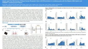 科学海报Simple Label-free Monitoring of Robust Human Pluripotent Stem Cell Differentiation to Cardiomyocytes with the Maestro MEA System and STEMdiff™ Cardiomyocyte Products

 沪公网安备31010102008431号