Tohyama S et al. (APR 2016)
Cell Metabolism 23 4 663--674
Glutamine Oxidation Is Indispensable for Survival of Human Pluripotent Stem Cells
Summary Human pluripotent stem cells (hPSCs) are uniquely dependent on aerobic glycolysis to generate ATP. However,the importance of oxidative phosphorylation (OXPHOS) has not been elucidated. Detailed amino acid profiling has revealed that glutamine is indispensable for the survival of hPSCs. Under glucose- and glutamine-depleted conditions,hPSCs quickly died due to the loss of ATP. Metabolome analyses showed that hPSCs oxidized pyruvate poorly and that glutamine was the main energy source for OXPHOS. hPSCs were unable to utilize pyruvate-derived citrate due to negligible expression of aconitase 2 (ACO2) and isocitrate dehydrogenase 2/3 (IDH2/3) and high expression of ATP-citrate lyase. Cardiomyocytes with mature mitochondria were not able to survive without glucose and glutamine,although they were able to use lactate to synthesize pyruvate and glutamate. This distinguishing feature of hPSC metabolism allows preparation of clinical-grade cell sources free of undifferentiated hPSCs,which prevents tumor formation during stem cell therapy.
View Publication
产品类型:
产品号#:
05850
05857
05870
05875
85850
85857
85870
85875
产品名:
mTeSR™1
mTeSR™1
Huijskens MJAJ et al. (DEC 2014)
Journal of leukocyte biology 96 6 1165--75
Technical advance: ascorbic acid induces development of double-positive T cells from human hematopoietic stem cells in the absence of stromal cells.
The efficacy of donor HSCT is partly reduced as a result of slow post-transplantation immune recovery. In particular,T cell regeneration is generally delayed,resulting in high infection-related mortality in the first years post-transplantation. Adoptive transfer of in vitro-generated human T cell progenitors seems a promising approach to accelerate T cell recovery in immunocompromised patients. AA may enhance T cell proliferation and differentiation in a controlled,feeder-free environment containing Notch ligands and defined growth factors. Our experiments show a pivotal role for AA during human in vitro T cell development. The blocking of NOS diminished this effect,indicating a role for the citrulline/NO cycle. AA promotes the transition of proT1 to proT2 cells and of preT to DP T cells. Furthermore,the addition of AA to feeder cocultures resulted in development of DP and SP T cells,whereas without AA,a preT cell-stage arrest occurred. We conclude that neither DLL4-expressing feeder cells nor feeder cell conditioned media are required for generating DP T cells from CB and G-CSF-mobilized HSCs and that generation and proliferation of proT and DP T cells are greatly improved by AA. This technology could potentially be used to generate T cell progenitors for adoptive therapy.
View Publication
Microfluidic Image Cytometry for Single-Cell Phenotyping of Human Pluripotent Stem Cells
A microfluidic human pluripotent stem cell (hPSC) array has been developed for robust and reproducible hPSC culture methods to assess chemically defined serum- and feeder-free culture conditions. This microfluidic platform,combined with image cytometry,enables the systematic analysis of multiple simultaneously detected marker expression in individual cells,for screening of various chemically defined media across hPSC lines,and the study of phenotypic responses.
View Publication
产品类型:
产品号#:
05850
05857
05870
05875
85850
85857
85870
85875
产品名:
mTeSR™1
mTeSR™1
Haniffa M et al. (FEB 2009)
The Journal of experimental medicine 206 2 371--85
Differential rates of replacement of human dermal dendritic cells and macrophages during hematopoietic stem cell transplantation.
Animal models of hematopoietic stem cell transplantation have been used to analyze the turnover of bone marrow-derived cells and to demonstrate the critical role of recipient antigen-presenting cells (APC) in graft versus host disease (GVHD). In humans,the phenotype and lineage relationships of myeloid-derived tissue APC remain incompletely understood. It has also been proposed that the risk of acute GVHD,which extends over many months,is related to the protracted survival of certain recipient APC. Human dermis contains three principal subsets of CD45(+)HLA-DR(+) cells: CD1a(+)CD14(-) DC,CD1a(-)CD14(+) DC,and CD1a(-)CD14(+)FXIIIa(+) macrophages. In vitro,each subset has characteristic properties. After transplantation,both CD1a(+) and CD14(+) DC are rapidly depleted and replaced by donor cells,but recipient macrophages can be found in GVHD lesions and may persist for many months. Macrophages isolated from normal dermis secrete proinflammatory cytokines. Although they stimulate little proliferation of naive or memory CD4(+) T cells,macrophages induce cytokine expression in memory CD4(+) T cells and activation and proliferation of CD8(+) T cells. These observations suggest that dermal macrophages and DC are from distinct lineages and that persistent recipient macrophages,although unlikely to initiate alloreactivity,may contribute to GVHD by sustaining the responses of previously activated T cells.
View Publication
Siatskas C et al. (OCT 2005)
FASEB journal : official publication of the Federation of American Societies for Experimental Biology 19 12 1752--4
Specific pharmacological dimerization of KDR in lentivirally transduced human hematopoietic cells activates anti-apoptotic and proliferative mechanisms.
Selective and regulatable expansion of transduced cells could augment gene therapy for many disorders. The activation of modified growth factor receptors via synthetic chemical inducers of dimerization allows for the coordinated growth of transduced cells. This system can also provide information on specific receptor-mediated signaling without interference from other family members. Although several receptor subunits have been investigated in this context,little is known about the precise molecular events associated with dimerizer-initiated signaling. We have constructed and expressed an AP20187-regulated KDR chimeric receptor in human TF1 cells and analyzed activation of this gene switch using functional,biochemical,and microarray analyses. When deprived of natural ligands,GM-CSF,interleukin-3,or erythropoietin,AP20187 prevented apoptosis of transduced TF1 cells,induced dose-dependent proliferation,and supported long-term growth. In addition,AP20187 stimulation activated the signaling molecules associated with mitogen-activated protein kinase and phosphatidyl-inositol 3-kinase/Akt pathways. Microarray analysis determined that a number of transcripts involved in a variety of cellular processes were differentially expressed. Notably,mRNAs affiliated with heat stress,including Hsp70 and Hsp105,were up-regulated. Functional assays showed that Hsp70 and Hsp105 protected transduced TF1 cells from apoptosis and premature senescence,in part through regulation of Akt. These observations delineate specific roles for kinase insert domain-containing receptor,or KDR,signaling and suggest strategies to endow genetically modified cells with a survival advantage enabling the generation of adequate cell numbers for therapeutic outcomes.
View Publication
SOX10-Nano-Lantern Reporter Human iPS Cells; A Versatile Tool for Neural Crest Research.
The neural crest is a source to produce multipotent neural crest stem cells that have a potential to differentiate into diverse cell types. The transcription factor SOX10 is expressed through early neural crest progenitors and stem cells in vertebrates. Here we report the generation of SOX10-Nano-lantern (NL) reporter human induced pluripotent stem cells (hiPS) by using CRISPR/Cas9 systems,that are beneficial to investigate the generation and maintenance of neural crest progenitor cells. SOX10-NL positive cells are produced transiently from hiPS cells by treatment with TGFβ inhibitor SB431542 and GSK3 inhibitor CHIR99021. We found that all SOX10-NL-positive cells expressed an early neural crest marker NGFR,however SOX10-NL-positive cells purified from differentiated hiPS cells progressively attenuate their NL-expression under proliferation. We therefore attempted to maintain SOX10-NL-positive cells with additional signaling on the plane and sphere culture conditions. These SOX10-NL cells provide us to investigate mass culture with neural crest cells for stem cell research.
View Publication

 EasySep™小鼠TIL(CD45)正选试剂盒


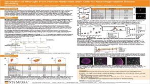 科学海报Generation of Microglia From Human Pluripotent Stem Cells for Neurodegenerative Disease Modeling
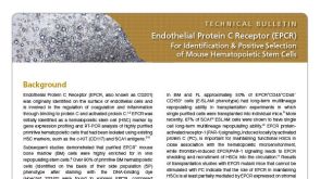 技术公告Endothelial Protein C Receptor (EPCR): A New Marker for Identification and Positive Selection of Mouse Hematopoietic Stem Cells
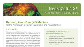 产品手册NeuroCult™-XF: Xeno-Free Culture Medium for the Proliferation of Human Neural Stem Cells

 沪公网安备31010102008431号