Weisberg E et al. (DEC 2008)
Blood 112 13 5161--70
Antileukemic effects of the novel, mutant FLT3 inhibitor NVP-AST487: effects on PKC412-sensitive and -resistant FLT3-expressing cells.
An attractive target for therapeutic intervention is constitutively activated,mutant FLT3,which is expressed in a subpopulation of patients with acute myelocyic leukemia (AML) and is generally a poor prognostic indicator in patients under the age of 65 years. PKC412 is one of several mutant FLT3 inhibitors that is undergoing clinical testing,and which is currently in late-stage clinical trials. However,the discovery of drug-resistant leukemic blast cells in PKC412-treated patients with AML has prompted the search for novel,structurally diverse FLT3 inhibitors that could be alternatively used to override drug resistance. Here,we report the potent and selective antiproliferative effects of the novel mutant FLT3 inhibitor NVP-AST487 on primary patient cells and cell lines expressing FLT3-ITD or FLT3 kinase domain point mutants. NVP-AST487,which selectively targets mutant FLT3 protein kinase activity,is also shown to override PKC412 resistance in vitro,and has significant antileukemic activity in an in vivo model of FLT3-ITD(+) leukemia. Finally,the combination of NVP-AST487 with standard chemotherapeutic agents leads to enhanced inhibition of proliferation of mutant FLT3-expressing cells. Thus,we present a novel class of FLT3 inhibitors that displays high selectivity and potency toward FLT3 as a molecular target,and which could potentially be used to override drug resistance in AML.
View Publication
产品类型:
产品号#:
04434
04444
产品名:
MethoCult™ H4434 Classic
MethoCult™ H4434 Classic
Balasubramaniam V et al. (MAR 2010)
American journal of physiology. Lung cellular and molecular physiology 298 3 L315--23
Bone marrow-derived angiogenic cells restore lung alveolar and vascular structure after neonatal hyperoxia in infant mice.
Neonatal hyperoxia impairs vascular and alveolar growth in mice and decreases endothelial progenitor cells. To determine the role of bone marrow-derived cells in restoration of neonatal lung structure after injury,we studied a novel bone marrow myeloid progenitor cell population from Tie2-green fluorescent protein (GFP) transgenic mice (bone marrow-derived angiogenic cells; BMDAC). We hypothesized that treatment with BMDAC would restore normal lung structure in infant mice during recovery from neonatal hyperoxia. Neonatal mice (1-day-old) were exposed to 80% oxygen for 10 days. BMDACs (1 x 10(5)),embryonic endothelial progenitor cells,mouse embryonic fibroblasts (control),or saline were then injected into the pulmonary circulation. At 21 days of age,saline-treated mice had enlarged alveoli,reduced septation,and a reduction in vascular density. In contrast,mice treated with BMDAC had complete restoration of lung structure that was indistinguishable from room air controls. BMDAC comprised 12% of distal lung cells localized to pulmonary vessels or alveolar type II (AT2) cells and persist (8.8%) for 8 wk postinjection. Coculture of AT2 cells or lung endothelial cells (luEC) with BMDAC augmented AT2 and luEC cell growth in vitro. We conclude that treatment with BMDAC after neonatal hyperoxia restores lung structure in this model of bronchopulmonary dysplasia.
View Publication
Genetic interaction of PGE2 and Wnt signaling regulates developmental specification of stem cells and regeneration.
Interactions between developmental signaling pathways govern the formation and function of stem cells. Prostaglandin (PG) E2 regulates vertebrate hematopoietic stem cells (HSC). Similarly,the Wnt signaling pathway controls HSC self-renewal and bone marrow repopulation. Here,we show that wnt reporter activity in zebrafish HSCs is responsive to PGE2 modulation,demonstrating a direct interaction in vivo. Inhibition of PGE2 synthesis blocked wnt-induced alterations in HSC formation. PGE2 modified the wnt signaling cascade at the level of beta-catenin degradation through cAMP/PKA-mediated stabilizing phosphorylation events. The PGE2/Wnt interaction regulated murine stem and progenitor populations in vitro in hematopoietic ES cell assays and in vivo following transplantation. The relationship between PGE2 and Wnt was also conserved during regeneration of other organ systems. Our work provides in vivo evidence that Wnt activation in stem cells requires PGE2,and suggests the PGE2/Wnt interaction is a master regulator of vertebrate regeneration and recovery.
View Publication
产品类型:
产品号#:
72372
产品名:
16,16-二甲基前列腺素E2
Nagai K-i et al. (APR 2010)
Biochemical and biophysical research communications 395 2 258--263
Long-term culture following ES-like gene-induced reprogramming elicits an aggressive phenotype in mutated cholangiocellular carcinoma cells.
BACKGROUND: We recently reported that gastrointestinal (GI) cancer cells can be reprogrammed to a pluripotent state by the ectopic expression of defined embryonic stem (ES)-like transcriptional factors. The induced pluripotent cancer (iPC) cells from GI cancer were sensitized to chemotherapeutic agents and differentiation-inducing treatment during a short-term culture,although a phenotype induced by long-term culture needs to be studied. METHODS: A long-term cultured (Lc)-iPC cells were produced in GI cancer cell lines by virus-mediated introduction of four ES-like genes-c-MYC,SOX2,OCT3/4,and KLF4-followed by a culture more than three months after iPC cells induction. An acquired state was studied by expression of immature-related surface antigens,Tra-1-60,Tra-1-81,Tra-2-49,and Ssea-4; and epigenetic trimethyl modification at lysine 4 of histone H3. Sensitivity to chemotherapeutic agents and tumorigenicity were studied in Lc-iPC cells. RESULTS: Whereas the introduction of defined factors of iPC cells once induced an immature state and sensitized cells to therapeutic reagents,the endogenous expression of the ES-like genes except for activated endogenous c-MYC was down-regulated in a long-term culture,suggesting a high magnitude of the reprogramming induction by defined factors and the requirement of therapeutic maintenance in Lc-iPC cells from cholangiocellular carcinoma HuCC-T1 cells,which harbor TP53(R175H) and KRAS(G12D). The Lc-iPC cells showed resistance to 5-fluorouracil in culture,and high tumorigenic ability with activated endogenous c-MYC in immunodeficient mice. CONCLUSION: The Lc-iPC cells from HuCC-T1 might be prone to an undesirable therapeutic response because of an association with the activated endogenous c-MYC. To consider the possible therapeutic approach in GI cancer,it would be necessary to develop a predictive method for evaluating the improper reprogramming-associated aggressive phenotype of iPC cells.
View Publication
产品类型:
产品号#:
05850
05857
05870
05875
85850
85857
85870
85875
产品名:
mTeSR™1
mTeSR™1
Zhou L et al. (AUG 2010)
Breast cancer research and treatment 122 3 795--801
The prognostic role of cancer stem cells in breast cancer: a meta-analysis of published literatures.
CD44+/CD24-/low tumor cells or aldehyde dehydrogenase 1 (ALDH1) positive tumor cells are considered cancer stem cells (CSCs) that possess the properties of self-renewal and tumorigenicity. However,their clinical value and significance in breast cancer remain controversial. A meta-analysis based on published studies was performed with the aim of obtaining an accurate evaluation of the association between the presence of CSCs in clinical samples and clinical outcome. A total of 12 eligible studies with 898 cases and 1,853 controls were included. CSC positive breast cancers,in particular those positive for ALDH1,were significantly associated with high histological grade,estrogen receptor (ER) negativity,progesterone receptor (PR) negativity,and human epidermal growth factor receptor type 2 (HER2) positivity. However,the presence of cancer stem cells was not associated with tumor size or nodal status. ALDH1 positive (RR = 2.83,95% CI: 2.16-3.67,P textless 0.001) and CD44+/CD24-/low tumor cells (RR = 2.32,95% CI: 1.51-3.60,P textless 0.001) were significantly associated with poor overall survival (OS). The stem cell markers are prognostic factors in breast cancer. Larger clinical studies are required to further evaluate the role of these markers in clinical practice.
View Publication
产品类型:
产品号#:
01700
01705
01701
01702
产品名:
ALDEFLUOR™ 试剂盒
ALDEFLUOR™ DEAB试剂, 1.5 mM, 1 mL
ALDEFLUOR™检测缓冲液
Yang J et al. (DEC 2010)
Journal of Biological Chemistry 285 51 40303--11
Induced pluripotent stem cells can be used to model the genomic imprinting disorder Prader-Willi syndrome.
The recent discovery of induced pluripotent stem cell (iPSC) technology provides an invaluable tool for creating in vitro representations of human genetic conditions. This is particularly relevant for those diseases that lack adequate animal models or where the species comparison is difficult,e.g. imprinting diseases such as the neurogenetic disorder Prader-Willi syndrome (PWS). However,recent reports have unveiled transcriptional and functional differences between iPSCs and embryonic stem cells that in cases are attributable to imprinting errors. This has suggested that human iPSCs may not be useful to model genetic imprinting diseases. Here,we describe the generation of iPSCs from a patient with PWS bearing a partial translocation of the paternally expressed chromosome 15q11-q13 region to chromosome 4. The resulting iPSCs match all standard criteria of bona fide reprogramming and could be readily differentiated into tissues derived from the three germ layers,including neurons. Moreover,these iPSCs retain a high level of DNA methylation in the imprinting center of the maternal allele and show concomitant reduced expression of the disease-associated small nucleolar RNA HBII-85/SNORD116. These results indicate that iPSCs may be a useful tool to study PWS and perhaps other genetic imprinting diseases as well.
View Publication
产品类型:
产品号#:
05850
05857
05870
05875
85850
85857
85870
85875
产品名:
mTeSR™1
mTeSR™1
Awad O et al. (JAN 2010)
PloS one 5 11 e13943
High ALDH activity identifies chemotherapy-resistant Ewing's sarcoma stem cells that retain sensitivity to EWS-FLI1 inhibition.
BACKGROUND: Cancer stem cells are a chemotherapy-resistant population capable of self-renewal and of regenerating the bulk tumor,thereby causing relapse and patient death. Ewing's sarcoma,the second most common form of bone tumor in adolescents and young adults,follows a clinical pattern consistent with the Cancer Stem Cell model - remission is easily achieved,even for patients with metastatic disease,but relapse remains frequent and is usually fatal. METHODOLOGY/PRINCIPAL FINDINGS: We have isolated a subpopulation of Ewing's sarcoma cells,from both human cell lines and human xenografts grown in immune deficient mice,which express high aldehyde dehydrogenase (ALDH(high)) activity and are enriched for clonogenicity,sphere-formation,and tumor initiation. The ALDH(high) cells are resistant to chemotherapy in vitro,but this can be overcome by the ATP binding cassette transport protein inhibitor,verapamil. Importantly,these cells are not resistant to YK-4-279,a small molecule inhibitor of EWS-FLI1 that is selectively toxic to Ewing's sarcoma cells both in vitro and in vivo. CONCLUSIONS/SIGNIFICANCE: Ewing's sarcoma contains an ALDH(high) stem-like population of chemotherapy-resistant cells that retain sensitivity to EWS-FLI1 inhibition. Inhibiting the EWS-FLI1 oncoprotein may prove to be an effective means of improving patient outcomes by targeting Ewing's sarcoma stem cells that survive standard chemotherapy.
View Publication
产品类型:
产品号#:
01700
01705
01702
产品名:
ALDEFLUOR™ 试剂盒
ALDEFLUOR™ DEAB试剂, 1.5 mM, 1 mL
ALDEFLUOR™检测缓冲液
Koh S et al. (MAR 2013)
Stem cells and development 22 6 951--63
Growth requirements and chromosomal instability of induced pluripotent stem cells generated from adult canine fibroblasts.
In mice and humans,it has been shown that embryonic and adult fibroblasts can be reprogrammed into pluripotency by introducing 4 transcription factors,Oct3/4,Klf4,Sox2,and c-Myc (OKSM). Here,we report the derivation of induced pluripotent stem cells (iPSCs) from adult canine fibroblasts by retroviral OKSM transduction. The isolated canine iPSCs (ciPSCs) were expanded in 3 different culture media [fibroblast growth factor 2 (FGF2),leukemia inhibitory factor (LIF),or FGF2 plus LIF]. Cells cultured in both FGF2 and LIF expressed pluripotency markers [POU5F1 (OCT4),SOX2,NANOG,and LIN28] and embryonic stem cell (ESC)-specific genes (PODXL,DPPA5,FGF5,REX1,and LAMP1) and showed strong levels of alkaline phosphatase expression. In vitro differentiation by formation of embryoid bodies and by directed differentiation generated cell derivatives of all 3 germ layers as confirmed by mRNA and protein expression. In vivo,the ciPSCs created solid tumors,which failed to reach epithelial structure formation,but expressed markers for all 3 germ layers. Array comparative genomic hybridization and chromosomal fluorescence in situ hybridization analyses revealed that while retroviral transduction per se did not result in significant DNA copy number imbalance,there was evidence for the emergence of low-level aneuploidy during prolonged culture or tumor formation. In summary,we were able to derive ciPSCs from adult fibroblasts by using 4 transcription factors. The isolated iPSCs have similar characteristics to ESCs from other species,but the exact cellular mechanisms behind their unique co-dependency on both FGF2 and LIF are still unknown.
View Publication
产品类型:
产品号#:
05850
05857
05870
05875
85850
85857
85870
85875
产品名:
mTeSR™1
mTeSR™1
Lan F et al. (JAN 2013)
Cell Stem Cell 12 1 101--113
Familial hypertrophic cardiomyopathy (HCM) is a prevalent hereditary cardiac disorder linked to arrhythmia and sudden cardiac death. While the causes of HCM have been identified as genetic mutations in the cardiac sarcomere,the pathways by which sarcomeric mutations engender myocyte hypertrophy and electrophysiological abnormalities are not understood. To elucidate the mechanisms underlying HCM development,we generated patient-specific induced pluripotent stem cell cardiomyocytes (iPSC-CMs) from a ten-member family cohort carrying a hereditary HCM missense mutation (Arg663His) in the MYH7 gene. Diseased iPSC-CMs recapitulated numerous aspects of the HCM phenotype including cellular enlargement and contractile arrhythmia at the single-cell level. Calcium (Ca2+) imaging indicated dysregulation of Ca2+ cycling and elevation in intracellular Ca2+ ([Ca2+] i) are central mechanisms for disease pathogenesis. Pharmacological restoration of Ca2+ homeostasis prevented development of hypertrophy and electrophysiological irregularities. We anticipate that these findings will help elucidate the mechanisms underlying HCM development and identify novel therapies for the disease. textcopyright 2013 Elsevier Inc.
View Publication
产品类型:
产品号#:
05850
05857
05870
05875
85850
85857
85870
85875
产品名:
mTeSR™1
mTeSR™1
Maltsev VA et al. (NOV 1993)
Mechanisms of development 44 1 41--50
Embryonic stem cells differentiate in vitro into cardiomyocytes representing sinusnodal, atrial and ventricular cell types.
Pluripotent embryonic stem cells (ESC,ES cells) of line D3 were differentiated in vitro and via embryo-like aggregates (embryoid bodies) of defined cell number into spontaneously beating cardiomyocytes. By using RT-PCR technique,alpha- and beta-cardiac myosin heavy chain (MHC) genes were found to be expressed in embryoid bodies of early to terminal differentiation stages. The exclusive expression of the beta-cardiac MHC gene detected in very early differentiated embryoid bodies proved to be dependent on the number of ES cells developing in the embryoid body. Cardiomyocytes enzymatically isolated from embryoid body outgrowths at different stages of development were further characterized by immunocytological and electrophysiological techniques. All cardiomyocytes appeared to be positive in immunofluorescence assays with monoclonal antibodies against cardiac-specific alpha-cardiac MHC,as well as muscle-specific sarcomeric myosin heavy chain and desmin. The patch-clamp technique allowed a more detailed characterization of the in vitro differentiated cardiomyocytes which were found to represent phenotypes corresponding to sinusnode,atrium or ventricle of the heart. The cardiac cells of early differentiated stage expressed pacemaker-like action potentials similar to those described for embryonic cardiomyocytes. The action potentials of terminally differentiated cells revealed shapes,pharmacological characteristics and hormonal regulation inherent to adult sinusnodal,atrial or ventricular cells. In cardiomyocytes of intermediate differentiation state,action potentials of very long duration (0.3-1 s) were found,which may represent developmentally controlled transitions between different types of action potentials. Therefore,the presented ES cell differentiation system permits the investigation of commitment and differentiation of embryonic cells into the cardiomyogenic lineage in vitro.
View Publication

 EasySep™小鼠TIL(CD45)正选试剂盒


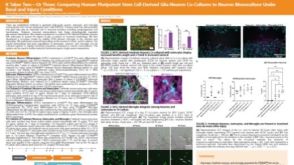 科学海报It Takes Two—Or Three: Comparing Human Pluripotent Stem Cell Derived Glia-Neuron Co-Cultures to Neuron Monoculture Under Basal and Injury Conditions
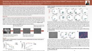 科学海报Generation and Characterization of a Homogenous Population of Early Mesoderm Cells Using STEMdiff Mesoderm Induction Medium
 沪公网安备31010102008431号