Dienelt A and zur Nieden NI (MAR 2011)
Stem cells and development 20 3 465--474
Hyperglycemia impairs skeletogenesis from embryonic stem cells by affecting osteoblast and osteoclast differentiation.
High maternal blood glucose levels caused by diabetes mellitus can irreversibly lead to maldevelopment of the growing fetus with specific effects on the skeleton. To date,it remains controversial at which stage embryonic development is affected. Specifically during embryonic bone development,it is unclear whether diminished bone mineral density is caused by reduced osteoblast or rather enhanced osteoclast function. Therefore,the aim of this study was to characterize the growth as well as the skeletal differentiation capability of pluripotent embryonic stem cells (ESCs),which may serve as an in vitro model for all stages of embryonic development,when cultured in diabetic levels of D-glucose (4.5 g/L) versus physiological levels (1.0 g/L). Results showed that cells cultivated in physiological glucose gave rise to a higher number of colonies with an undifferentiated character as compared to cells grown in diabetic glucose concentrations. In contrast,these cultures were characterized by slightly decreased expression of proteins associated with the stem cell state. Furthermore,differentiation of ESCs into osteoblasts and osteoclasts was favored in physiological glucose concentrations,demonstrated by an increased matrix calcification,enhanced expression of cell-type-specific mRNAs,as well as activity of the cell-type-specific enzymes,alkaline,and tartrate resistant acidic phosphatase. In fact,this pattern was noted in murine as well as in primate ESCs. Our study suggests that an interplay between both the osteoblast and the osteoclast lineage is needed for proper skeletal development to occur,which seems impaired in hyperglycemic conditions.
View Publication
产品类型:
产品号#:
05850
05857
05870
05875
85850
85857
85870
85875
产品名:
mTeSR™1
mTeSR™1
Park S et al. (APR 2017)
Stem cell reports 8 4 1076--1085
A Comprehensive, Ethnically Diverse Library of Sickle Cell Disease-Specific Induced Pluripotent Stem Cells.
Sickle cell anemia affects millions of people worldwide and is an emerging global health burden. As part of a large NIH-funded NextGen Consortium,we generated a diverse,comprehensive,and fully characterized library of sickle-cell-disease-specific induced pluripotent stem cells (iPSCs) from patients of different ethnicities,β-globin gene (HBB) haplotypes,and fetal hemoglobin (HbF) levels. iPSCs stand to revolutionize the way we study human development,model disease,and perhaps eventually,treat patients. Here,we describe this unique resource for the study of sickle cell disease,including novel haplotype-specific polymorphisms that affect disease severity,as well as for the development of patient-specific therapeutics for this phenotypically diverse disorder. As a complement to this library,and as proof of principle for future cell- and gene-based therapies,we also designed and employed CRISPR/Cas gene editing tools to correct the sickle hemoglobin (HbS) mutation.
View Publication
产品类型:
产品号#:
05850
05857
05870
05875
85850
85857
85870
85875
产品名:
mTeSR™1
mTeSR™1
Bartel MA and Schaffer DV ( 2014)
1114 169--179
Enhanced gene targeting of adult and pluripotent stem cells using evolved adeno-Associated virus
Efficient approaches for the precise genetic engineering of stem cells can enhance both basic and applied stem cell research. Adeno-associated virus (AAV) vectors have demonstrated high-efficiency gene delivery and gene targeting to numerous cell types,and AAV vectors developed specifically for gene delivery to stem cells have further increased gene targeting frequency compared to plasmid construct techniques. This chapter details the production and purification techniques necessary to generate adeno-associated viral vectors for use in high-efficiency gene targeting of adult or pluripotent stem cell applications. Culture conditions used to achieve high gene targeting frequencies in rat neural stem cells and human pluripotent stem cells are also described.
View Publication
产品类型:
产品号#:
05850
05857
05870
05875
85850
85857
85870
85875
产品名:
mTeSR™1
mTeSR™1
van den Akker E et al. (SEP 2010)
Haematologica 95 9 1594--8
The majority of the in vitro erythroid expansion potential resides in CD34(-) cells, outweighing the contribution of CD34(+) cells and significantly increasing the erythroblast yield from peripheral blood samples.
The study of human erythropoiesis in health and disease requires a robust culture system that consistently and reliably generates large numbers of immature erythroblasts that can be induced to differentiate synchronously. We describe a culture method modified from Leberbauer et al. (2005) and obtain a homogenous population of erythroblasts from peripheral blood mononuclear cells (PBMC) without prior purification of CD34(+) cells. This pure population of immature erythroblasts can be expanded to obtain 4x10(8) erythroblasts from 1x10(8) PBMC after 13-14 days in culture. Upon synchronized differentiation,high levels of enucleation (80-90%) and low levels of cell death (textless10%) are achieved. We compared the yield of erythroblasts obtained from PBMC,CD34(+) cells or PBMC depleted of CD34(+) cells and show that CD34(-) cells represent the most significant early erythroid progenitor population. This culture system may be particularly useful for investigating the pathophysiology of anemic patients where only small blood volumes are available.
View Publication
A bioengineered niche promotes in vivo engraftment and maturation of pluripotent stem cell derived human lung organoids.
Human pluripotent stem cell (hPSC) derived tissues often remain developmentally immature in vitro,and become more adult-like in their structure,cellular diversity and function following transplantation into immunocompromised mice. Previously we have demonstrated that hPSC-derived human lung organoids (HLOs) resembled human fetal lung tissue in vitro (Dye et al.,2015). Here we show that HLOs required a bioartificial microporous poly(lactide-co-glycolide) (PLG) scaffold niche for successful engraftment,long-term survival,and maturation of lung epithelium in vivo. Analysis of scaffold-grown transplanted tissue showed airway-like tissue with enhanced epithelial structure and organization compared to HLOs grown in vitro. By further comparing in vitro and in vivo grown HLOs with fetal and adult human lung tissue,we found that in vivo transplanted HLOs had improved cellular differentiation of secretory lineages that is reflective of differences between fetal and adult tissue,resulting in airway-like structures that were remarkably similar to the native adult human lung.
View Publication
产品类型:
产品号#:
05850
05857
05870
05875
85850
85857
85870
85875
产品名:
mTeSR™1
mTeSR™1
Ovchinnikov DA et al. (JUL 2012)
World journal of stem cells 4 7 71--9
Generation of a human embryonic stem cell line stably expressing high levels of the fluorescent protein mCherry.
AIM: The generation and characterization of a human embryonic stem cell (hESC) line stably expressing red fluorescent mCherry protein.backslashnbackslashnMETHODS: Lentiviral transduction of a ubiquitously-expressed human EF-1α promoter driven mCherry transgene was performed in MEL2 hESC. Red fluore-scence was assessed by immunofluorescence and flow cytometry. Pluripotency of stably transduced hESC was determined by immunofluorescent pluripotency marker expression,flow cytometry,teratoma assays and embryoid body-based differentiation followed by reverse transcriptase-polymerase chain reaction. Quantification of cell motility and survival was performed with time lapse microscopy.backslashnbackslashnRESULTS: Constitutively fluorescently-labeled hESCs are useful tools for facile in vitro and in vivo tracking of survival,motility and cell spreading on various surfaces before and after differentiation. Here we describe the generation and characterization of a hESC line (MEL2) stably expressing red fluorescent protein,mCherry. This line was generated by random integration of a fluorescent protein-expressing cassette,driven by the ubiquitously-expressed human EF-1α promoter. Stably transfected MEL2-mCherry hESC were shown to express pluripotency markers in the nucleus (POU5F1/OCT4,NANOG and SOX2) and on the cell surface (SSEA4,TRA1-60 and TG30/CD9) and were shown to maintain a normal karyotype in long-term (for at least 35 passages) culture. MEL2-mCherry hESC further readily differentiated into representative cell types of the three germ layers in embryoid body and teratoma based assays and,importantly,maintained robust mCherry expression throughout differentiation. The cell line was next adapted to single-cell passaging,rendering it compatible with numerous bioengineering applications such as measurement of cell motility and cell spreading on various protein modified surfaces,quantification of cell attachment to nanoparticles and rapid estimation of cell survival.backslashnbackslashnCONCLUSION: The MEL2-mCherry hESC line conforms to the criteria of bona fide pluripotent stem cells and maintains red fluorescence throughout differentiation,making it a useful tool for bioengineering and in vivo tracking experiments.
View Publication
产品类型:
产品号#:
05850
05857
05870
05875
85850
85857
85870
85875
产品名:
mTeSR™1
mTeSR™1
Kumagai T et al. (JUN 2003)
Journal of the National Cancer Institute 95 12 896--905
Vitamin D2 analog 19-nor-1,25-dihydroxyvitamin D2: antitumor activity against leukemia, myeloma, and colon cancer cells.
BACKGROUND: 1,25-Dihydroxyvitamin D(3) inhibits growth of several types of human cancer cells in vitro,but its therapeutic use is hampered because it causes hypercalcemia. 19-nor-1,25-Dihydroxyvitamin D(2) (paricalcitol) is a noncalcemic vitamin D analog that is approved by the Food and Drug Administration for the treatment of secondary hyperparathyroidism. We investigated the antitumor activity and mechanism of action of paricalcitol in vitro and in vivo. METHODS: Effects of paricalcitol on proliferation,the cell cycle,differentiation,and apoptosis were examined in cancer cell lines. Effects on tumor growth were examined with colon cancer cell xenografts in nude mice (five in the experimental group and five in the control group). The interaction of paricalcitol with the vitamin D receptor (VDR) in mononuclear spleen cells and myeloid stem cells from wild-type and VDR knockout mice was examined. All statistical tests were two-sided. RESULTS: Paricalcitol inhibited the proliferation of myeloid leukemia cell lines HL-60,NB-4,and THP-1 cells at an effective dose that inhibited growth 50% (ED(50)) of 2.4-5.8 x 10(-9) M by inducing cell cycle arrest and differentiation. Paricalcitol inhibited the proliferation of NCI-H929 myeloma cells at an ED(50) of 2.0 x 10(-10) M by inducing cell cycle arrest and apoptosis. Paricalcitol also inhibited the proliferation of colon cancer cell lines HT-29 (ED(50) = 1.7 x 10(-8) M) and SW837 (ED(50) = 3.2 x 10(-8) M). HT-29 colon cancer xenografts in paricalcitol-treated nude mice were smaller (1044 mm(3) and 1752 mm(3),difference = 708 mm(3),95% confidence interval = 311 to 1104 mm(3); P =.03) and weighed less (1487 mg and 4162 mg,difference = 2675 mg,95% confidence interval = 2103 to 3248 mg; Ptextless.001) than those in vehicle-treated mice. Paricalcitol induced committed myeloid hematopoietic stem cells from wild-type but not from VDR knockout mice to differentiate as macrophages. CONCLUSION: Paricalcitol has anticancer activity against myeloid leukemia,myeloma,and colon cancer cells that may be mediated through the VDR. Because it has been approved by the Food and Drug Administration,clinical trials of this agent in certain cancers are reasonable.
View Publication
产品类型:
产品号#:
03234
产品名:
MethoCult™ M3234
Carotta S et al. (SEP 2004)
Blood 104 6 1873--80
Directed differentiation and mass cultivation of pure erythroid progenitors from mouse embryonic stem cells.
Differentiating embryonic stem (ES) cells are an increasingly important source of hematopoietic progenitors,useful for both basic research and clinical applications. Besides their characterization in colony assays,protocols exist for the cultivation of lymphoid,myeloid,and erythroid cells. With the possible exception of mast cells,however,long-term expansion of pure hematopoietic progenitors from ES cells has not been possible without immortalization caused by overexpression of exogenous genes. Here,we describe for the first time an efficient yet easy strategy to generate mass cultures of pure,immature erythroid progenitors from mouse ES cells (ES-EPs),using serum-free medium plus recombinant cytokines and hormones. ES-EPs represent long-lived,adult,definitive erythroid progenitors that resemble immature erythroid cells expanding in vivo during stress erythropoiesis. When exposed to terminal differentiation conditions,ES-EPs differentiated into mature,enucleated erythrocytes. Importantly,ES-EPs injected into mice did not exhibit tumorigenic potential but differentiated into normal erythrocytes. Both the virtually unlimited supply of cells and the defined culture conditions render our system a valuable tool for the analysis of factors influencing proliferation and maturation of erythroid progenitors. In addition,the system allows detailed characterization of processes during erythroid proliferation and differentiation using wild-type (wt) and genetically modified ES cells.
View Publication
产品类型:
产品号#:
03234
03434
03444
产品名:
MethoCult™ M3234
MethoCult™ GF M3434
MethoCult™ GF M3434
Cohen-Haguenauer O et al. (FEB 2006)
Proceedings of the National Academy of Sciences of the United States of America 103 7 2340--5
In vivo repopulation ability of genetically corrected bone marrow cells from Fanconi anemia patients.
Fanconi anemia (FA) is a rare inherited genomic instability syndrome representing one of the best examples of hematopoietic stem cell deficiency. Although FA might be an excellent candidate for bone marrow (BM) genetic correction ex vivo,knockout animal models are not sufficient to guide preclinical steps,and gene therapy attempts have proven disappointing so far. Contributing to these poor results is a characteristic and dramatic early BM-cells die-off when placed in culture. We show here that human primary FA BM cell survival can be ameliorated by using specific culture conditions that limit oxidative stress. When coupled with retrovirus-mediated transfer of the main complementation group FANCA-cDNA,we could achieve long-term reconstitution of the stem cell compartment both in vitro and in vivo. Gene-corrected BM cultures grew for textgreater120 days,and after cultured cell transplantation into NOD/SCID mice,clonogenic human cells carrying the FANCA transgene could be detected 6 months after transduction. By comparison,untransduced cells died in culture by 15 days. Of necessity for ethical reasons,experiments were conducted on a very limited number of primary BM cells. By using low cytokine regimen and conditions matching regulatory requirements,a contingent of gene-corrected cells slowly emerges with an unmet potential for in vivo engraftment. Future therapeutic applications of stem cells might be expanding from these data. In addition,we provide a model of gene-corrected human primary cell growth that carries the potential to better delineate the combined role of both DNA damage and oxidative stress in the pathogenesis of FA.
View Publication
产品类型:
产品号#:
04436
产品名:
MethoCult™ SF H4436
Ma Y et al. (OCT 2006)
Blood 108 8 2726--35
SALL4, a novel oncogene, is constitutively expressed in human acute myeloid leukemia (AML) and induces AML in transgenic mice.
SALL4,a human homolog to Drosophila spalt,is a novel zinc finger transcriptional factor essential for development. We cloned SALL4 and its isoforms (SALL4A and SALL4B). Through immunohistochemistry and real-time reverse-transcription-polymerase chain reaction (RT-PCR),we demonstrated that SALL4 was constitutively expressed in human primary acute myeloid leukemia (AML,n = 81),and directly tested the leukemogenic potential of constitutive expression of SALL4 in a murine model. SALL4B transgenic mice developed myelodysplastic syndrome (MDS)-like features and subsequently AML that was transplantable. Increased apoptosis associated with dysmyelopoiesis was evident in transgenic mouse marrow and colony-formation (CFU) assays. Both isoforms could bind to beta-catenin and synergistically enhanced the Wnt/beta-catenin signaling pathway. Our data suggest that the constitutive expression of SALL4 causes MDS/AML,most likely through the Wnt/beta-catenin pathway. Our murine model provides a useful platform to study human MDS/AML transformation,as well as the Wnt/beta-catenin pathway's role in the pathogenesis of leukemia stem cells.
View Publication

 EasySep™小鼠TIL(CD45)正选试剂盒


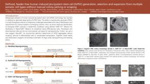 科学海报Defined, Feeder-Free Human Induced Pluripotent Stem Cell (hiPSC) Generation, Selection and Expansion from Multiple Somatic Cell Types
 沪公网安备31010102008431号