Mapping the Pairwise Choices Leading from Pluripotency to Human Bone, Heart, and Other Mesoderm Cell Types
Stem-cell differentiation to desired lineages requires navigating alternating developmental paths that often lead to unwanted cell types. Hence,comprehensive developmental roadmaps are crucial to channel stem-cell differentiation toward desired fates. To this end,here,we map bifurcating lineage choices leading from pluripotency to 12 human mesodermal lineages,including bone,muscle,and heart. We defined the extrinsic signals controlling each binary lineage decision,enabling us to logically block differentiation toward unwanted fates and rapidly steer pluripotent stem cells toward 80%???99% pure human mesodermal lineages at most branchpoints. This strategy enabled the generation of human bone and heart progenitors that could engraft in respective in??vivo models. Mapping stepwise chromatin and single-cell gene expression changes in mesoderm development uncovered somite segmentation,a previously unobservable human embryonic event transiently marked by HOPX expression. Collectively,this roadmap enables navigation of mesodermal development to produce transplantable human tissue progenitors and uncover developmental processes. Video Abstract
View Publication
产品类型:
产品号#:
05850
05857
05870
05875
85850
85857
85870
85875
产品名:
mTeSR™1
mTeSR™1
Srinivasakumar N et al. (DEC 2013)
PeerJ 1 e224
Gammaretroviral vector encoding a fluorescent marker to facilitate detection of reprogrammed human fibroblasts during iPSC generation.
Induced pluripotent stem cells (iPSCs) are becoming mainstream tools to study mechanisms of development and disease. They have a broad range of applications in understanding disease processes,in vitro testing of novel therapies,and potential utility in regenerative medicine. Although the techniques for generating iPSCs are becoming more straightforward,scientists can expend considerable resources and time to establish this technology. A major hurdle is the accurate determination of valid iPSC-like colonies that can be selected for further cloning and characterization. In this study,we describe the use of a gammaretroviral vector encoding a fluorescent marker,mRFP1,to not only monitor the efficiency of initial transduction but also to identify putative iPSC colonies through silencing of mRFP1 gene as a consequence of successful reprogramming.
View Publication
产品类型:
产品号#:
05854
05855
05850
05857
05870
05875
85850
85857
85870
85875
产品名:
mFreSR™
mFreSR™
mTeSR™1
mTeSR™1
Rawat VPS et al. (SEP 2010)
Proceedings of the National Academy of Sciences of the United States of America 107 39 16946--51
The vent-like homeobox gene VENTX promotes human myeloid differentiation and is highly expressed in acute myeloid leukemia.
Recent data indicate that a variety of regulatory molecules active in embryonic development may also play a role in the regulation of early hematopoiesis. Here we report that the human Vent-like homeobox gene VENTX,a putative homolog of the Xenopus xvent2 gene,is a unique regulatory hematopoietic gene that is aberrantly expressed in CD34(+) leukemic stem-cell candidates in human acute myeloid leukemia (AML). Quantitative RT-PCR documented expression of the gene in lineage positive hematopoietic subpopulations,with the highest expression in CD33(+) myeloid cells. Notably,expression levels of VENTX were negligible in normal CD34(+)/CD38(-) or CD34(+) human progenitor cells. In contrast to this,leukemic CD34(+)/CD38(-) cells from AML patients with translocation t(8,21) and normal karyotype displayed aberrantly high expression of VENTX. Gene expression and pathway analysis demonstrated that in normal CD34(+) cells enforced expression of VENTX initiates genes associated with myeloid development and down-regulates genes involved in early lymphoid development. Functional analyses confirmed that aberrant expression of VENTX in normal CD34(+) human progenitor cells perturbs normal hematopoietic development,promoting generation of myeloid cells and impairing generation of lymphoid cells in vitro and in vivo. Stable knockdown of VENTX expression inhibited the proliferation of human AML cell lines. Taken together,these data extend our insights into the function of embryonic mesodermal factors in human postnatal hematopoiesis and indicate a role for VENTX in normal and malignant myelopoiesis.
View Publication
Raju R et al. (FEB 2017)
Stem cells and development 26 4 274--284
Cell Expansion During Directed Differentiation of Stem Cells Toward the Hepatic Lineage.
The differentiation of human pluripotent stem cells toward the hepatocyte lineage can potentially provide an unlimited source of functional hepatocytes for transplantation and extracorporeal bioartificial liver applications. It is anticipated that the quantities of cells needed for these applications will be in the order of 10(9)-10(10) cells,because of the size of the liver. An ideal differentiation protocol would be to enable directed differentiation to the hepatocyte lineage with simultaneous cell expansion. We introduced a cell expansion stage after the commitment of human embryonic stem cells to the endodermal lineage,to allow for at least an eightfold increase in cell number,with continuation of cell maturation toward the hepatocyte lineage. The progressive changes in the transcriptome were measured by expression array,and the expression dynamics of certain lineage markers was measured by mass cytometry during the differentiation and expansion process. The findings revealed that while cells were expanding they were also capable of progressing in their differentiation toward the hepatocyte lineage. In addition,our transcriptome,protein and functional studies,including albumin secretion,drug-induced CYP450 expression and urea production,all indicated that the hepatocyte-like cells obtained with or without cell expansion are very similar. This method of simultaneous cell expansion and hepatocyte differentiation should facilitate obtaining large quantities of cells for liver cell applications.
View Publication
产品类型:
产品号#:
05850
05857
05870
05875
85850
85857
85870
85875
产品名:
mTeSR™1
mTeSR™1
Cho SK et al. (AUG 1999)
Proceedings of the National Academy of Sciences of the United States of America 96 17 9797--802
Functional characterization of B lymphocytes generated in vitro from embryonic stem cells.
To study molecular events involved in B lymphocyte development and V(D)J rearrangement,we have established an efficient system for the differentiation of embryonic stem (ES) cells into mature Ig-secreting B lymphocytes. Here,we show that B lineage cells generated in vitro from ES cells are functionally analogous to normal fetal liver-derived or bone marrow-derived B lineage cells at three important developmental stages: first,they respond to Flt-3 ligand during an early lymphopoietic progenitor stage; second,they become targets for Abelson murine leukemia virus (A-MuLV) infection at a pre-B cell stage; third,they secrete Ig upon stimulation with lipopolysaccharide at a mature mitogen-responsive stage. Moreover,the ES cell-derived A-MuLV-transformed pre-B (EAB) cells are phenotypically and functionally indistinguishable from standard A-MuLV-transformed pre-B cells derived from infection of mouse fetal liver or bone marrow. Notably,EAB cells possess functional V(D)J recombinase activity. In particular,the generation of A-MuLV transformants from ES cells will provide an advantageous system to investigate genetic modifications that will help to elucidate molecular mechanisms in V(D)J recombination and in A-MuLV-mediated transformation.
View Publication
产品类型:
产品号#:
06902
06952
00321
00322
00323
00324
00325
产品名:
Krishnamurthy S et al. (DEC 2010)
Cancer research 70 23 9969--78
Endothelial cell-initiated signaling promotes the survival and self-renewal of cancer stem cells.
Recent studies have demonstrated that cancer stem cells play an important role in the pathobiology of head and neck squamous cell carcinomas (HNSCC). However,little is known about functional interactions between head and neck cancer stem-like cells (CSC) and surrounding stromal cells. Here,we used aldehyde dehydrogenase activity and CD44 expression to sort putative stem cells from primary human HNSCC. Implantation of 1,000 CSC (ALDH+CD44+Lin-) led to tumors in 13 (out of 15) mice,whereas 10,000 noncancer stem cells (ALDH-CD44-Lin-) resulted in 2 tumors in 15 mice. These data demonstrated that ALDH and CD44 select a subpopulation of cells that are highly tumorigenic. The ability to self-renew was confirmed by the observation that ALDH+CD44+Lin- cells sorted from human HNSCC formed more spheroids (orospheres) in 3-D agarose matrices or ultra-low attachment plates than controls and were serially passaged in vivo. We observed that approximately 80% of the CSC were located in close proximity (within 100-μm radius) of blood vessels in human tumors,suggesting the existence of perivascular niches in HNSCC. In vitro studies demonstrated that endothelial cell-secreted factors promoted self-renewal of CSC,as demonstrated by the upregulation of Bmi-1 expression and the increase in the number of orospheres as compared with controls. Notably,selective ablation of tumor-associated endothelial cells stably transduced with a caspase-based artificial death switch (iCaspase-9) caused a marked reduction in the fraction of CSC in xenograft tumors. Collectively,these findings indicate that endothelial cell-initiated signaling can enhance the survival and self-renewal of head and neck CSC.
View Publication
产品类型:
产品号#:
01700
01705
01702
产品名:
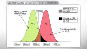
ALDEFLUOR™ 试剂盒
ALDEFLUOR™ DEAB试剂, 1.5 mM, 1 mL
ALDEFLUOR™检测缓冲液
Li W et al. (JAN 2012)
Human Molecular Genetics 21 1 32--45
Modeling abnormal early development with induced pluripotent stem cells from aneuploid syndromes
Many human diseases share a developmental origin that manifests during childhood or maturity. Aneuploid syndromes are caused by supernumerary or reduced number of chromosomes and represent an extreme example of developmental disease,as they have devastating consequences before and after birth. Investigating how alterations in gene dosage drive these conditions is relevant because it might help treat some clinical aspects. It may also provide explanations as to how quantitative differences in gene expression determine phenotypic diversity and disease susceptibility among natural populations. Here,we aimed to produce induced pluripotent stem cell (iPSC) lines that can be used to improve our understanding of aneuploid syndromes. We have generated iPSCs from monosomy X [Turner syndrome (TS)],trisomy 8 (Warkany syndrome 2),trisomy 13 (Patau syndrome) and partial trisomy 11;22 (Emanuel syndrome),using either skin fibroblasts from affected individuals or amniocytes from antenatal diagnostic tests. These cell lines stably maintain the karyotype of the donors and behave like embryonic stem cells in all tested assays. TS iPSCs were used for further studies including global gene expression analysis and tissue-specific directed differentiation. Multiple clones displayed lower levels of the pseudoautosomal genes ASMTL and PPP2R3B than the controls. Moreover,they could be transformed into neural-like,hepatocyte-like and heart-like cells,but displayed insufficient up-regulation of the pseudoautosomal placental gene CSF2RA during embryoid body formation. These data support that abnormal organogenesis and early lethality in TS are not caused by a tissue-specific differentiation blockade,but rather involves other abnormalities including impaired placentation.
View Publication
ROCK Inhibition Promotes Attachment, Proliferation, and Wound Closure in Human Embryonic Stem Cell-Derived Retinal Pigmented Epithelium.
PURPOSE Nonexudative (dry) age-related macular degeneration (AMD),a leading cause of blindness in the elderly,is associated with the loss of retinal pigmented epithelium (RPE) cells and the development of geographic atrophy,which are areas devoid of RPE cells and photoreceptors. One possible treatment option would be to stimulate RPE attachment and proliferation to replace dying/dysfunctional RPE and bring about wound repair. Clinical trials are underway testing injections of RPE cells derived from pluripotent stem cells to determine their safety and efficacy in treating AMD. However,the factors regulating RPE responses to AMD-associated lesions are not well understood. Here,we use cell culture to investigate the role of RhoA coiled coil kinases (ROCKs) in human embryonic stem cell-derived RPE (hESC-RPE) attachment,proliferation,and wound closure. METHODS H9 hESC were spontaneously differentiated into RPE cells. hESC-RPE cells were treated with a pan ROCK1/2 or a ROCK2 only inhibitor; attachment,and proliferation and cell size within an in vitro scratch assay were examined. RESULTS Pharmacological inhibition of ROCKs promoted hESC-RPE attachment and proliferation,and increased the rate of closure of in vitro wounds. ROCK inhibition decreased phosphorylation of cofilin and myosin light chain,suggesting that regulation of the cytoskeleton underlies the mechanism of action of ROCK inhibition. CONCLUSIONS ROCK inhibition promotes attachment,proliferation,and wound closure in H9 hESC-RPE cells. ROCK isoforms may have different roles in wound healing. TRANSLATIONAL RELEVANCE Modulation of the ROCK-cytoskeletal axis has potential in stimulating wound repair in transplanted RPE cells and attachment in cellular therapies.
View Publication
产品类型:
产品号#:
05850
05857
05870
05875
85850
85857
85870
85875
产品名:
mTeSR™1
mTeSR™1
Costa V et al. (APR 2016)
Cell reports 15 1 86--95
mTORC1 Inhibition Corrects Neurodevelopmental and Synaptic Alterations in a Human Stem Cell Model of Tuberous Sclerosis.
Hyperfunction of the mTORC1 pathway has been associated with idiopathic and syndromic forms of autism spectrum disorder (ASD),including tuberous sclerosis,caused by loss of either TSC1 or TSC2. It remains largely unknown how developmental processes and biochemical signaling affected by mTORC1 dysregulation contribute to human neuronal dysfunction. Here,we have characterized multiple stages of neurogenesis and synapse formation in human neurons derived from TSC2-deleted pluripotent stem cells. Homozygous TSC2 deletion causes severe developmental abnormalities that recapitulate pathological hallmarks of cortical malformations in patients. Both TSC2(+/-) and TSC2(-/-) neurons display altered synaptic transmission paralleled by molecular changes in pathways associated with autism,suggesting the convergence of pathological mechanisms in ASD. Pharmacological inhibition of mTORC1 corrects developmental abnormalities and synaptic dysfunction during independent developmental stages. Our results uncouple stage-specific roles of mTORC1 in human neuronal development and contribute to a better understanding of the onset of neuronal pathophysiology in tuberous sclerosis.
View Publication
产品类型:
产品号#:
05850
05857
05870
05875
85850
85857
85870
85875
产品名:
mTeSR™1
mTeSR™1
Darabi R et al. (MAY 2012)
Cell stem cell 10 5 610--619
Human ES- and iPS-derived myogenic progenitors restore DYSTROPHIN and improve contractility upon transplantation in dystrophic mice.
A major obstacle in the application of cell-based therapies for the treatment of neuromuscular disorders is obtaining the appropriate number of stem/progenitor cells to produce effective engraftment. The use of embryonic stem (ES) or induced pluripotent stem (iPS) cells could overcome this hurdle. However,to date,derivation of engraftable skeletal muscle precursors that can restore muscle function from human pluripotent cells has not been achieved. Here we applied conditional expression of PAX7 in human ES/iPS cells to successfully derive large quantities of myogenic precursors,which,upon transplantation into dystrophic muscle,are able to engraft efficiently,producing abundant human-derived DYSTROPHIN-positive myofibers that exhibit superior strength. Importantly,transplanted cells also seed the muscle satellite cell compartment,and engraftment is present over 11 months posttransplant. This study provides the proof of principle for the derivation of functional skeletal myogenic progenitors from human ES/iPS cells and highlights their potential for future therapeutic application in muscular dystrophies.
View Publication
产品类型:
产品号#:
05850
05857
05870
05875
85850
85857
85870
85875
产品名:
mTeSR™1
mTeSR™1
Liu W et al. (DEC 2014)
Cell death and differentiation 4 12 1950--1960
BRD4 regulates Nanog expression in mouse embryonic stem cells and preimplantation embryos.
Bromodomain-containing protein 4 (BRD4) is an important epigenetic reader implicated in the pathogenesis of a number of different cancers and other diseases. Brd4-null mouse embryos die shortly after implantation and are compromised in their ability to maintain the inner cell mass,which gives rise to embryonic stem cells (ESCs). Here we report that BRD4 regulates expression of the pluripotency factor Nanog in mouse ESCs and preimplantation embryos,as well as in human ESCs and embryonic cancer stem cells. Inhibition of BRD4 function using a chemical inhibitor,small interfering RNAs,or a dominant-negative approach suppresses Nanog expression,and abolishes the self-renewal ability of ESCs. We also find that BRD4 associates with BRG1 (brahma-related gene 1,aka Smarca4 (SWI/SNF-related,matrix-associated,actin-dependent regulator of chromatin,subfamily a,member 4)),a key regulator of ESC self-renewal and pluripotency,in the Nanog regulatory regions to regulate Nanog expression. Our study identifies Nanog as a novel BRD4 target gene,providing new insights for the biological function of BRD4 in stem cells and mouse embryos. Knowledge gained from these non-cancerous systems will facilitate future investigations of how Brd4 dysfunction leads to cancers.Cell Death and Differentiation advance online publication,22 August 2014; doi:10.1038/cdd.2014.124.
View Publication

 EasySep™小鼠TIL(CD45)正选试剂盒


 沪公网安备31010102008431号