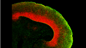
An atlas of transcriptional, chromatin accessibility, and surface marker changes in human mesoderm development.
Mesoderm is the developmental precursor to myriad human tissues including bone,heart,and skeletal muscle. Unravelling the molecular events through which these lineages become diversified from one another is integral to developmental biology and understanding changes in cellular fate. To this end,we developed an in vitro system to differentiate human pluripotent stem cells through primitive streak intermediates into paraxial mesoderm and its derivatives (somites,sclerotome,dermomyotome) and separately,into lateral mesoderm and its derivatives (cardiac mesoderm). Whole-population and single-cell analyses of these purified populations of human mesoderm lineages through RNA-seq,ATAC-seq,and high-throughput surface marker screens illustrated how transcriptional changes co-occur with changes in open chromatin and surface marker landscapes throughout human mesoderm development. This molecular atlas will facilitate study of human mesoderm development (which cannot be interrogated in vivo due to restrictions on human embryo studies) and provides a broad resource for the study of gene regulation in development at the single-cell level,knowledge that might one day be exploited for regenerative medicine.
View Publication
Matsumoto K et al. (JAN 2000)
Stem cells (Dayton,Ohio) 18 3 196--203
In vitro proliferation potential of AC133 positive cells in peripheral blood.
AC133 antigen is a novel marker for human hematopoietic stem/progenitor cells. In this study,we examined the expression and proliferation potential of AC133(+) cells obtained from steady-state peripheral blood (PB). The proportion of AC133(+) cells in the CD34(+) subpopulation of steady-state PB was significantly lower than that of cord blood (CB),although that of cytokine-mobilized PB was higher than that of CB. The proliferation potential of AC133(+)CD34(+) and AC133(-)CD34(+) cells was examined by colony-forming analysis and analysis of long-term culture-initiating cells (LTC-IC). Although the total number of colony-forming cells was essentially the same in the AC133(+)CD34(+) fraction as in the AC133(-)CD34(+) fraction,the proportion of LTC-IC was much higher in the AC133(+)CD34(+) fraction. Virtually no LTC-IC were detected in the AC133(-)CD34(+) fraction. In addition,the features of the colonies grown from these two fractions were quite different. Approximately 70% of the colonies derived from the AC133(+)CD34(+) fraction were granulocyte-macrophage colonies,whereas more than 90% of the colonies derived from the AC133(-)CD34(+) fraction were erythroid colonies. Furthermore,an ex vivo expansion study observed expansion of colony-forming cells only in the AC133(+)CD34(+) population,and not in the AC133(-)CD34(+) population. These findings suggest that to isolate primitive hematopoietic cells from steady-state PB,selection by AC133 expression is better than selection by CD34 expression.
View Publication
产品类型:
产品号#:
04034
04044
产品名:
MethoCult™ H4034 Optimum
MethoCult™ H4034 Optimum
Schwieger M et al. (SEP 2009)
Blood 114 12 2476--88
Homing and invasiveness of MLL/ENL leukemic cells is regulated by MEF2C.
Acute myelogenous leukemia is driven by leukemic stem cells (LSCs) generated by mutations that confer (or maintain) self-renewal potential coupled to an aberrant differentiation program. Using retroviral mutagenesis,we identified genes that generate LSCs in collaboration with genetic disruption of the gene encoding interferon response factor 8 (Irf8),which induces a myeloproliferation in vivo. Among the targeted genes,we identified Mef2c,encoding a MCM1-agamous-deficiens-serum response factor transcription factor,and confirmed that overexpression induced a myelomonocytic leukemia in cooperation with Irf8 deficiency. Strikingly,several of the genes identified in our screen have been reported to be up-regulated in the mixed-lineage leukemia (MLL) subtype. High MEF2C expression levels were confirmed in acute myelogenous leukemia patient samples with MLL gene disruptions,prompting an investigation of the causal interplay. Using a conditional mouse strain,we demonstrated that Mef2c deficiency does not impair the establishment or maintenance of LSCs generated in vitro by MLL/ENL fusion proteins; however,its loss led to compromised homing and invasiveness of the tumor cells. Mef2c-dependent targets included several genes encoding matrix metalloproteinases and chemokine ligands and receptors,providing a mechanistic link to increased homing and motility. Thus,MEF2C up-regulation may be responsible for the aggressive nature of this leukemia subtype.
View Publication
产品类型:
产品号#:
03434
03444
09600
09650
产品名:
MethoCult™ GF M3434
MethoCult™ GF M3434
StemSpan™ SFEM
StemSpan™ SFEM
Eguchi M et al. (JAN 2005)
Proceedings of the National Academy of Sciences of the United States of America 102 4 1133--8
Directing oncogenic fusion genes into stem cells via an SCL enhancer.
TEL-TRKC is a fusion gene generated by chromosomal translocation and encodes an activated tyrosine kinase. Uniquely,it is found in both solid tumors and leukemia. However,a single exon difference (in TEL) in TEL-TRKC fusions is associated with the two sets of cancer phenotypes. We expressed the two TEL-TRKC variants in vivo by using the 3' regulatory element of SCL that is selectively active in a subset of mesodermal cell lineages,including endothelial and hematopoietic stem cells and progenitors. The leukemia form of TEL-TRKC (-exon 5 of TEL) enhanced hematopoietic stem cell renewal and initiated leukemia. In contrast,the TEL-TRKC solid tumor variant (+ TEL exon 5) elicited an embryonic lethal phenotype with impairment of both angiogenesis and hematopoiesis indicative of an effect at the level of the hemangioblasts. The ability of TEL-TRKC to repress expression of Flk1,a critical regulator of early endothelial and hematopoietic cells,depended on TEL exon 5. These data indicate that related oncogenic fusion proteins similarly expressed in a hierarchy of early stem cells can have selective,cell type-specific developmental impacts.
View Publication
产品类型:
产品号#:
03231
产品名:
MethoCult™ M3231
Nayak RC et al. (AUG 2015)
The Journal of clinical investigation 125 8 3103--3116
Pathogenesis of ELANE-mutant severe neutropenia revealed by induced pluripotent stem cells.
Severe congenital neutropenia (SCN) is often associated with inherited heterozygous point mutations in ELANE,which encodes neutrophil elastase (NE). However,a lack of appropriate models to recapitulate SCN has substantially hampered the understanding of the genetic etiology and pathobiology of this disease. To this end,we generated both normal and SCN patient-derived induced pluripotent stem cells (iPSCs),and performed genome editing and differentiation protocols that recapitulate the major features of granulopoiesis. Pathogenesis of ELANE point mutations was the result of promyelocyte death and differentiation arrest,and was associated with NE mislocalization and activation of the unfolded protein response/ER stress (UPR/ER stress). Similarly,high-dose G-CSF (or downstream signaling through AKT/BCL2) rescues the dysgranulopoietic defect in SCN patient-derived iPSCs through C/EBP$$-dependent emergency granulopoiesis. In contrast,sivelestat,an NE-specific small-molecule inhibitor,corrected dysgranulopoiesis by restoring normal intracellular NE localization in primary granules; ameliorating UPR/ER stress; increasing expression of CEBPA,but not CEBPB; and promoting promyelocyte survival and differentiation. Together,these data suggest that SCN disease pathogenesis includes NE mislocalization,which in turn triggers dysfunctional survival signaling and UPR/ER stress. This paradigm has the potential to be clinically exploited to achieve therapeutic responses using lower doses of G-CSF combined with targeting to correct NE mislocalization.
View Publication
产品类型:
产品号#:
05850
05857
05870
05875
04034
04044
85850
85857
85870
85875
产品名:
MethoCult™ H4034 Optimum
MethoCult™ H4034 Optimum
mTeSR™1
mTeSR™1
Dotsenko O et al. (DEC 2010)
The Annals of thoracic surgery 90 6 1944--51
Bone marrow resident and circulating progenitor cells in patients undergoing cardiac surgery.
BACKGROUND: Vascular trauma induced by surgical revascularization stimulates mobilization of hematopoietic and nonhematopoietic progenitor cells. However,it is not clear whether mobilized progenitors are functionally active and participate in peripheral homing. We have found no clinical studies available regarding the reaction of bone marrow to surgical revascularization. METHODS: This was an observational prospective study of 76 patients undergoing elective coronary artery bypass graft surgery. Bone marrow aspirates and blood samples were collected at baseline,at the end of surgery,and 24 hours postoperatively (blood samples only). The CD34+,CD34+CD133+,and CD34+CXCR4+ progenitor cell counts,CXCR4+ mononuclear cell counts,and CXCR4 expression on CD34+ cells were measured by flow cytometry. Progenitor cell functions were studied in vitro by clonogenic and migration assays. RESULTS: In response to coronary revascularization there was mobilization of CD34+ progenitors,having increased migratory and clonogenic function. The CD34+CXCR4+ subsets and CXCR4 expression on CD34+ cells in peripheral blood increased significantly 24 hours postoperatively. The CXCR4 expression on mobilized progenitors at the end of surgery was independently related to baseline CXCR4 expression on bone marrow resident CD34+ cells and duration of cardiopulmonary bypass in a multivariate model. At the end of surgery there was a significant fall in the expression of CXCR4 on CD34+ bone marrow cells,suggesting egress into peripheral circulation of the most active CXCR4-expressing progenitors. CONCLUSIONS: Coronary artery bypass graft surgery is associated with bone marrow release of functionally active progenitor cells. Further studies are needed to verify whether mobilized progenitors participate in regeneration of injured tissues.
View Publication
产品类型:
产品号#:
04434
04444
产品名:
MethoCult™ H4434 Classic
MethoCult™ H4434 Classic
Flores-Figueroa E et al. (FEB 2005)
Leukemia research 29 2 215--24
Mesenchymal stem cells in myelodysplastic syndromes: phenotypic and cytogenetic characterization.
Bone marrow-derived mesenchymal stem cells (MSC) have been defined as primitive,undifferentiated cells,capable of self-renewal and with the ability to give rise to different cell lineages,including adipocytes,osteocytes,fibroblasts,chondrocytes,and myoblasts. MSC are key components of the hematopoietic microenvironment. Several studies,including some from our own group,suggest that important quantitative and functional alterations are present in the stroma of patients with myelodysplasia (MDS). However,in most of such studies the stroma has been analyzed as a complex network of different cell types and molecules,thus it has been difficult to identify and characterize the cell(s) type(s) that is (are) altered in MDS. In the present study,we have focused on the biological characterization of MSC from MDS. As a first approach,we have quantified their numbers in bone marrow,and have worked on their phenotypic (morphology and immunophenotype) and cytogenetic properties. MSC were obtained by a negative selection procedure and cultured in a MSC liquid culture medium. In terms of morphology,as well as the expression of certain cell markers,no differences were observed between MSC from MDS patients and those derived from normal marrow. In both cases,MSC expressed CD29,CD90,CD105 and Prolyl-4-hydroxylase; in contrast,they did not express CD14,CD34,CD68,or alkaline phosphatase. Interestingly,in five out of nine MDS patients,MSC developed in culture showed cytogenetic abnormalities,usually involving the loss of chromosomal material. All those five cases also showed cytogenetic abnormalities in their hematopoietic cells. Interestingly,in some cases there was a complete lack of overlap between the karyotypes of hematopoietic cells and MSC. To the best of our knowledge,the present study is the first in which a pure population of MSC from MDS patients is analyzed in terms of their whole karyotype and demonstrates that in a significant proportion of patients,MSC are cytogenetically abnormal. Although the reason of this is still unclear,such alterations may have an impact on the physiology of these cells. Further studies are needed to assess the functional integrity of MDS-derived MSC.
View Publication
产品类型:
产品号#:
06902
06952
00321
00322
00323
00324
00325
产品名:
Koga C et al. (DEC 2014)
Annals of surgical oncology 21 Suppl 4 4 591--600
Reprogramming Using microRNA-302 Improves Drug Sensitivity in Hepatocellular Carcinoma Cells.
BACKGROUND Although studies have shown that Oct4,Sox2,Klf4,and c-Myc (OKSM)-mediated induced pluripotent stem cell (iPSC) technology sensitizes cancer cells to drugs,the potential risk of inserting c-Myc and random insertions of exogenous sequences into the genome persists. Several authors,including us,have presented microRNA (miRNA)-mediated reprogramming as an alternative approach. Herein,we evaluated the efficacy of miRNA-mediated reprogramming on hepatocellular carcinoma (HCC) cells. METHODS Among three miRNAs (miR-200c,miR-302s,and miR-369s) that were previously presented for miRNA-mediated reprogramming,miR-302 was expressed at low levels in HCC cells. After transfecting three times with miR-302,the cells were incubated in ES medium for 3 weeks and then characterized. RESULTS iPSC-like spheres were obtained after the 3-week incubation. Spheres presented high NANOG and OCT4 expression,low proliferation,high apoptosis,low epithelial-mesenchymal transition marker expression (N-cadherin,TGFBR2),and sensitization to drugs. Several miRNAs were changed (e.g.,low oncomiR miR-21,high miR-29b). cMyc was decreased,and methylation was elevated on histone 3 at lysine 4 (H3K4). Differentiated cells expressed markers of each germ layer (GFAP,FABP4,and ALB). AOF2 (also known as LSD1 or KDM1),one of the targets for miR-302,was repressed in iPSC-like-spheres. Silencing of AOF2 resulted in similar features of iPSC-like-spheres,including cMyc down-regulation and H3K4 methylation. In drug-resistant cells,sensitization was achieved through miR-302-mediated reprogramming. CONCLUSIONS miR-302-mediated iPSC technology reprogrammed HCC cells and improved drug sensitivity through AOF2 down-regulation,which caused H3K4 methylation and c-Myc repression.
View Publication
产品类型:
产品号#:
05850
05857
05870
05875
85850
85857
85870
85875
产品名:
mTeSR™1
mTeSR™1
Lin H et al. (JAN 2003)
Stem cells (Dayton,Ohio) 21 2 152--61
Multilineage potential of homozygous stem cells derived from metaphase II oocytes.
Human stem cells derived from human fertilized oocytes,fetal primordial germ cells,umbilical cord blood,and adult tissues provide potential cell-based therapies for repair of degenerating or damaged tissues. However,the diversity of major histocompatibility complex (MHC) antigens in the general population and the resultant risk of immune-mediated rejection complicates the allogenic use of established stem cells. We assessed an alternative approach,employing chemical activation of nonfertilized metaphase II oocytes for producing stem cells homozygous for MHC. By using F1 hybrid mice (H-2-B/D),we established stem cell lines homozygous for H-2-B and H-2-D,respectively. The undifferentiated cells retained a normal karyotype,expressed stage-specific embryonic antigen-1 and Oct4,and were positive for alkaline phosphatase and telomerase. Teratomatous growth of these cells displayed the development of a variety of tissue types encompassing all three germ layers. In addition,these cells demonstrated the potential for in vitro differentiation into endoderm,neuronal,and hematopoietic lineages. We also evaluated this homozygous stem cell approach in human tissue. Five unfertilized blastocysts were derived from a total of 25 human oocytes,and cells from one of the five hatched blastocysts proliferated and survived beyond two passages. Our studies demonstrate a plausible homozygous stem cell" approach for deriving pluripotent stem cells that can overcome the immune-mediated rejection response common in allotransplantation�
View Publication
产品类型:
产品号#:
03434
03444
产品名:
MethoCult™ GF M3434
MethoCult™ GF M3434
Traggiai E et al. (FEB 2008)
Stem cells (Dayton,Ohio) 26 2 562--9
Bone marrow-derived mesenchymal stem cells induce both polyclonal expansion and differentiation of B cells isolated from healthy donors and systemic lupus erythematosus patients.
Human bone marrow multipotent mesenchymal stromal cells are progenitor cells that can be expanded in vitro and differentiate into various cells of mesodermal origin. They contribute to the bone marrow reticular niche,where mature B cells and long-lived plasma cells are maintained. Multipotent mesenchymal stromal cells were recently shown to modulate T- and B-cell proliferation and differentiation,dendritic cell maturation,and natural killer activity. These immunoregulatory properties encouraged a possible use of these cells to modulate autoimmune responses in humans. We studied the influence of bone marrow mesenchymal stem cells on highly purified B-cell subsets isolated from healthy donors and total B cells from pediatric systemic lupus erythematosus patients. Bone marrow mesenchymal stem cells promoted proliferation and differentiation into immunoglobulin-secreting cells of transitional and naive B cells stimulated with an agonist of Toll-like receptor 9,in the absence of B cell receptor triggering. They strongly enhanced proliferation and differentiation into plasma cells of memory B-cell populations. A similar effect was observed in response to polyclonal stimulation of B cells isolated from pediatric patients with systemic lupus erythematosus. This study casts important questions on bone marrow mesenchymal stem cells as a therapeutic tool in autoimmune diseases in which B-cell activation is crucially implicated in the pathogenesis of the disease.
View Publication
产品类型:
产品号#:
05401
05402
05411
产品名:
MesenCult™ MSC 基础培养基(人)
MesenCult™ MSC 刺激补充剂(人)
MesenCult™ 增殖试剂盒(人)
Xu C et al. (NOV 2016)
Nature communications 7 13287
Long non-coding RNA GAS5 controls human embryonic stem cell self-renewal by maintaining NODAL signalling.
Long non-coding RNAs (lncRNAs) are known players in the regulatory circuitry of the self-renewal in human embryonic stem cells (hESCs). However,most hESC-specific lncRNAs remain uncharacterized. Here we demonstrate that growth-arrest-specific transcript 5 (GAS5),a known tumour suppressor and growth arrest-related lncRNA,is highly expressed and directly regulated by pluripotency factors OCT4 and SOX2 in hESCs. Phenotypic analysis shows that GAS5 knockdown significantly impairs hESC self-renewal,but its overexpression significantly promotes hESC self-renewal. Using RNA sequencing and functional analysis,we demonstrate that GAS5 maintains NODAL signalling by protecting NODAL expression from miRNA-mediated degradation. Therefore,we propose that the above pluripotency factors,GAS5 and NODAL form a feed-forward signalling loop that maintains hESC self-renewal. As this regulatory function of GAS5 is stem cell specific,our findings also indicate that the functions of lncRNAs may vary in different cell types due to competing endogenous mechanisms.
View Publication

 EasySep™小鼠TIL(CD45)正选试剂盒


 沪公网安备31010102008431号