Jiang T et al. (FEB 2009)
Cancer research 69 3 845--54
Achaete-scute complex homologue 1 regulates tumor-initiating capacity in human small cell lung cancer.
The basic helix-loop-helix transcription factor achaete-scute complex homologue 1 (ASCL1) is essential for the development of normal lung neuroendocrine cells as well as other endocrine and neural tissues. Small cell lung cancer (SCLC) and non-SCLC with neuroendocrine features express ASCL1,where the factor may play a role in the virulence and primitive neuroendocrine phenotype of these tumors. In this study,RNA interference knockdown of ASCL1 in cultured SCLC resulted in inhibition of soft agar clonogenic capacity and induction of apoptosis. cDNA microarray analyses bolstered by expression studies,flow cytometry,and chromatin immunoprecipitation identified two candidate stem cell marker genes,CD133 and aldehyde dehydrogenase 1A1 (ALDH1A1),to be directly regulated by ASCL1 in SCLC. In SCLC direct xenograft tumors,we detected a relatively abundant CD133(high)-ASCL1(high)-ALDH1(high) subpopulation with markedly enhanced tumorigenicity compared with cells with weak CD133 expression. Tumorigenicity in the CD133(high) subpopulation depended on continued ASCL1 expression. Whereas CD133(high) cells readily reconstituted the range of CD133 expression seen in the original xenograft tumor,CD133(low) cells could not. Our findings suggest that a broad range of SCLC cells has tumorigenic capacity rather than a small discrete population. Intrinsic tumor cell heterogeneity,including variation in key regulatory factors such as ASCL1,can modulate tumorigenicity in SCLC.
View Publication
产品类型:
产品号#:
01700
01705
01702
产品名:
ALDEFLUOR™ 试剂盒
ALDEFLUOR™ DEAB试剂, 1.5 mM, 1 mL
ALDEFLUOR™检测缓冲液
Hansen A et al. (JUN 2014)
Advanced Healthcare Materials 3 6 848--853
High-Density Polymer Microarrays: Identifying Synthetic Polymers that Control Human Embryonic Stem Cell Growth
The fabrication of high-density polymer microarray is described,allowing the simultaneous and efficient evaluation of more than 7000 different polymers in a single-cellular-based screen. These high-density polymer arrays are applied in the search for synthetic substrates for hESCs culture. Up-scaling of the identified hit polymers enables long-term cellular cultivation and promoted successful stem-cell maintenance.
View Publication
产品类型:
产品号#:
05850
05857
05870
05875
85850
85857
85870
85875
27845
27945
27840
27865
27940
27965
产品名:
mTeSR™1
mTeSR™1
Hussain I et al. (JUL 2012)
Cell biology international 36 7 595--600
New approach to isolate mesenchymal stem cell (MSC) from human umbilical cord blood.
HUCB (human umbilical cord blood) has been frequently used in clinical allogeneic HSC (haemopoietic stem cell) transplant. However,HUCB is poorly recognized as a rich source of MSC (mesenchymal stem cell). The aim of this study has been to establish a new method for isolating large number of MSC from HUCB to recognize it as a good source of MSC. HUCB samples were collected from women following their elective caesarean section. The new method (Clot Spot method) was carried out by explanting HUCB samples in mesencult complete medium and maintained in 37°C,in a 5% CO2 and air incubator. MSC presence was established by quantitative and qualitative immunophenotyping of cells and using FITC attached to MSC phenotypic markers (CD29,CD73,CD44 and CD105). Haematopoietic antibodies (CD34 and CD45) were used as negative control. MSC differentiation was examined in neurogenic and adipogenic media. Immunocytochemistry was carried out for the embryonic markers: SOX2 (sex determining region Y-box 2),OLIG-4 (oligodendrocyte-4) and FABP-4 (fatty acid binding protein-4). The new method was compared with the conventional Rosset Sep method. MSC cultures using the Clot Spot method showed 3-fold increase in proliferation rate compared with conventional method. Also,the cells showed high expression of MSC markers CD29,CD73,CD44 and CD105,but lacked the expression of specific HSC markers (CD34 and CD45). The isolated MSC showed some differentiation by expressing the neurogenic (SOX2 and Olig4) and adipogenic (FABP-4) markers respectively. In conclusion,HUCB is a good source of MSC using this new technique.
View Publication
产品类型:
产品号#:
05401
05402
05411
15128
15168
产品名:
MesenCult™ MSC 基础培养基(人)
MesenCult™ MSC刺激添加物(人)
MesenCult™ 增殖试剂盒(人)
RosetteSep™人间充质干细胞富集抗体混合物
RosetteSep™人间充质干细胞富集抗体混合物
Deville L et al. (MAY 2011)
Molecular cancer therapeutics 10 5 711--9
Imatinib mesylate has shown remarkable efficacy in the treatment of patients in the chronic phase of chronic myeloid leukemia. However,despite an overall significant hematological and cytogenetic response,imatinib therapy may favor the emergence of drug-resistant clones,ultimately leading to relapse. Some imatinib resistance mechanisms had not been fully elucidated yet. In this study we used sensitive and resistant sublines from a Bcr-Abl positive cell line to investigate the putative involvement of telomerase in the promotion of imatinib resistance. We showed that sensitivity to imatinib can be partly restored in imatinib-resistant cells by targeting telomerase expression,either by the introduction of a dominant-negative form of the catalytic protein subunit of the telomerase (hTERT) or by the treatment with all-trans-retinoic acid,a clinically used drug. Furthermore,we showed that hTERT overexpression favors the development of imatinib resistance through both its antiapoptotic and telomere maintenance functions. Therefore,combining antitelomerase strategies to imatinib treatment at the beginning of the treatment should be promoted to reduce the risk of imatinib resistance development and increase the probability of eradicating the disease.
View Publication
Enhanced in vivo homing of uncultured and selectively amplified cord blood CD34+ cells by cotransplantation with cord blood-derived unrestricted somatic stem cells.
Mesenchymal stem cells have been implicated as playing an important role in stem cell engraftment. Recently,a new pluripotent population of umbilical cord blood (UCB) cells,unrestricted somatic stem cells (USSCs),with intrinsic and directable potential to develop into mesodermal,endodermal,and ectodermal fates,has been identified. In this study,we evaluated the capacity of ex vivo expanded USSCs to influence the homing of UCB-derived CD34(+) cells into the marrow and spleen of nonobese diabetic/severe combined immunodeficient (NOD/SCID) mice. USSCs induced a significant enhancement of CD34(+) cell homing to both bone marrow and spleen (2.2 +/- 0.3- and 2.4 +/- 0.6-fold,respectively; p textless .05),with a magnitude similar to that induced by USSCs that had been thawed prior to transplantation. The effect of USSCs was dose-dependent and detectable at USSC:CD34(+) ratios of 1:1 and above. Enhanced marrow homing by USSCs was unaltered by extensive culture passaging of the cells,as similar enhancement was observed for both early-passage (passage 5 [p5]) and late-passage (p10) USSCs. The homing effect of USSCs was also reflected in an increased proportion of NOD/SCID mice exhibiting significant human cell engraftment 6 weeks after transplantation,with a similar distribution of myeloid and lymphoid components. USSCs enhanced the homing of cellular products of ex vivo expanded UCB lineage-negative (lin(-)) cells,generated in 14-day cultures by Selective Amplification. The relative proportion of homing CD34(+) cells within the culture-expanded cell population was unaltered by USSC cotransplantation. Production of stromal-derived factor-1 (SDF-1) by USSCs was detected by both gene expression and protein released into culture media of these cells. Knockdown of SDF-1 production by USSCs using lentiviral-SiRNA led to a significant (p textless .05) reduction in USSC-mediated enhancement of CD34(+) homing. Our findings thus suggest a clinical potential for using USSCs in facilitating homing and engraftment for cord blood transplant recipients.
View Publication
Rathjen J and Rathjen PD (OCT 2001)
Current opinion in genetics & development 11 5 587--94
Mouse ES cells: experimental exploitation of pluripotent differentiation potential.
Pluripotent ES cells can be used to generate a wide variety of cell populations in vitro in a manner resembling embryonic development. Recent advances in controlling ES cell differentiation,combined with the power of genetic and biochemical manipulation,are providing insights into cell biology and the determination of cell fate.
View Publication
产品类型:
产品号#:
06902
06952
00321
00322
00323
00324
00325
产品名:
Chagraoui J et al. (APR 2003)
Blood 101 8 2973--82
Fetal liver stroma consists of cells in epithelial-to-mesenchymal transition.
Liver becomes the predominant site of hematopoiesis by 11.5 dpc (days after coitus) in the mouse and 15 gestational weeks in humans and stays so until the end of gestation. The reason the liver is the major hematopoietic site during fetal life is not clear. In this work,we tried to define which of the fetal liver microenvironmental cell populations would be associated with the development of hematopoiesis and found that a population of cells with mixed endodermal and mesodermal features corresponded to hematopoietic-supportive fetal liver stroma. Stromal cells generated from primary cultures or stromal lines from mouse or human fetal liver in the hematopoietic florid phase expressed both mesenchymal markers (vimentin,osteopontin,collagen I,alpha smooth muscle actin,thrombospondin-1,EDa fibronectin,calponin,Stro-1 antigens,myocyte-enhancer factor 2C) and epithelial (alpha-fetoprotein,cytokeratins 8 and 18,albumin,E-cadherin,hepatocyte nuclear factor 3 alpha) markers. Such a cell population fits with the description of cells in epithelial-to-mesenchymal transition (EMT),often observed during development,including that of the liver. The hematopoietic supportive capacity of EMT cells was lost after hepatocytic maturation,induced by oncostatin M in the cell line AFT024. EMT cells were observed in the fetal liver microenvironment during the hematopoietic phase but not in nonhematopoietic liver by the end of gestation and in the adult. EMT cells represent a novel stromal cell type that may be generated from hepatic endodermal or mesenchymal stem cells or even from circulating hematopoietic stem cells (HSCs) seeding the liver rudiment.
View Publication
产品类型:
产品号#:
05150
产品名:
MyeloCult™ H5100
Roelandt P et al. (JAN 2013)
34 4 141--147
Directed Differentiation of Pluripotent Stem Cells to Functional Hepatocytes
Differentiation of human stem cells to hepatocytes is crucial for industrial applications as well as to develop new therapeutic strategies for liver disease. The protocol described here,using sequentially growth factors known to play a role in liver embryonic development,efficiently differentiates human embryonic stem cells (hESC) as well as human-induced pluripotent stem cells (hiPSC) to hepatocytes by directing them through defined embryonic intermediates,namely,mesendoderm/definitive endoderm and hepatoblast and hepatocyte phenotype. After 28 days,the final differentiated progeny is a mixture of cells,comprising cells with characteristics of hepatoblasts and a smaller cell fraction with morphological and phenotypical features of mature hepatocytes. An extensive functional characterization of the stem cell progeny should be used to confirm that differentiated cells display functional characteristics of mature hepatocytes including albumin secretion,glycogen storage,and several detoxifying functions such as urea production,bilirubin conjugation,glutathione S-transferase activity,cytochrome activity and drug transporter activity.
View Publication
产品类型:
产品号#:
05850
05857
05870
05875
产品名:
Naujok O et al. ( 2015)
1341 67--85
Gene transfer into pluripotent stem cells via lentiviral transduction
Recombinant lentiviral vectors are powerful tools to stably manipulate human pluripotent stem cells. They can be used to deliver ectopic genes,shRNAs,miRNAs,or any possible genetic DNA sequence into diving and nondividing cells. Here we describe a general protocol for the production of self-inactivating lentiviral vector particles and their purification to high titers by either ultracentrifugation or ultrafiltration. Next we provide a basic procedure to transduce human pluripotent stem cells and propagate clonal cell lines.
View Publication
产品类型:
产品号#:
05850
05857
05870
05875
07923
85850
85857
85870
85875
产品名:
Dispase (1 U/mL)
mTeSR™1
mTeSR™1
Keller KC et al. (MAR 2016)
Stem Cells and Development 25 13 scd.2015.0367
Wnt5a Supports Osteogenic Lineage Decisions in Embryonic Stem Cells
The specification of pluripotent stem cells into the bone-forming osteoblasts has been explored in a number of studies. However,the current body of literature has yet to adequately address the role of Wnt glycoproteins in the differentiation of pluripotent stem cells along the osteogenic lineage. During mouse embryonic stem cell (ESC) in vitro osteogenesis,the non-canonical WNT5a is expressed early on. Cells either sorted by their positive WNT5a expression or when supplemented with recombinant WNT5a (rWNT5a) during a two-day window showed significantly enhanced osteogenic yield. Mechanistically,rWNT5a supplementation up-regulated PKC,CamKII and JNK activity while antagonizing the key effector of canonical Wnt signaling: beta-catenin. Conversely,when recombinant WNT3a (rWNT3a) or other positive regulators of �?�-catenin were employed during this same time-window there was a decrease in osteogenic marker expression. However,if rWNT3a was supplemented during a time-window following rWNT5a treatment,osteogenic differentiation was enhanced both in murine and human ESCs. Elucidating the role of these WNT ligands in directing the early stages of osteogenesis has the potential to considerably improve tissue engineering protocols and applications for regenerative medicine.
View Publication

 EasySep™小鼠TIL(CD45)正选试剂盒


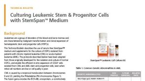 技术公告Culturing Leukemic Stem & Progenitor Cells with StemSpan™ Medium

 沪公网安备31010102008431号