Orellana DI et al. (OCT 2016)
EMBO molecular medicine 8 10 1197--1211
Coenzyme A corrects pathological defects in human neurons of PANK2-associated neurodegeneration.
Pantothenate kinase-associated neurodegeneration (PKAN) is an early onset and severely disabling neurodegenerative disease for which no therapy is available. PKAN is caused by mutations in PANK2,which encodes for the mitochondrial enzyme pantothenate kinase 2. Its function is to catalyze the first limiting step of Coenzyme A (CoA) biosynthesis. We generated induced pluripotent stem cells from PKAN patients and showed that their derived neurons exhibited premature death,increased ROS production,mitochondrial dysfunctions-including impairment of mitochondrial iron-dependent biosynthesis-and major membrane excitability defects. CoA supplementation prevented neuronal death and ROS formation by restoring mitochondrial and neuronal functionality. Our findings provide direct evidence that PANK2 malfunctioning is responsible for abnormal phenotypes in human neuronal cells and indicate CoA treatment as a possible therapeutic intervention.
View Publication
产品类型:
产品号#:
05850
05857
05870
05875
05872
05873
85850
85857
85870
85875
100-0483
100-0484
产品名:
mTeSR™1
mTeSR™1
Hausser Scientificᵀᴹ 明线血球计数板
ReLeSR™
Kumagai H et al. (MAY 2013)
Biochemical and Biophysical Research Communications 434 4 710--716
Identification of small molecules that promote human embryonic stem cell self-renewal
Human embryonic stem cells (hESCs) and induced pluripotent cells have the potential to provide an unlimited source of tissues for regenerative medicine. For this purpose,development of defined/xeno-free culture systems under feeder-free conditions is essential for the expansion of hESCs. Most defined/xeno-free media for the culture of hESCs contain basic fibroblast growth factor (bFGF). Therefore,bFGF is thought to have an almost essential role for the expansion of hESCs in an undifferentiated state. Here,we report identification of small molecules,some of which were neurotransmitter antagonists (trimipramine and ethopropazine),which promote long-term hESC self-renewal without bFGF in the medium. The hESCs maintained high expression levels of pluripotency markers,had a normal karyotype after 20 passages,and could differentiate into all three germ layers. ?? 2013 Elsevier Inc.
View Publication
Castro-Diaz N et al. (JUL 2014)
Genes and Development 28 13 1397--1409
Evolutionally dynamic L1 regulation in embryonic stem cells
Mobile elements are important evolutionary forces that challenge genomic integrity. Long interspersed element-1 (L1,also known as LINE-1) is the only autonomous transposon still active in the human genome. It displays an unusual pattern of evolution,with,at any given time,a single active L1 lineage amplifying to thousands of copies before getting replaced by a new lineage,likely under pressure of host restriction factors,which act notably by silencing L1 expression during early embryogenesis. Here,we demonstrate that in human embryonic stem (hES) cells,KAP1 (KRAB [Kruppel-associated box domain]-associated protein 1),the master cofactor of KRAB-containing zinc finger proteins (KRAB-ZFPs) previously implicated in the restriction of endogenous retroviruses,represses a discrete subset of L1 lineages predicted to have entered the ancestral genome between 26.8 million and 7.6 million years ago. In mice,we documented a similar chronologically conditioned pattern,albeit with a much contracted time scale. We could further identify an L1-binding KRAB-ZFP,suggesting that this rapidly evolving protein family is more globally responsible for L1 recognition. KAP1 knockdown in hES cells induced the expression of KAP1-bound L1 elements,but their younger,human-specific counterparts (L1Hs) were unaffected. Instead,they were stimulated by depleting DNA methyltransferases,consistent with recent evidence demonstrating that the PIWI-piRNA (PIWI-interacting RNA) pathway regulates L1Hs in hES cells. Altogether,these data indicate that the early embryonic control of L1 is an evolutionarily dynamic process and support a model in which newly emerged lineages are first suppressed by DNA methylation-inducing small RNA-based mechanisms before KAP1-recruiting protein repressors are selected.
View Publication
产品类型:
产品号#:
05850
05857
05870
05875
85850
85857
85870
85875
产品名:
mTeSR™1
mTeSR™1
West FD et al. (AUG 2010)
Stem cells and development 19 8 1211--1220
Porcine induced pluripotent stem cells produce chimeric offspring.
Ethical and moral issues rule out the use of human induced pluripotent stem cells (iPSCs) in chimera studies that would determine the full extent of their reprogrammed state,instead relying on less rigorous assays such as teratoma formation and differentiated cell types. To date,only mouse iPSC lines are known to be truly pluripotent. However,initial mouse iPSC lines failed to form chimeric offspring,but did generate teratomas and differentiated embryoid bodies,and thus these specific iPSC lines were not completely reprogrammed or truly pluripotent. Therefore,there is a need to address whether the reprogramming factors and process used eventually to generate chimeric mice are universal and sufficient to generate reprogrammed iPSC that contribute to chimeric offspring in additional species. Here we show that porcine mesenchymal stem cells transduced with 6 human reprogramming factors (POU5F1,SOX2,NANOG,KLF4,LIN28,and C-MYC) injected into preimplantation-stage embryos contributed to multiple tissue types spanning all 3 germ layers in 8 of 10 fetuses. The chimerism rate was high,85.3% or 29 of 34 live offspring were chimeras based on skin and tail biopsies harvested from 2- to 5-day-old pigs. The creation of pluripotent porcine iPSCs capable of generating chimeric offspring introduces numerous opportunities to study the facets significantly affecting cell therapies,genetic engineering,and other aspects of stem cell and developmental biology.
View Publication
产品类型:
产品号#:
05850
05857
05870
05875
85850
85857
85870
85875
27845
27945
27840
27865
27940
27965
产品名:
mTeSR™1
mTeSR™1
Rubin MR et al. (JAN 2011)
The Journal of clinical endocrinology and metabolism 96 1 176--86
Parathyroid hormone stimulates circulating osteogenic cells in hypoparathyroidism.
CONTEXT: The osteoanabolic properties of PTH may be due to increases in the number and maturity of circulating osteogenic cells. Hypoparathyroidism is a useful clinical model because this hypothesis can be tested by administering PTH. OBJECTIVE: The objective of the study was to characterize circulating osteogenic cells in hypoparathyroid subjects during 12 months of PTH (1-84) administration. DESIGN: Osteogenic cells were characterized using flow cytometry and antibodies against osteocalcin,an osteoblast-specific protein product,and stem cell markers CD34 and CD146. Changes in bone formation from biochemical markers and quadruple-labeled transiliac crest bone biopsies (0 and 3 month time points) were correlated with measurements of circulating osteogenic cells. SETTING: The study was conducted at a clinical research center. PATIENTS: Nineteen control and 19 hypoparathyroid patients were included in the study. INTERVENTION: Intervention included the administration of PTH (1-84). RESULTS: Osteocalcin-positive cells were lower in hypoparathyroid subjects than controls (0.7 ± 0.1 vs. 2.0 ± 0.1%; P textless 0.0001),with greater coexpression of the early cell markers CD34 and CD146 among the osteocalcin-positive cells in the hypoparathyroid subjects (11.0 ± 1.0 vs. 5.6 ± 0.7%; P textless 0.001). With PTH (1-84) administration,the number of osteogenic cells increased 3-fold (P textless 0.0001),whereas the coexpression of the early cell markers CD34 and CD146 decreased. Increases in osteogenic cells correlated with circulating and histomorphometric indices of osteoblast function: N-terminal propeptide of type I procollagen (R(2) = 0.4,P ≤ 0.001),bone-specific alkaline phosphatase (R(2) = 0.3,P textless 0.001),osteocalcin (R(2) = 0.4,P textless 0.001),mineralized perimeter (R(2) = 0.5,P textless 0.001),mineral apposition rate (R(2) = 0.4,P = 0.003),and bone formation rate (R(2) = 0.5,P textless 0.001). CONCLUSIONS: It is likely that PTH stimulates bone formation by stimulating osteoblast development and maturation. Correlations between circulating osteogenic cells and histomorphometric indices of bone formation establish that osteoblast activity is being identified by this methodology.
View Publication
产品类型:
产品号#:
05404
产品名:
Ioannidis P et al. (MAY 2005)
The Journal of biological chemistry 280 20 20086--93
CRD-BP/IMP1 expression characterizes cord blood CD34+ stem cells and affects c-myc and IGF-II expression in MCF-7 cancer cells.
The coding region determinant-binding protein/insulin-like growth factor II mRNA-binding protein (CRD-BP/IMP1) is an RNA-binding protein specifically recognizing c-myc,leader 3' IGF-II and tau mRNAs,and the H19 RNA. CRD-BP/IMP1 is predominantly expressed in embryonal tissues but is de novo activated and/or overexpressed in various human neoplasias. To address the question of whether CRD-BP/IMP1 expression characterizes certain cell types displaying distinct proliferation and/or differentiation properties (i.e. stem cells),we isolated cell subpopulations from human bone marrow,mobilized peripheral blood,and cord blood,all sources known to contain stem cells,and monitored for its expression. CRD-BP/IMP1 was detected only in cord blood-derived CD34(+) stem cells and not in any other cell type of either adult or cord blood origin. Adult BM CD34(+) cells cultured in the presence of 5'-azacytidine expressed de novo CRD-BP/IMP1,suggesting that epigenetic modifications may be responsible for its silencing in adult non-expressing cells. Furthermore,by applying the short interfering RNA methodology in MCF-7 cells,we observed,subsequent to knocking down CRD-BP/IMP1,decreased c-myc expression,increased IGF-II mRNA levels,and reduced cell proliferation rates. These data 1) suggest a normal role for CRD-BP/IMP1 in pluripotent stem cells with high renewal capacity,like the CB CD34(+) cells,2) indicate that altered methylation may directly or indirectly affect its expression in adult cells,3) imply that its de novo activation in cancer cells may affect the expression of c-Myc and insulin-like growth factor II,and 4) indicate that the inhibition of CRD-BP/IMP1 expression might affect cancer cell proliferation.
View Publication
产品类型:
产品号#:
09850
产品名:
Thein SL et al. (JUL 2007)
Proceedings of the National Academy of Sciences of the United States of America 104 27 11346--51
Intergenic variants of HBS1L-MYB are responsible for a major quantitative trait locus on chromosome 6q23 influencing fetal hemoglobin levels in adults.
Individual variation in fetal hemoglobin (HbF,alpha(2)gamma(2)) response underlies the remarkable diversity in phenotypic severity of sickle cell disease and beta thalassemia. HbF levels and HbF-associated quantitative traits (e.g.,F cell levels) are highly heritable. We have previously mapped a major quantitative trait locus (QTL) controlling F cell levels in an extended Asian-Indian kindred with beta thalassemia to a 1.5-Mb interval on chromosome 6q23,but the causative gene(s) are not known. The QTL encompasses several genes including HBS1L,a member of the GTP-binding protein family that is expressed in erythroid progenitor cells. In this high-resolution association study,we have identified multiple genetic variants within and 5' to HBS1L at 6q23 that are strongly associated with F cell levels in families of Northern European ancestry (P = 10(-75)). The region accounts for 17.6% of the F cell variance in northern Europeans. Although mRNA levels of HBS1L and MYB in erythroid precursors grown in vitro are positively correlated,only HBS1L expression correlates with high F cell alleles. The results support a key role for the HBS1L-related genetic variants in HbF control and illustrate the biological complexity of the mechanism of 6q QTL as a modifier of fetal hemoglobin levels in the beta hemoglobinopathies.
View Publication
产品类型:
产品号#:
09600
09650
产品名:
StemSpan™ SFEM
StemSpan™ SFEM
Malerba I et al. (OCT 2002)
Toxicological sciences : an official journal of the Society of Toxicology 69 2 433--8
In vitro myelotoxicity of propanil and 3,4-dichloroaniline on murine and human CFU-E/BFU-E progenitors.
Because of the wide use of pesticides for domestic and industrial purposes,the evaluation of their potential effects is of major concern for public health. The myelotoxicity of the herbicide propanil (3,4-dichloroproprioanilide) and its metabolite 3,4-dichloroaniline (DCA) is well documented in mice,but evidence that pesticides may severely compromise hematopoiesis in humans is lacking. In this study,an interspecies comparison of in vitro toxicity of these two compounds on murine and human burst- and colony-forming unit-erythrocyte (BFU-E,CFU-E) and colony-forming unit-granulocyte/macrophage (CFU-GM) progenitors,has been carried out. Murine bone marrow progenitors and human cord blood cells were exposed to propanil or DCA in doses ranging from 10 micro M to 1000 micro M,and the toxic effect was detected by a clonogenic assay with continuous exposure to the compounds. The results on murine cells indicate that the erythrocytic lineage is the most sensitive target for propanil and DCA. On the other hand,human progenitors seem to be less sensitive to the toxic effects of both compounds than murine progenitors at the same concentrations (IC(50) values are 305.2 +/- 22.6 micro M [total erythroid colonies] and textgreater500 micro M [CFU-GM] for propanil). Propanil was significantly more toxic to human erythroid progenitors than to human CFU-GM progenitors,as was found for the murine cells,emphasizing the role of the heme pathway as the target for propanil. These data confirm the evidence that the compounds investigated interfere with erythroid colony formation at different stages of the differentiation pathway and have different effects according to the dose.
View Publication
产品类型:
产品号#:
04564
04534
04544
产品名:
MethoCult™ H4534 Classic 无 EPO 入门试剂盒
MethoCult™ H4534 Classic(不含 EPO)
MethoCult™ H4534 Classic(不含 EPO)
Verma R et al. (AUG 2014)
The Journal of experimental medicine 211 9 1715--22
RHEX, a novel regulator of human erythroid progenitor cell expansion and erythroblast development.
Ligation of erythropoietin (EPO) receptor (EPOR) JAK2 kinase complexes propagates signals within erythroid progenitor cells (EPCs) that are essential for red blood cell production. To reveal hypothesized novel EPOR/JAK2 targets,a phosphotyrosine (PY) phosphoproteomics approach was applied. Beyond known signal transduction factors,32 new targets of EPO-modulated tyrosine phosphorylation were defined. Molecular adaptors comprised one major set including growth factor receptor-bound protein 2 (GRB2)-associated binding proteins 1-3 (GAB1-3),insulin receptor substrate 2 (IRS2),docking protein 1 (DOK1),Src homology 2 domain containing transforming protein 1 (SHC1),and sprouty homologue 1 (SPRY1) as validating targets,and SPRY2,SH2 domain containing 2A (SH2D2A),and signal transducing adaptor molecule 2 (STAM2) as novel candidate adaptors together with an ORF factor designated as regulator of human erythroid cell expansion (RHEX). RHEX is well conserved in Homo sapiens and primates but absent from mouse,rat,and lower vertebrate genomes. Among tissues and lineages,RHEX was elevated in EPCs,occurred as a plasma membrane protein,was rapidly PY-phosphorylated textgreater20-fold upon EPO exposure,and coimmunoprecipitated with the EPOR. In UT7epo cells,knockdown of RHEX inhibited EPO-dependent growth. This was associated with extracellular signal-regulated kinase 1,2 (ERK1,2) modulation,and RHEX coupling to GRB2. In primary human EPCs,shRNA knockdown studies confirmed RHEX regulation of erythroid progenitor expansion and further revealed roles in promoting the formation of hemoglobinizing erythroblasts. RHEX therefore comprises a new EPO/EPOR target and regulator of human erythroid cell expansion that additionally acts to support late-stage erythroblast development.
View Publication
产品类型:
产品号#:
04434
04444
22001
22005
22006
22007
22008
22009
22011
22012
22013
产品名:
MethoCult™ H4434 Classic
MethoCult™ H4434 Classic
STEMvision™ 人脐带血7-天CFU分析包
STEMvision™人脐带血14天CFU分析套装
STEMvision™人骨髓14天CFU分析套装
STEMvision™人动员外周血14天CFU分析套装
STEMvision™ 小鼠总CFU分析包
STEMvision™ 小鼠髓系CFU分析包
STEMvision™ 小鼠红系CFU分析包
STEMvision™小鼠CFU分析套装组合
Laurent B et al. (JAN 2010)
Blood 115 3 687--95
High-mobility group protein HMGB2 regulates human erythroid differentiation through trans-activation of GFI1B transcription.
Gfi-1B is a transcriptional repressor that is crucial for erythroid differentiation: inactivation of the GFI1B gene in mice leads to embryonic death due to failure to produce differentiated red cells. Accordingly,GFI1B expression is tightly regulated during erythropoiesis,but the mechanisms involved in such regulation remain partially understood. We here identify HMGB2,a high-mobility group HMG protein,as a key regulator of GFI1B transcription. HMGB2 binds to the GFI1B promoter in vivo and up-regulates its trans-activation most likely by enhancing the binding of Oct-1 and,to a lesser extent,of GATA-1 and NF-Y to the GFI1B promoter. HMGB2 expression increases during erythroid differentiation concomitantly to the increase of GfI1B transcription. Importantly,knockdown of HMGB2 in immature hematopoietic progenitor cells leads to decreased Gfi-1B expression and impairs their erythroid differentiation. We propose that HMGB2 potentiates GATA-1-dependent transcription of GFI1B by Oct-1 and thereby controls erythroid differentiation.
View Publication

 EasySep™小鼠TIL(CD45)正选试剂盒


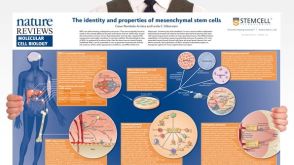 挂图The Identity and Properties of Mesenchymal Stem Cells Overview of MSC expansion, differentiation, immunoregulatory properties and therapeutic potential

 沪公网安备31010102008431号