West FD et al. (OCT 2011)
Stem Cells 29 10 1640--1643
Brief report: chimeric pigs produced from induced pluripotent stem cells demonstrate germline transmission and no evidence of tumor formation in young pigs.
The recent development of porcine induced pluripotent stem cells (piPSCs) capable of generating chimeric animals,a feat not previously accomplished with embryonic stem cells or iPSCs in a species outside of rodents,has opened the doors for in-depth study of iPSC tumorigenicity,autologous transplantation,and other key aspects to safely move iPSC therapies to the clinic. The study of iPSC tumorigenicity is critical as previous research in the mouse showed that iPSC-derived chimeras possessed large numbers of tumors,rising significant concerns about the safety of iPSC therapies. Additionally,piPSCs capable of generating germline chimeras could revolutionize the transgenic animal field by enabling complex genetic manipulations (e.g.,knockout or knockin of genes) to produce biomedically important large animal models or improve livestock production. In this study,we demonstrate for the first time in a nonrodent species germline transmission of iPSCs with the live birth of a transgenic piglet that possessed genome integration of the human POU5F1 and NANOG genes. In addition,gross and histological examination of necropsied porcine chimeras at 2,7,and 9 months showed that these animals lacked tumor formation and demonstrated normal development. Tissue samples positive for human POU5F1 DNA showed no C-MYC gene expression,further implicating C-MYC as a cause of tumorigenicity. The development of germline-competent porcine iPSCs that do not produce tumors in young chimeric animals presents an attractive and powerful translational model to study the efficacy and safety of stem cell therapies and perhaps to efficiently produce complex transgenic animals.
View Publication
产品类型:
产品号#:
05850
05857
05870
05875
85850
85857
85870
85875
产品名:
mTeSR™1
mTeSR™1
Meenhuis A et al. (JUL 2011)
Blood 118 4 916--25
MiR-17/20/93/106 promote hematopoietic cell expansion by targeting sequestosome 1-regulated pathways in mice.
MicroRNAs (miRNAs) are pivotal for regulation of hematopoiesis but their critical targets remain largely unknown. Here,we show that ectopic expression of miR-17,-20,-93 and -106,all AAAGUGC seed-containing miRNAs,increases proliferation,colony outgrowth and replating capacity of myeloid progenitors and results in enhanced P-ERK levels. We found that these miRNAs are endogenously and abundantly expressed in myeloid progenitors and down-regulated in mature neutrophils. Quantitative proteomics identified sequestosome 1 (SQSTM1),an ubiquitin-binding protein and regulator of autophagy-mediated protein degradation,as a major target for these miRNAs in myeloid progenitors. In addition,we found increased expression of Sqstm1 transcripts during CSF3-induced neutrophil differentiation of 32D-CSF3R cells and an inverse correlation of SQSTM1 protein levels and miR-106 expression in AML samples. ShRNA-mediated silencing of Sqstm1 phenocopied the effects of ectopic miR-17/20/93/106 expression in hematopoietic progenitors in vitro and in mice. Further,SQSTM1 binds to the ligand-activated colony-stimulating factor 3 receptor (CSF3R) mainly in the late endosomal compartment,but not in LC3 positive autophagosomes. SQSTM1 regulates CSF3R stability and ligand-induced mitogen-activated protein kinase signaling. We demonstrate that AAAGUGC seed-containing miRNAs promote cell expansion,replating capacity and signaling in hematopoietic cells by interference with SQSTM1-regulated pathways.
View Publication
产品类型:
产品号#:
03231
产品名:
MethoCult™ M3231
Lidonnici MR et al. (OCT 2010)
Cancer research 70 20 7949--59
Expression of the transcriptional repressor Gfi-1 is regulated by C/EBPalpha and is involved in its proliferation and colony formation-inhibitory effects in p210BCR/ABL-expressing cells.
Ectopic expression of CAAT/enhancer binding protein α (C/EBPα) in p210BCR/ABL-expressing cells induces granulocytic differentiation,inhibits proliferation,and suppresses leukemogenesis. To dissect the molecular mechanisms underlying these biological effects,C/EBPα-regulated genes were identified by microarray analysis in 32D-p210BCR/ABL cells. One of the genes whose expression was activated by C/EBPα in a DNA binding-dependent manner in BCR/ABL-expressing cells is the transcriptional repressor Gfi-1. We show here that C/EBPα interacts with a functional C/EBP binding site in the Gfi-1 5'-flanking region and enhances the promoter activity of Gfi-1. Moreover,in K562 cells,RNA interference-mediated downregulation of Gfi-1 expression partially rescued the proliferation-inhibitory but not the differentiation-inducing effect of C/EBPα. Ectopic expression of wild-type Gfi-1,but not of a transcriptional repressor mutant (Gfi-1P2A),inhibited proliferation and markedly suppressed colony formation but did not induce granulocytic differentiation of BCR/ABL-expressing cells. By contrast,Gfi-1 short hairpin RNA-tranduced CD34(+) chronic myeloid leukemia cells were markedly more clonogenic than the scramble-transduced counterpart. Together,these studies indicate that Gfi-1 is a direct target of C/EBPα required for its proliferation and survival-inhibitory effects in BCR/ABL-expressing cells.
View Publication
产品类型:
产品号#:
02690
09600
09650
产品名:
StemSpan™ CC100
StemSpan™ SFEM
StemSpan™ SFEM
Rodrí et al. (MAY 2004)
Blood 103 9 3349--54
Interleukin-6 deficiency affects bone marrow stromal precursors, resulting in defective hematopoietic support.
Interleukin-6 (IL-6) is a critical factor in the regulation of stromal function and hematopoiesis. In vivo bromodeoxyuridine incorporation analysis indicates that the percentage of Lin(-)Sca-1(+) hematopoietic progenitors undergoing DNA synthesis is diminished in IL-6-deficient (IL-6(-/-)) bone marrow (BM) compared with wild-type BM. Reduced proliferation of IL-6(-/-) BM progenitors is also observed in IL-6(-/-) long-term BM cultures,which show defective hematopoietic support as measured by production of total cells,granulocyte macrophage-colony-forming units (CFU-GMs),and erythroid burst-forming units (BFU-Es). Seeding experiments of wild-type and IL-6(-/-) BM cells on irradiated wild-type or IL-6-deficient stroma indicate that the hematopoietic defect can be attributed to the stromal and not to the hematopoietic component. In IL-6(-/-) BM,stromal mesenchymal precursors,fibroblast CFUs (CFU-Fs),and stroma-initiating cells (SICs) are reduced to almost 50% of the wild-type BM value. Moreover,IL-6(-/-) stromata show increased CD34 and CD49e expression and reduced expression of the membrane antigens vascular cell adhesion molecule-1 (VCAM-1),Sca-1,CD49f,and Thy1. These data strongly suggest that IL-6 is an in vivo growth factor for mesenchymal precursors,which are in part implicated in the reduced longevity of the long-term repopulating stem cell compartment of IL-6(-/-) mice.
View Publication
产品类型:
产品号#:
03534
05501
05502
05350
28600
产品名:
MethoCult™ GF M3534
L-Calc™有限稀释软件
Selleri C et al. (MAR 2005)
Blood 105 5 2198--205
Involvement of the urokinase-type plasminogen activator receptor in hematopoietic stem cell mobilization.
We investigated the involvement of the urokinase-type plasminogen-activator receptor (uPAR) in granulocyte-colony-stimulating factor (G-CSF)-induced mobilization of CD34+ hematopoietic stem cells (HSCs) from 16 healthy donors. Analysis of peripheral blood mononuclear cells (PBMNCs) showed an increased uPAR expression after G-CSF treatment in CD33+ myeloid and CD14+ monocytic cells,whereas mobilized CD34+ HSCs remained uPAR negative. G-CSF treatment also induced an increase in serum levels of soluble uPAR (suPAR). Cleaved forms of suPAR (c-suPAR) were released in vitro by PBMNCs and were also detected in the serum of G-CSF-treated donors. c-suPAR was able to chemoattract CD34+ KG1 leukemia cells and CD34+ HSCs,as documented by their in vitro migratory response to a chemotactic suPAR-derived peptide (uPAR84-95). uPAR84-95 induced CD34+ KG1 and CD34+ HSC migration by activating the high-affinity fMet-Leu-Phe (fMLP) receptor (FPR). In addition,uPAR84-95 inhibited CD34+ KG1 and CD34+ HSC in vitro migration toward the stromal-derived factor 1 (SDF1),thus suggesting the heterologous desensitization of its receptor,CXCR4. Finally,uPAR84-95 treatment significantly increased the output of clonogenic progenitors from long-term cultures of CD34+ HSCs. Our findings demonstrate that G-CSF-induced upregulation of uPAR on circulating CD33+ and CD14+ cells is associated with increased uPAR shedding,which leads to the appearance of serum c-suPAR. c-suPAR could contribute to the mobilization of HSCs by promoting their FPR-mediated migration and by inducing CXCR4 desensitization.
View Publication
产品类型:
产品号#:
05150
产品名:
MyeloCult™ H5100
Capron C et al. (AUG 2010)
Blood 116 8 1244--53
A major role of TGF-beta1 in the homing capacities of murine hematopoietic stem cell/progenitors.
Transforming growth factor-beta1 (TGF-beta1) is a pleiotropic cytokine with major in vitro effects on hematopoietic stem cells (HSCs) and lymphocyte development. Little is known about hematopoiesis from mice with constitutive TGF-beta1 inactivation largely because of important embryonic lethality and development of a lethal inflammatory disorder in TGF-beta1(-/-) pups,making these studies difficult. Here,we show that no sign of the inflammatory disorder was detectable in 8- to 10-day-old TGF-beta1(-/-) neonates as judged by both the number of T-activated and T-regulator cells in secondary lymphoid organs and the level of inflammatory cytokines in sera. After T-cell depletion,the inflammatory disease was not transplantable in recipient mice. Bone marrow cells from 8- to 10-day-old TGF-beta1(-/-) neonates showed strikingly impaired short- and long-term reconstitutive activity associated with a parallel decreased in vivo homing capacity of lineage negative (Lin(-)) cells. In addition an in vitro-reduced survival of immature progenitors (Lin(-) Kit(+) Sca(+)) was observed. Similar defects were found in liver cells from TGF-beta1(-/-) embryos on day 14 after vaginal plug. These data indicate that TGF-beta1 is a critical regulator for in vivo homeostasis of the HSCs,especially for their homing potential.
View Publication
产品类型:
产品号#:
03234
09600
09650
产品名:
MethoCult™ M3234
StemSpan™ SFEM
StemSpan™ SFEM
Cheng E-C et al. (MAR 2009)
Blood 113 12 2826--34
Role for MKL1 in megakaryocytic maturation.
Megakaryoblastic leukemia 1 (MKL1),identified as part of the t(1;22) translocation specific to acute megakaryoblastic leukemia,is highly expressed in differentiated muscle cells and promotes muscle differentiation by activating serum response factor (SRF). Here we show that Mkl1 expression is up-regulated during murine megakaryocytic differentiation and that enforced overexpression of MKL1 enhances megakaryocytic differentiation. When the human erythroleukemia (HEL) cell line is induced to differentiate with 12-O-tetradecanoylphorbol 13-acetate,overexpression of MKL1 results in an increased number of megakaryocytes with a concurrent increase in ploidy. MKL1 overexpression also promotes megakaryocytic differentiation of primary human CD34(+) cells cultured in the presence of thrombopoietin. The effect of MKL1 is abrogated when SRF is knocked down,suggesting that MKL1 acts through SRF. Consistent with these findings in human cells,knockout of Mkl1 in mice leads to reduced platelet counts in peripheral blood,and reduced ploidy in bone marrow megakaryocytes. In conclusion,MKL1 promotes physiologic maturation of human and murine megakaryocytes.
View Publication
产品类型:
产品号#:
09500
09600
09650
04960
04902
04900
04963
04962
04970
04971
04901
产品名:
BIT 9500血清替代物
StemSpan™ SFEM
StemSpan™ SFEM
MegaCult™-C胶原和无细胞因子培养基
胶原蛋白溶液
MegaCult™-C无细胞因子培养基
双室载玻片套件
MegaCult™-C CFU-Mk染色试剂盒
MegaCult™-C无细胞因子全套试剂盒
MegaCult™-C含细胞因子全套试剂盒
MegaCult™-C含细胞因子培养基
Yang Q et al. (MAR 2011)
Blood 117 13 3529--38
E47 regulates hematopoietic stem cell proliferation and energetics but not myeloid lineage restriction.
The immune system is replenished by self-renewing hematopoietic stem cells (HSCs) that produce multipotent progenitors (MPPs) with little renewal capacity. E-proteins,the widely expressed basic helix-loop-helix transcription factors,contribute to HSC and MPP activity,but their specific functions remain undefined. Using quantitative in vivo and in vitro approaches,we show that E47 is dispensable for the short-term myeloid differentiation of HSCs but regulates their long-term capabilities. E47-deficient progenitors show competent myeloid production in short-term assays in vitro and in vivo. However,long-term myeloid and lymphoid differentiation is compromised because of a progressive loss of HSC self-renewal that is associated with diminished p21 expression and hyperproliferation. The activity of E47 is shown to be cell-intrinsic. Moreover,E47-deficient HSCs and MPPs have altered expression of genes associated with cellular energy metabolism,and the size of the MPP pool but not downstream lymphoid precursors in bone marrow or thymus is rescued in vivo by antioxidant. Together,these observations suggest a role for E47 in the tight control of HSC proliferation and energy metabolism,and demonstrate that E47 is not required for short-term myeloid differentiation.
View Publication
产品类型:
产品号#:
03434
03444
产品名:
MethoCult™ GF M3434
MethoCult™ GF M3434
Zhu HH et al. (MAY 2011)
Blood 117 20 5350--61
Kit-Shp2-Kit signaling acts to maintain a functional hematopoietic stem and progenitor cell pool.
The stem cell factor (SCF)/Kit system has served as a classic model in deciphering molecular signaling events in the hematopoietic compartment,and Kit expression is a most critical marker for hematopoietic stem cells (HSCs) and progenitors. However,it remains to be elucidated how Kit expression is regulated in HSCs. Herein we report that a cytoplasmic tyrosine phosphatase Shp2,acting downstream of Kit and other RTKs,promotes Kit gene expression,constituting a Kit-Shp2-Kit signaling axis. Inducible ablation of PTPN11/Shp2 resulted in severe cytopenia in BM,spleen,and peripheral blood in mice. Shp2 removal suppressed the functional pool of HSCs/progenitors,and Shp2-deficient HSCs failed to reconstitute lethally irradiated recipients because of defects in homing,self-renewal,and survival. We show that Shp2 regulates coordinately multiple signals involving up-regulation of Kit expression via Gata2. Therefore,this study reveals a critical role of Shp2 in maintenance of a functional HSC/progenitor pool in adult mammals,at least in part through a kinase-phosphatase-kinase cascade.
View Publication
产品类型:
产品号#:
03434
03444
产品名:
MethoCult™ GF M3434
MethoCult™ GF M3434
Rybtsov S et al. (JUN 2011)
The Journal of experimental medicine 208 6 1305--15
Hierarchical organization and early hematopoietic specification of the developing HSC lineage in the AGM region.
The aorta-gonad-mesonephros region plays an important role in hematopoietic stem cell (HSC) development during mouse embryogenesis. The vascular endothelial cadherin�?� CD45�?� (VE-cad�?�CD45�?�) population contains the major type of immature pre-HSCs capable of developing into long-term repopulating definitive HSCs. In this study,we developed a new coaggregation culture system,which supports maturation of a novel population of CD45-negative (VE-cad�?�CD45�?�CD41�?�) pre-HSCs into definitive HSCs. The appearance of these pre-HSCs precedes development of the VE-cad�?�CD45�?� pre-HSCs (termed here type I and type II pre-HSCs,respectively),thus establishing a hierarchical directionality in the developing HSC lineage. By labeling the luminal surface of the dorsal aorta,we show that both type I and type II pre-HSCs are distributed broadly within the endothelial and subendothelial aortic layers,in contrast to mature definitive HSCs which localize to the aortic endothelial layer. In agreement with expression of CD41 in pre-HSCs,in vivo CD41-Cre-mediated genetic tagging occurs in embryonic pre-HSCs and persists in all lymphomyeloid lineages of the adult animal.
View Publication
A role for thrombopoietin in hemangioblast development.
Vascular endothelial growth factor (VEGF) and stem cell factor (SCF) act as growth factors for the hemangioblast,an embryonic progenitor of the hematopoietic and endothelial lineages. Because thrombopoietin (TPO) and its receptor,c-Mpl,regulate primitive hematopoietic populations,including bone marrow hematopoietic stem cells,we investigated whether TPO acts on the hemangioblasts that derive from differentiation of embryonic stem cells in vitro. Reverse transcriptase polymerase chain reaction analysis detected expression of c-Mpl beginning on day 3 of embryoid body differentiation when the hemangioblast first arises. In assays of the hemangioblast colony-forming cell (BL-CFC),TPO alone supported BL-CFC formation and nearly doubled the number of BL-CFC when added together with VEGF and SCF. When replated under the appropriate conditions,TPO-stimulated BL-CFC gave rise to secondary hematopoietic colonies,as well as endothelial cells,confirming their nature as hemangioblasts. Addition of a neutralizing anti-VEGF antibody did not block TPO enhancement of BL-CFC formation,suggesting that TPO acts independently of VEGF. These results establish that Mpl signaling plays a role in the earliest stages of hematopoietic development and that TPO represents a third growth factor influencing hemangioblast formation.
View Publication

 EasySep™小鼠TIL(CD45)正选试剂盒


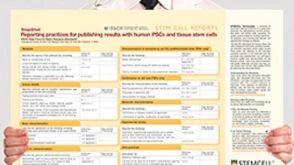 挂图Reporting Practices for Publishing Results with hPSCs Learn how to plan and conduct your human pluripotent stem cell (hPSC)-based research following the ISSCR’s Standards for Human Stem Cell Use in Research
 沪公网安备31010102008431号