Smad4 binds Hoxa9 in the cytoplasm and protects primitive hematopoietic cells against nuclear activation by Hoxa9 and leukemia transformation.
We studied leukemic stem cells (LSCs) in a Smad4(-/-) mouse model of acute myelogenous leukemia (AML) induced either by the HOXA9 gene or by the fusion oncogene NUP98-HOXA9. Although Hoxa9-Smad4 complexes accumulate in the cytoplasm of normal hematopoietic stem cells and progenitor cells (HSPCs) transduced with these oncogenes,there is no cytoplasmic stabilization of HOXA9 in Smad4(-/-) HSPCs,and as a consequence increased levels of Hoxa9 is observed in the nucleus leading to increased immortalization in vitro. Loss of Smad4 accelerates the development of leukemia in vivo because of an increase in transformation of HSPCs. Therefore,the cytoplasmic binding of Hoxa9 by Smad4 is a mechanism to protect Hoxa9-induced transformation of normal HSPCs. Because Smad4 is a potent tumor suppressor involved in growth control,we developed a strategy to modify the subcellular distribution of Smad4. We successfully disrupted the interaction between Hoxa9 and Smad4 to activate the TGF-β pathway and apoptosis,leading to a loss of LSCs. Together,these findings reveal a major role for Smad4 in the negative regulation of leukemia initiation and maintenance induced by HOXA9/NUP98-HOXA9 and provide strong evidence that antagonizing Smad4 stabilization by these oncoproteins might be a promising novel therapeutic approach in leukemia.
View Publication
产品类型:
产品号#:
03434
03444
03236
产品名:
MethoCult™ GF M3434
MethoCult™ GF M3434
MethoCult™ SF M3236
Renz PF and Beyer TA (FEB 2016)
Methods in molecular biology (Clifton,N.J.) 1341 369--376
A Concise Protocol for siRNA-Mediated Gene Suppression in Human Embryonic Stem Cells.
Human embryonic stem cells hold great promise for future biomedical applications such as disease modeling and regenerative medicine. However,these cells are notoriously difficult to culture and are refractory to common means of genetic manipulation,thereby limiting their range of applications. In this protocol,we present an easy and robust method of gene repression in human embryonic stem cells using lipofection of small interfering RNA (siRNA).
View Publication
产品类型:
产品号#:
05850
05857
05870
05875
05872
05873
07909
07920
85850
85857
85870
85875
100-0483
100-0484
07922
产品名:
IV型胶原酶(1mg /mL)
ACCUTASE™
mTeSR™1
mTeSR™1
Hausser Scientificᵀᴹ 明线血球计数板
ReLeSR™
ACCUTASE™
Irwin EF et al. (OCT 2011)
Biomaterials 32 29 6912--6919
Engineered polymer-media interfaces for the long-term self-renewal of human embryonic stem cells.
We have developed a synthetic polymer interface for the long-term self-renewal of human embryonic stem cells (hESCs) in defined media. We successfully cultured hESCs on hydrogel interfaces of aminopropylmethacrylamide (APMAAm) for over 20 passages in chemically-defined mTeSR™1 media and demonstrated pluripotency of multiple hESC lines with immunostaining and quantitative RT-PCR studies. Results for hESC proliferation and pluripotency markers were both qualitatively and quantitatively similar to cells cultured on Matrigel™-coated substrates. Mechanistically,it was resolved that bovine serum albumin (BSA) in the mTeSR™1 media was critical for cell adhesion on APMAAm hydrogel interfaces. This study uniquely identified a robust long-term culture surface for the self-renewal of hESCs without the use of biologic coatings (e.g.,peptides,proteins,or Matrigel™) in completely chemically-defined media that employed practical culturing techniques amenable to clinical-scale cell expansion.
View Publication
Sumitomo A et al. (OCT 2010)
Molecular and cellular biology 30 20 4818--27
The transcriptional mediator subunit MED1/TRAP220 in stromal cells is involved in hematopoietic stem/progenitor cell support through osteopontin expression.
MED1/TRAP220,a subunit of the transcriptional Mediator/TRAP complex,is crucial for various biological events through its interaction with distinct activators,such as nuclear receptors and GATA family activators. In hematopoiesis,MED1 plays a pivotal role in optimal nuclear receptor-mediated myelomonopoiesis and GATA-1-induced erythropoiesis. In this study,we present evidence that MED1 in stromal cells is involved in supporting hematopoietic stem and/or progenitor cells (HSPCs) through osteopontin (OPN) expression. We found that the proliferation of bone marrow (BM) cells cocultured with MED1 knockout (Med1(-/-)) mouse embryonic fibroblasts (MEFs) was significantly suppressed compared to the control. Furthermore,the number of long-term culture-initiating cells (LTC-ICs) was attenuated for BM cells cocultured with Med1(-/-) MEFs. The vitamin D receptor (VDR)- and Runx2-mediated expression of OPN,as well as Mediator recruitment to the Opn promoter,was specifically attenuated in the Med1(-/-) MEFs. Addition of OPN to these MEFs restored the growth of cocultured BM cells and the number of LTC-ICs,both of which were attenuated by the addition of the anti-OPN antibody to Med1(+/+) MEFs and to BM stromal cells. Consequently,MED1 in niche appears to play an important role in supporting HSPCs by upregulating VDR- and Runx2-mediated transcription on the Opn promoter.
View Publication
Chemically defined serum-free conditions for cartilage regeneration from human embryonic stem cells.
AIMS The aim of this study was to improve a method that induce cartilage differentiation of human embryoid stem cells (hESCs) in vitro,and test the effect of in vivo environments on the further maturation of hESCs derived cells. MAIN METHODS Embryoid bodies (EBs) formed from hESCs,with serum-free KSR-based medium and mesodermal specification related factors,CHIR,and Noggin for first 8days. Then cells were digested and cultured as micropellets in serum-free KSR-based chondrogenic medium that was supplemented with PDGF-BB,TGF β3,BMP4 in sequence for 24days. The morphology,FACS,histological staining as well as the expression of chondrogenic specific genes were detected in each stage,and further in vivo experiments,cell injections and tissue transplantations,further verified the formation of chondrocytes. KEY FINDINGS We were able to obtain chondrocyte/cartilage from hESCs using serum-free KSR-based conditioned medium. qPCR analysis showed that expression of the chondroprogenitor genes and the chondrocyte/cartilage matrix genes. Morphology analysis demonstrated we got PG+COL2+COL1-particles. It indicated we obtained hyaline cartilage-like particles. 32-Day differential cells were injected subcutaneous. Staining results showed grafts developed further mature in vivo. But when transplanted in subrenal capsule,their effect was not good as in subcutaneous. Microenvironment might affect the cartilage formation. SIGNIFICANCE The results of this study provide an absolute serum-free and efficient approach for generation of hESC-derived chondrocytes,and cells will become further maturation in vivo. It provides evidence and technology for the hypothesis that hESCs may be a promising therapy for the treatment of cartilage disease.
View Publication
产品类型:
产品号#:
05850
05857
05870
05875
85850
85857
85870
85875
产品名:
mTeSR™1
mTeSR™1
Mortensen M et al. (MAR 2011)
The Journal of experimental medicine 208 3 455--67
The autophagy protein Atg7 is essential for hematopoietic stem cell maintenance.
The role of autophagy,a lysosomal degradation pathway which prevents cellular damage,in the maintenance of adult mouse hematopoietic stem cells (HSCs) remains unknown. Although normal HSCs sustain life-long hematopoiesis,malignant transformation of HSCs leads to leukemia. Therefore,mechanisms protecting HSCs from cellular damage are essential to prevent hematopoietic malignancies. In this study,we crippled autophagy in HSCs by conditionally deleting the essential autophagy gene Atg7 in the hematopoietic system. This resulted in the loss of normal HSC functions,a severe myeloproliferation,and death of the mice within weeks. The hematopoietic stem and progenitor cell compartment displayed an accumulation of mitochondria and reactive oxygen species,as well as increased proliferation and DNA damage. HSCs within the Lin(-)Sca-1(+)c-Kit(+) (LSK) compartment were significantly reduced. Although the overall LSK compartment was expanded,Atg7-deficient LSK cells failed to reconstitute the hematopoietic system of lethally irradiated mice. Consistent with loss of HSC functions,the production of both lymphoid and myeloid progenitors was impaired in the absence of Atg7. Collectively,these data show that Atg7 is an essential regulator of adult HSC maintenance.
View Publication
产品类型:
产品号#:
03434
03444
产品名:
MethoCult™ GF M3434
MethoCult™ GF M3434
Christopher MJ et al. (FEB 2011)
The Journal of experimental medicine 208 2 251--60
Expression of the G-CSF receptor in monocytic cells is sufficient to mediate hematopoietic progenitor mobilization by G-CSF in mice.
Granulocyte colony-stimulating factor (G-CSF),the prototypical mobilizing cytokine,induces hematopoietic stem and progenitor cell (HSPC) mobilization from the bone marrow in a cell-nonautonomous fashion. This process is mediated,in part,through suppression of osteoblasts and disruption of CXCR4/CXCL12 signaling. The cellular targets of G-CSF that initiate the mobilization cascade have not been identified. We use mixed G-CSF receptor (G-CSFR)-deficient bone marrow chimeras to show that G-CSF-induced mobilization of HSPCs correlates poorly with the number of wild-type neutrophils. We generated transgenic mice in which expression of the G-CSFR is restricted to cells of the monocytic lineage. G-CSF-induced HSPC mobilization,osteoblast suppression,and inhibition of CXCL12 expression in the bone marrow of these transgenic mice are intact,demonstrating that G-CSFR signals in monocytic cells are sufficient to induce HSPC mobilization. Moreover,G-CSF treatment of wild-type mice is associated with marked loss of monocytic cells in the bone marrow. Finally,we show that bone marrow macrophages produce factors that support the growth and/or survival of osteoblasts in vitro. Together,these data suggest a model in which G-CSFR signals in bone marrow monocytic cells inhibit the production of trophic factors required for osteoblast lineage cell maintenance,ultimately leading to HSPC mobilization.
View Publication
产品类型:
产品号#:
03434
03444
产品名:
MethoCult™ GF M3434
MethoCult™ GF M3434
Salvagiotto G et al. (JAN 2011)
PLoS ONE 6 3 e17829
A defined, feeder-free, serum-free system to generate In Vitro hematopoietic progenitors and differentiated blood cells from hESCs and hiPSCs
Human ESC and iPSC are an attractive source of cells of high quantity and purity to be used to elucidate early human development processes,for drug discovery,and in clinical cell therapy applications. To efficiently differentiate pluripotent cells into a pure population of hematopoietic progenitors we have developed a new 2-dimensional,defined and highly efficient protocol that avoids the use of feeder cells,serum or embryoid body formation. Here we showed that a single matrix protein in combination with growth factors and a hypoxic environment is sufficient to generate from pluripotent cells hematopoietic progenitors capable of differentiating further in mature cell types of different lineages of the blood system. We tested the differentiation method using hESCs and 9 iPSC lines generated from different tissues. These data indicate the robustness of the protocol providing a valuable tool for the generation of clinical-grade hematopoietic cells from pluripotent cells.
View Publication
产品类型:
产品号#:
05850
05857
05870
05875
85850
85857
85870
85875
产品名:
mTeSR™1
mTeSR™1
Wognum AW et al. ( )
Archives of medical research 34 6 461--75
Identification and isolation of hematopoietic stem cells.
Hematopoietic stem cells (HSCs) are defined by their ability to repopulate all of the hematopoietic lineages in vivo and sustain the production of these cells for the life span of the individual. In the absence of reliable direct markers for HSCs,their identification and enumeration depends on functional long-term,multilineage,in vivo repopulation assays. The extremely low frequency of HSCs in any tissue and the absence of a specific HSC phenotype have made their purification and characterization a highly challenging goal. HSCs and primitive hematopoietic cells can be distinguished from mature blood cells by their lack of lineage-specific markers and presence of certain other cell-surface antigens,such as CD133 (for human cells) and c-kit and Sca-1 (for murine cells). Functional analyses of purified subpopulations of primitive hematopoietic cells have led to the development of several procedures for isolating cell populations that are highly enriched in cells with in vivo stem cell activity. Simplified methods for obtaining these cells at high yield have been important to the practical exploitation of such advances. This article reviews recent progress in identifying human and mouse HSCs and current techniques for their purification.
View Publication
产品类型:
产品号#:
18056
18056RF
产品名:
Kang M et al. (APR 2014)
International journal of molecular sciences 15 5 7139--7157
Generation of bladder urothelium from human pluripotent stem cells under chemically defined serum- and feeder-free system.
Human stem cells are promising sources for bladder regeneration. Among several possible sources,pluripotent stem cells are the most fascinating because they can differentiate into any cell type,and proliferate limitlessly in vitro. Here,we developed a protocol for differentiation of human pluripotent stem cells (hPSCs) into bladder urothelial cells (BUCs) under a chemically defined culture system. We first differentiated hPSCs into definitive endoderm (DE),and further specified DE cells into BUCs by treating retinoic acid under a keratinocyte-specific serum free medium. hPSC-derived DE cells showed significantly expressed DE-specific genes,but did not express mesodermal or ectodermal genes. After DE cells were specified into BUCs,they notably expressed urothelium-specific genes such as UPIb,UPII,UPIIIa,P63 and CK7. Immunocytochemistry showed that BUCs expressed UPII,CK8/18 and P63 as well as tight junction molecules,E-CADHERIN and ZO-1. Additionally,hPSCs-derived BUCs exhibited low permeability in a FITC-dextran permeability assay,indicating BUCs possessed the functional units of barrier on their surfaces. However,BUCs did not express the marker genes of other endodermal lineage cells (intestine and liver) as well as mesodermal or ectodermal lineage cells. In summary,we sequentially differentiated hPSCs into DE and BUCs in a serum- and feeder-free condition. Our differentiation protocol will be useful for producing cells for bladder regeneration and studying normal and pathological development of the human bladder urothelium in vitro.
View Publication

 EasySep™小鼠TIL(CD45)正选试剂盒


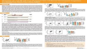 科学海报Generation of T and NK Cells From Pluripotent Stem Cell-Derived Hematopoietic Progenitors in a Stroma-Free, Serum-Free Culture System

 沪公网安备31010102008431号