Wiedemann A et al. (DEC 2012)
Cellular reprogramming 14 6 485--496
Induced pluripotent stem cells generated from adult bone marrow-derived cells of the nonhuman primate (Callithrix jacchus) using a novel quad-cistronic and excisable lentiviral vector.
Regenerative medicine is in need of solid,large animal models as a link between rodents and humans to evaluate the functionality,immunogenicity,and clinical safety of stem cell-derived cell types. The common marmoset (Callithrix jacchus) is an excellent large animal model,genetically close to humans and readily used worldwide in clinical research. Until now,only two groups showed the generation of induced pluripotent stem cells (iPSCs) from the common marmoset using integrating retroviral vectors. Therefore,we reprogrammed bone marrow-derived mesenchymal cells (MSCs) of adult marmosets in the presence of TAV,SB431542,PD0325901,and ascorbic acid via a novel,excisable lentiviral spleen focus-forming virus (SFFV)-driven quad-cistronic vector system (OCT3/4,KLF4,SOX2,C-MYC). Endogenous pluripotency markers like OCT3/4,KLF4,SOX2,C-MYC,LIN28,NANOG,and strong alkaline phosphatase signals were detected. Exogenous genes were silenced and additionally the cassette was removed with a retroviral Gag precursor system. The cell line could be cultured in absence of leukemia inhibitory factor (LIF) and basic fibroblast growth factor (bFGF) and could be successfully differentiated into embryoid bodies and teratomas with presence of all three germ layers. Directed differentiation generated neural progenitors,megakaryocytes,adipocytes,chondrocytes,and osteogenic cells. Thus,all criteria for fully reprogrammed bone marrow-MSCs of a nonhuman primate with a genetically sophisticated construct could be demonstrated. These cells will be a promising tool for future autologous transplantations.
View Publication
Meng G et al. (APR 2009)
Stem cells and development 19 4 1--31
Extra-cellular Matrix Isolated from Foreskin Fibroblasts Supports Long Term Xeno-Free Human Embryonic Stem Cell Culture.
Human embryonic stem (hES) cells hold great promise for application of human cell and tissue replacement therapy. However,the overwhelming majority of currently available hES cell lines have been directly or indirectly exposed to materials containing animal-derived components during their derivation,propagation,and cryopreservation. Unlike feeder based cultures,which require the simultaneous growth of feeder and stem cells,resulting in mixed cell populations,stem cells grown on feeder-free systems are easily separated from the surface,presenting a pure population of cells for downstream applications. In this study we have developed a novel method to expand hES cells in xeno-free,feeder-free conditions using two different matrices derived from xeno-free human foreskin fibroblasts (XF-HFFs). Using XF-HFF-derived extracellular matrix,together with 100ng/ml recombinant bFGF supplemented HEScGRO Basal Medium,long term xeno-free expansion of hES cells is possible. Resulting hES cells were subjected to stringent tests and were found to maintain ES cell features,including morphology,pluripotency,stable karyotype,and expression of cell surface markers,for at least 20 passages. Xeno-free culturing practices are essential for the translation of basic hES cell research into the clinic. Therefore,the method presented in this study demonstrates that hES cells can be cultured in complete xeno-free conditions without the loss of pluripotency and furthermore,without the possibility of contamination from exogenous sources.
View Publication
产品类型:
产品号#:
05850
05857
05870
05875
85850
85857
85870
85875
产品名:
mTeSR™1
mTeSR™1
Li Y et al. (MAY 2010)
Clinical cancer research : an official journal of the American Association for Cancer Research 16 9 2580--90
Sulforaphane, a dietary component of broccoli/broccoli sprouts, inhibits breast cancer stem cells.
PURPOSE: The existence of cancer stem cells (CSCs) in breast cancer has profound implications for cancer prevention. In this study,we evaluated sulforaphane,a natural compound derived from broccoli/broccoli sprouts,for its efficacy to inhibit breast CSCs and its potential mechanism. EXPERIMENTAL DESIGN: Aldefluor assay and mammosphere formation assay were used to evaluate the effect of sulforaphane on breast CSCs in vitro. A nonobese diabetic/severe combined immunodeficient xenograft model was used to determine whether sulforaphane could target breast CSCs in vivo,as assessed by Aldefluor assay,and tumor growth upon cell reimplantation in secondary mice. The potential mechanism was investigated using Western blotting analysis and beta-catenin reporter assay. RESULTS: Sulforaphane (1-5 micromol/L) decreased aldehyde dehydrogenase-positive cell population by 65% to 80% in human breast cancer cells (P textless 0.01) and reduced the size and number of primary mammospheres by 8- to 125-fold and 45% to 75% (P textless 0.01),respectively. Daily injection with 50 mg/kg sulforaphane for 2 weeks reduced aldehyde dehydrogenase-positive cells by textgreater50% in nonobese diabetic/severe combined immunodeficient xenograft tumors (P = 0.003). Sulforaphane eliminated breast CSCs in vivo,thereby abrogating tumor growth after the reimplantation of primary tumor cells into the secondary mice (P textless 0.01). Western blotting analysis and beta-catenin reporter assay showed that sulforaphane downregulated the Wnt/beta-catenin self-renewal pathway. CONCLUSIONS: Sulforaphane inhibits breast CSCs and downregulates the Wnt/beta-catenin self-renewal pathway. These findings support the use of sulforaphane for the chemoprevention of breast cancer stem cells and warrant further clinical evaluation.
View Publication
产品类型:
产品号#:
01700
01705
01702
产品名:
ALDEFLUOR™ 试剂盒
ALDEFLUOR™ DEAB试剂, 1.5 mM, 1 mL
ALDEFLUOR™检测缓冲液
Xing Q et al. (AUG 2014)
Digestive and liver disease : official journal of the Italian Society of Gastroenterology and the Italian Association for the Study of the Liver 46 8 731--737
Hepatectomised patient sera promote hepatocyte differentiation of human-induced pluripotent stem cells.
Background: Human induced pluripotent stem cells,which can be differentiated into hepatocyte-like cells,could provide a source for liver regeneration and bio-artificial liver devices. However,the functionality of hepatocyte-like cells is significantly lower than that of primary hepatocytes. Aims: To investigate whether serum from patients undergoing hepatectomy might promote differentiation from human induced pluripotent stem cells to hepatocyte-like cells. Methods: Serum from patients undergoing hepatectomy (acquired pre-hepatectomy and 3. hours,1 day and 3 days post-hepatectomy) was used to replace foetal bovine serum when differentiating human induced pluripotent stem cells into hepatocyte-like cells. Properties of hepatocyte-like cells were assessed and compared with cells cultured in foetal bovine serum. Results: The differentiation efficiency and functionality of hepatocyte-like cells cultured in human serum 3. hours and 1 day post-hepatectomy were superior to those cultured in foetal bovine serum and human serum pre-hepatectomy. Human serum 3 days post-hepatectomy had an equal effect to that of human serum pre-hepatectomy. Some cytochrome P450 isozyme transcript levels of hepatocyte-like cells cultured in human serum were higher than those cultured in foetal bovine serum. Conclusion: Human serum,particularly that acquired relatively soon after hepatectomy,can enhance the differentiation efficiency and functionality of hepatocyte-like cells derived from human induced pluripotent stem cells. textcopyright 2014 Editrice Gastroenterologica Italiana S.r.l.
View Publication
产品类型:
产品号#:
05850
05857
05870
05875
07920
85850
85857
85870
85875
27845
27945
27840
27865
27940
27965
07922
产品名:
ACCUTASE™
mTeSR™1
mTeSR™1
ACCUTASE™
Lee Y-KK et al. (JAN 2016)
International journal of cardiology 203 964--971
Efficient attenuation of Friedreich's ataxia (FRDA) cardiomyopathy by modulation of iron homeostasis-human induced pluripotent stem cell (hiPSC) as a drug screening platform for FRDA.
BACKGROUND Friedreich's ataxia (FRDA),a recessive neurodegenerative disorder commonly associated with hypertrophic cardiomyopathy,is caused by silencing of the frataxin (FXN) gene encoding the mitochondrial protein involved in iron-sulfur cluster biosynthesis. METHODS Application of our previously established FRDA human induced pluripotent stem cell (hiPSC) derived cardiomyocytes model as a platform to assess the efficacy of treatment with either the antioxidant coenzyme Q10 analog,idebenone (IDE) or the iron chelator,deferiprone (DFP),which are both under clinical trial. RESULTS DFP was able to more significantly suppress synthesis of reactive oxygen species (ROS) than IDE at the dosages of 25 $\$ and 10nM respectively which agreed with the reduced rate of intracellular accumulation of iron by DFP treatment from 25 to 50 $\$ With regard to cardiac electrical-contraction (EC) coupling function,decay velocity of calcium handling kinetics in FRDA-hiPSC-cardiomyocytes was significantly improved by DFP treatment but not by IDE. Further mechanistic studies revealed that DFP also modulated iron induced mitochondrial stress as reflected by mitochondria network disorganization and decline level of respiratory chain protein,succinate dehydrogenase (CxII) and cytochrome c oxidase (COXIV). In addition,iron-response protein (IRP-1) regulatory loop was overridden by DFP as reflected by resumed level of ferritin (FTH) back to basal level and the attenuated transferrin receptor (TSFR) mRNA level suppression thereby reducing further iron uptake. CONCLUSIONS DFP modulated iron homeostasis in FRDA-hiPSC-cardiomyocytes and effectively relieved stress-stimulation related to cardiomyopathy. The resuming of redox condition led to the significantly improved cardiac prime events,cardiac electrical-coupling during contraction.
View Publication
产品类型:
产品号#:
05850
05857
05870
05875
85850
85857
85870
85875
产品名:
mTeSR™1
mTeSR™1
Dí et al. (DEC 2010)
Cardiovascular research 88 3 502--11
Endothelial progenitor cells undergo an endothelial-to-mesenchymal transition-like process mediated by TGFbetaRI.
AIMS: Endothelial progenitor cells (EPC) have been shown to repair pulmonary endothelium,although they can also migrate into the arterial intima and differentiate into smooth muscle-like (mesenchymal) cells contributing to intimal hyperplasia. The molecular mechanisms by which this process proceeds have not been fully elucidated. Here,we study whether genes involved in the endothelial-to-mesenchymal transition (EnMT) may contribute to the mesenchymal phenotype acquisition of EPC and we evaluate whether transforming growth factor β1 (TGFβ1) is involved in this process. METHODS AND RESULTS: Our results show that co-culture of EPC with smooth muscle cells (SMC) increases the expression of the mesenchymal cell markers α-smooth muscle actin,sm22-α,and myocardin,and decreases the expression of the endothelial cell marker CD31. In the same conditions,we also observed a concomitant increase in the gene expression of the EnMT-related transcription factors: slug,snail,zeb1,and endothelin-1. This indicates that mesenchymal phenotype acquisition occurred through an EnMT-like process. Inhibition of TGFβ receptor I (TGFβRI) downregulated snail gene expression,blocked the EnMT,and facilitated the differentiation of EPC to the endothelial cell lineage. Furthermore,TGFβRI inhibition decreased migration of EPC stimulated by SMC without affecting their functionality and adhesion capacity. CONCLUSION: These results indicate that EPC may differentiate into SMC-like cells through an EnMT-like process and that TGFβI plays an important role in the fate of EPC.
View Publication
产品类型:
产品号#:
09600
09650
产品名:
StemSpan™ SFEM
StemSpan™ SFEM
Raynaud CM et al. (JAN 2013)
PLoS ONE 8 1 e54524
Human Embryonic Stem Cell Derived Mesenchymal Progenitors Express Cardiac Markers but Do Not Form Contractile Cardiomyocytes
Mesenchymal progenitors or stromal cells have shown promise as a therapeutic strategy for a range of diseases including heart failure. In this context,we explored the growth and differentiation potential of mesenchymal progenitors (MPs) derived in vitro from human embryonic stem cells (hESCs). Similar to MPs isolated from bone marrow,hESC derived MPs (hESC-MPs) efficiently differentiated into archetypical mesenchymal derivatives such as chondrocytes and adipocytes. Upon treatment with 5-Azacytidine or TGF-β1,hESC-MPs modified their morphology and up-regulated expression of key cardiac transcription factors such as NKX2-5,MEF2C,HAND2 and MYOCD. Nevertheless,NKX2-5+ hESC-MP derivatives did not form contractile cardiomyocytes,raising questions concerning the suitability of these cells as a platform for cardiomyocyte replacement therapy. Gene profiling experiments revealed that,although hESC-MP derived cells expressed a suite of cardiac related genes,they lacked the complete repertoire of genes associated with bona fide cardiomyocytes. Our results suggest that whilst agents such as TGF-β1 and 5-Azacytidine can induce expression of cardiac related genes,but treated cells retain a mesenchymal like phenotype.
View Publication
The longevity of organisms is maintained by stem cells. If an organism loses the ability to maintain a balance between quiescence and differentiation in the stem/progenitor cell compartment due to aging and/or stress,this may result in death or age-associated diseases,including cancer. Ewing sarcoma is the most lethal bone tumor in young patients and arises from primitive stem cells. Here,we demonstrated that endogenous Ewing sarcoma gene (Ews) is indispensable for stem cell quiescence,and that the ablation of Ews promotes the early onset of senescence in hematopoietic stem progenitor cells. The phenotypic and functional changes in Ews-deficient stem cells were accompanied by an increase in senescence-associated β-galactosidase staining and a marked induction of p16(INK4a) compared with wild-type counterparts. With its relevance to cancer and possibly aging,EWS is likely to play a significant role in maintaining the functional capacity of stem cells and may provide further insight into the complexity of Ewing sarcoma in the context of stem cells.
View Publication
产品类型:
产品号#:
03434
03444
产品名:
MethoCult™ GF M3434
MethoCult™ GF M3434
Crist SA et al. (APR 2008)
Blood 111 7 3553--61
Nuclear factor of activated T cells (NFAT) mediates CD154 expression in megakaryocytes.
Platelets are an abundant source of CD40 ligand (CD154),an immunomodulatory and proinflammatory molecule implicated in the onset and progression of several inflammatory diseases,including systemic lupus erythematosus (SLE),diabetes,and cardiovascular disease. Heretofore considered largely restricted to activated T cells,we initiated studies to investigate the source and regulation of platelet-associated CD154. We found that CD154 is abundantly expressed in platelet precursor cells,megakaryocytes. We show that CD154 is expressed in primary human CD34+ and murine hematopoietic precursor cells only after cytokine-driven megakaryocyte differentiation. Furthermore,using several established megakaryocyte-like cells lines,we performed promoter analysis of the CD154 gene and found that NFAT,a calcium-dependent transcriptional regulator associated with activated T cells,mediated both differentiation-dependent and inducible megakaryocyte-specific CD154 expression. Overall,these data represent the first investigation of the regulation of a novel source of CD154 and suggests that platelet-associated CD154 can be biochemically modulated.
View Publication
产品类型:
产品号#:
09600
09650
产品名:
StemSpan™ SFEM
StemSpan™ SFEM
Tian L et al. ( 2016)
1353 271--283
In Vitro Modeling of Alcohol-Induced Liver Injury using Human-Induced Pluripotent Stem Cells
Alcohol consumption has long been associated with a majority of liver diseases and has been found to influence both fetal and adult liver functions. In spite of being one of the major causes of morbidity and mortality in the world,currently,there are no effective strategies that can prevent or treat alcoholic liver disease (ALD),due to a lack of human-relevant research models. Recent success in generation of functionally active mature hepatocyte-like cells from human-induced pluripotent cells (iPSCs) enables us to better understand the effects of alcohol on liver functions. Here,we describe the method and effect of alcohol exposure on multistage hepatic cell types derived from human iPSCs,in an attempt to recapitulate the early stages of liver tissue injury associated with ALD. We exposed different stages of iPSC-induced hepatic cells to ethanol at a pathophysiological concentration. In addition to stage-specific molecular markers,we measured several key cellular parameters of hepatocyte injury,including apoptosis,proliferation,and lipid accumulation.
View Publication
产品类型:
产品号#:
产品名:
Basma H et al. (MAR 2014)
American journal of physiology. Lung cellular and molecular physiology 306 6 L552--65
Reprogramming of COPD lung fibroblasts through formation of induced pluripotent stem cells.
Reprogramming somatic cells to induced pluripotent stem cells (iPSCs) eliminates many epigenetic modifications that characterize differentiated cells. In this study,we tested whether functional differences between chronic obstructive pulmonary disease (COPD) and non-COPD fibroblasts could be reduced utilizing this approach. Primary fibroblasts from non-COPD and COPD patients were reprogrammed to iPSCs. Reprogrammed iPSCs were positive for oct3/4,nanog,and sox2,formed embryoid bodies in vitro,and induced teratomas in nonobese diabetic/severe combined immunodeficient mice. Reprogrammed iPSCs were then differentiated into fibroblasts (non-COPD-i and COPD-i) and were assessed either functionally by chemotaxis and gel contraction or for gene expression by microarrays and compared with their corresponding primary fibroblasts. Primary COPD fibroblasts contracted three-dimensional collagen gels and migrated toward fibronectin less robustly than non-COPD fibroblasts. In contrast,redifferentiated fibroblasts from iPSCs derived from the non-COPD and COPD fibroblasts were similar in response in both functional assays. Microarray analysis identified 1,881 genes that were differentially expressed between primary COPD and non-COPD fibroblasts,with 605 genes differing by more than twofold. After redifferentiation,112 genes were differentially expressed between COPD-i and non-COPD-i with only three genes by more than twofold. Similar findings were observed with microRNA (miRNA) expression: 56 miRNAs were differentially expressed between non-COPD and COPD primary cells; after redifferentiation,only 3 miRNAs were differentially expressed between non-COPD-i and COPD-i fibroblasts. Interestingly,of the 605 genes that were differentially expressed between COPD and non-COPD fibroblasts,293 genes were changed toward control after redifferentiation. In conclusion,functional and epigenetic alterations of COPD fibroblasts can be reprogrammed through formation of iPSCs.
View Publication

 EasySep™小鼠TIL(CD45)正选试剂盒


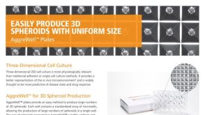 产品手册AggreWell™ for Spheroids
 沪公网安备31010102008431号