Reutershan J et al. (MAR 2006)
The Journal of clinical investigation 116 3 695--702
Critical role of endothelial CXCR2 in LPS-induced neutrophil migration into the lung.
In models of acute lung injury,CXC chemokine receptor 2 (CXCR2) mediates migration of polymorphonuclear leukocytes (PMNs) into the lung. Since CXCR2 ligands,including CXCL1 and CXCL2/3,are chemotactic for PMNs,CXCR2 is thought to recruit PMNs by inducing chemotactic migration. In a model of PMN recruitment to the lung,aerosolized bacterial LPS inhalation induced PMN recruitment to the lung in wild-type mice,but not in littermate CXCR2-/- mice. Surprisingly,lethally irradiated wild-type mice reconstituted with CXCR2-/- BM still showed about 50% PMN recruitment into bronchoalveolar lavage fluid and into lung interstitium,but CXCR2-/- mice reconstituted with CXCR2-/- BM showed no PMN recruitment. Conversely,CXCR2-/- mice reconstituted with wild-type BM showed a surprisingly large defect in PMN recruitment,inconsistent with a role of CXCR2 on PMNs alone. Cell culture,immunohistochemistry,flow cytometry,and real-time RT-PCR were used to show expression of CXCR2 on pulmonary endothelial and bronchial epithelial cells. The LPS-induced increase in lung microvascular permeability as measured by Evans blue extravasation required CXCR2 on nonhematopoietic cells. Our data revealed what we believe to be a previously unrecognized role of endothelial and epithelial CXCR2 in LPS-induced PMN recruitment and lung injury.
View Publication
产品号#:
18556
18556RF
产品名:
Yang X et al. (NOV 2010)
Cancer research 70 22 9463--72
Double-negative feedback loop between reprogramming factor LIN28 and microRNA let-7 regulates aldehyde dehydrogenase 1-positive cancer stem cells.
A relatively rare aldehyde dehydrogenase 1 (ALDH1)-positive stem cell-like" subpopulation of tumor cells has the unique ability to initiate and perpetuate tumor growth; moreover�
View Publication
产品号#:
01700
01705
01702
产品名:
ALDEFLUOR™ 试剂盒
ALDEFLUOR™ DEAB试剂, 1.5 mM, 1 mL
ALDEFLUOR™检测缓冲液
Song DH et al. (AUG 2000)
Journal of Biological Chemistry 275 31 23790--97
Endogenous protein kinase CK2 participates in Wnt signaling in mammary epithelial cells
Protein kinase CK2 (formerly casein kinase II) is a serine/threonine kinase overexpressed in many human tumors,transformed cell lines,and rapidly proliferating tissues. Recent data have shown that many cancers involve inappropriate reactivation of Wnt signaling through ectopic expression of Wnts themselves,as has been seen in a number of human breast cancers,or through mutation of intermediates in the Wnt pathway,such as adenomatous polyposis coli or beta-catenin,as described in colon and other cancers. Wnts are secreted factors that are important in embryonic development,but overexpression of certain Wnts,such as Wnt-1,leads to proliferation and transformation of cells. We report that upon stable transfection of Wnt-1 into the mouse mammary epithelial cell line C57MG,morphological changes and increased proliferation are accompanied by increased levels of CK2,as well as of beta-catenin. CK2 and beta-catenin co-precipitate with the Dvl proteins,which are Wnt signaling intermediates. A major phosphoprotein of the size of beta-catenin appears in in vitro kinase reactions performed on the Dvl immunoprecipitates. In vitro translated beta-catenin,Dvl-2,and Dvl-3 are phosphorylated by CK2. The selective CK2 inhibitor apigenin blocks proliferation of Wnt-1-transfected cells,abrogates phosphorylation of beta-catenin,and reduces beta-catenin and Dvl protein levels. These results demonstrate that endogenous CK2 is a positive regulator of Wnt signaling and growth of mammary epithelial cells.
View Publication
产品号#:
03800
03801
03802
03803
03804
03805
03806
产品名:
ClonaCell™-HY杂交瘤试剂盒
ClonaCell™-HY培养基A
ClonaCell™-HY 培养基 B
ClonaCell™-HY 培养基 C
ClonaCell™-HY 培养基 D
ClonaCell™-HY 培养基 E
ClonaCell™-HY PEG
Zhao X et al. (AUG 2010)
Proceedings of the National Academy of Sciences of the United States of America 107 32 14146--51
Telomerase-immortalized human mammary stem/progenitor cells with ability to self-renew and differentiate.
There is increasing evidence that breast and other cancers originate from and are maintained by a small fraction of stem/progenitor cells with self-renewal properties. Whether such cancer stem/progenitor cells originate from normal stem cells based on initiation of a de novo stem cell program,by reprogramming of a more differentiated cell type by oncogenic insults,or both remains unresolved. A major hurdle in addressing these issues is lack of immortal human stem/progenitor cells that can be deliberately manipulated in vitro. We present evidence that normal and human telomerase reverse transcriptase (hTERT)-immortalized human mammary epithelial cells (hMECs) isolated and maintained in Dana-Farber Cancer Institute 1 (DFCI-1) medium retain a fraction with progenitor cell properties. These cells coexpress basal (K5,K14,and vimentin),luminal (E-cadherin,K8,K18,or K19),and stem/progenitor (CD49f,CD29,CD44,and p63) cell markers. Clonal derivatives of progenitors coexpressing these markers fall into two distinct types--a K5(+)/K19(-) type and a K5(+)/K19(+) type. We show that both types of progenitor cells have self-renewal and differentiation ability. Microarray analyses confirmed the differential expression of components of stem/progenitor-associated pathways,such as Notch,Wnt,Hedgehog,and LIF,in progenitor cells compared with differentiated cells. Given the emerging evidence that stem/progenitor cells serve as precursors for cancers,these cellular reagents represent a timely and invaluable resource to explore unresolved questions related to stem/progenitor origin of breast cancer.
View Publication
Korkaya H et al. (OCT 2008)
Oncogene 27 47 6120--30
HER2 regulates the mammary stem/progenitor cell population driving tumorigenesis and invasion.
The cancer stem cell hypothesis proposes that cancers arise in stem/progenitor cells through disregulation of self-renewal pathways generating tumors,which are driven by a component of 'tumor-initiating cells' retaining stem cell properties. The HER2 gene is amplified in 20-30% of human breast cancers and has been implicated in mammary tumorigenesis as well as in mediating aggressive tumor growth and metastasis. We demonstrate that HER2 overexpression drives mammary carcinogenesis,tumor growth and invasion through its effects on normal and malignant mammary stem cells. HER2 overexpression in normal mammary epithelial cells (NMEC) increases the proportion of stem/progenitor cells as demonstrated by in vitro mammosphere assays and the expression of stem cell marker aldehyde dehydrogenase (ALDH) as well as by generation of hyperplastic lesions in humanized fat pads of NOD (nucleotide-binding oligomerization domain)/SCID (severe combined immunodeficient) mice. Overexpression of HER2 in a series of breast carcinoma cell lines increases the ALDH-expressing 'cancer stem cell' population which displays increased expression of stem cell regulatory genes,increased invasion in vitro and increased tumorigenesis in NOD/SCID mice. The effects of HER2 overexpression on breast cancer stem cells are blocked by trastuzumab in sensitive,but not resistant,cell lines,an effect mediated by the PI3-kinase/Akt pathway. These studies provide support for the cancer stem cell hypothesis by suggesting that the effects of HER2 amplification on carcinogenesis,tumorigenesis and invasion may be due to its effects on normal and malignant mammary stem/progenitor cells. Furthermore,the clinical efficacy of trastuzumab may relate to its ability to target the cancer stem cell population in HER2-amplified tumors.
View Publication
产品号#:
01700
01705
01702
产品名:
ALDEFLUOR™ 试剂盒
ALDEFLUOR™ DEAB试剂, 1.5 mM, 1 mL
ALDEFLUOR™检测缓冲液
Law JH et al. (JAN 2010)
PloS one 5 9
Molecular decoy to the Y-box binding protein-1 suppresses the growth of breast and prostate cancer cells whilst sparing normal cell viability.
The Y-box binding protein-1 (YB-1) is an oncogenic transcription/translation factor that is activated by phosphorylation at S102 whereby it induces the expression of growth promoting genes such as EGFR and HER-2. We recently illustrated by an in vitro kinase assay that a novel peptide to YB-1 was highly phosphorylated by the serine/threonine p90 S6 kinases RSK-1 and RSK-2,and to a lesser degree PKCα and AKT. Herein,we sought to develop this decoy cell permeable peptide (CPP) as a cancer therapeutic. This 9-mer was designed as an interference peptide that would prevent endogenous YB-1(S102) phosphorylation based on molecular docking. In cancer cells,the CPP blocked P-YB-1(S102) and down-regulated both HER-2 and EGFR transcript level and protein expression. Further,the CPP prevented YB-1 from binding to the EGFR promoter in a gel shift assay. Notably,the growth of breast (SUM149,MDA-MB-453,AU565) and prostate (PC3,LNCap) cancer cells was inhibited by ∼90% with the CPP. Further,treatment with this peptide enhanced sensitivity and overcame resistance to trastuzumab in cells expressing amplified HER-2. By contrast,the CPP had no inhibitory effect on the growth of normal immortalized breast epithelial (184htert) cells,primary breast epithelial cells,nor did it inhibit differentiation of hematopoietic progenitors. These data collectively suggest that the CPP is a novel approach to suppressing the growth of cancer cells while sparing normal cells and thereby establishes a proof-of-concept that blocking YB-1 activation is a new course of cancer therapeutics.
View Publication
产品号#:
05601
18056
18056RF
04435
04445
产品名:
EpiCult™-B 人培养基
MethoCult™ H4435 Enriched
MethoCult™ H4435 Enriched
Alison MR et al. (DEC 2010)
The Journal of pathology 222 4 335--44
Finding cancer stem cells: are aldehyde dehydrogenases fit for purpose?
Despite many years of intensive effort,there is surprisingly little consensus on the most suitable markers with which to locate and isolate stem cells from adult tissues. By comparison,the study of cancer stem cells is still in its infancy; so,unsurprisingly,there is great uncertainty as to the identity of these cells. Stem cell markers can be broadly categorized into molecular determinants of self-renewal,clonogenicity,multipotentiality,adherence to the niche,and longevity. This review assesses the utility of recognizing cancer stem cells by virtue of high expression of aldehyde dehydrogenases (ALDHs),probably significant determinants of cell survival through their ability to detoxify many potentially cytotoxic molecules,and contributing to drug resistance. Antibodies are available against the ALDH enzyme family,but the vast majority of studies have used cell sorting techniques to enrich for cells expressing these enzymes. Live cells expressing high ALDH activity are usually identified by the ALDEFLUOR kit and sorted by fluorescence activated cell sorting (FACS). For many human tumours,but notably breast cancer,cell selection based upon ALDH activity appears to be a useful marker for enriching for cells with tumour-initiating activity (presumed cancer stem cells) in immunodeficient mice,and indeed the frequency of so-called ALDH(bri) cells in many tumours can be an independent prognostic indicator.
View Publication
产品号#:
01700
01705
01702
产品名:
ALDEFLUOR™ 试剂盒
ALDEFLUOR™ DEAB试剂, 1.5 mM, 1 mL
ALDEFLUOR™检测缓冲液
McCune K et al. (NOV 2010)
Oncology reports 24 5 1233--9
Loss of ERα and FOXA1 expression in a progression model of luminal type breast cancer: insights from PyMT transgenic mouse model.
The classification of breast cancer into multiple molecular subtypes has necessitated the need for biomarkers that can assess tumor progression and the effects of chemopreventive agents on specific breast cancer subtypes. The goal of this study was to identify biomarkers whose expression are altered along with estrogen receptor α (ERα) in the polyoma middle-T antigen (PyMT) transgenic model of breast cancer and to investigate the chemopreventive activity of phenethyl isothiocyanate (PEITC). The diet of PyMT female mice was fortified with PEITC (8 mmol/kg) and the mammary streak and/or gross tumors and metastases in lungs were subjected to immunohistochemical analyses for ERα,FOXA1,and GATA-3. FOXA1 is associated with luminal type A cancers,while GATA-3 is a marker of luminal progenitor cell differentiation. In both control and PEITC-treated groups,there was a progressive loss of ERα and FOXA1 but persistence of GATA-3 expression indicating that the tumors retain luminal phenotype. Overall,the PyMT induced tumors exhibited the entire gamut of phenotypes from ERα+/FOXA1+/GATA-3+ tumors in the early stage to ERα±/FOXA1-/GATA-3+ in the late stage. Thus,PyMT model serves as an excellent model for studying progression of luminal subtype tumors. PEITC treated animals had multiple small tumors,indicating delay in tumor progression. Although these tumors were histologically similar to those in controls,there was a lower expression of these biomarkers in normal luminal cells indicating delay in tumor initiation. In in vitro studies,PEITC depleted AldeFluor-positive putative stem/progenitor cells,which may partly be responsible for the delay in tumor initiation.
View Publication
Stingl J et al. (MAY 2001)
Breast cancer research and treatment 67 2 93--109
Characterization of bipotent mammary epithelial progenitor cells in normal adult human breast tissue.
The purpose of the present study was to characterize primitive epithelial progenitor populations present in adult normal human mammary tissue using a combination of flow cytometry and in vitro colony assay procedures. Three types of human breast epithelial cell (HBEC) progenitors were identified: luminal-restricted,myoepithelial-restricted and bipotent progenitors. The first type expressed epithelial cell adhesion molecule (EpCAM),alpha6 integrin and MUC1 and generated colonies composed exclusively of cells positive for the luminal-associated markers keratin 8/18,keratin 19,EpCAM and MUC1. Bipotent progenitors produced colonies containing a central core of cells expressing luminal markers surrounded by keratin 14+ myoepithelial-like cells. Single cell cultures confirmed the bipotentiality of these progenitors. Their high expression of alpha6 integrin and low expression of MUC1 suggests a basal position of these cells in the mammary epithelium in vivo. Serial passage in vitro of an enriched population of bipotent progenitors demonstrated that only myoepithelial-restricted progenitors could be readily generated under the culture conditions used. These results support a hierarchical branching model of HBEC progenitor differentiation from a primitive uncommitted cell to luminal- and myoepithelial-restricted progenitors.
View Publication

 EasySep™小鼠TIL(CD45)正选试剂盒


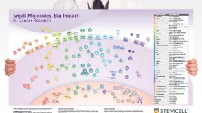 挂图Small Molecules, Big Impact in Cancer Research Overview of signaling pathways and small molecules in cancer research


 沪公网安备31010102008431号