Lippmann ES et al. (FEB 2014)
Scientific reports 4 February 2014 4160
A retinoic acid-enhanced, multicellular human blood-brain barrier model derived from stem cell sources.
Blood-brain barrier (BBB) models are often used to investigate BBB function and screen brain-penetrating therapeutics,but it has been difficult to construct a human model that possesses an optimal BBB phenotype and is readily scalable. To address this challenge,we developed a human in vitro BBB model comprising brain microvascular endothelial cells (BMECs),pericytes,astrocytes and neurons derived from renewable cell sources. First,retinoic acid (RA) was used to substantially enhance BBB phenotypes in human pluripotent stem cell (hPSC)-derived BMECs,particularly through adherens junction,tight junction,and multidrug resistance protein regulation. RA-treated hPSC-derived BMECs were subsequently co-cultured with primary human brain pericytes and human astrocytes and neurons derived from human neural progenitor cells (NPCs) to yield a fully human BBB model that possessed significant tightness as measured by transendothelial electrical resistance (˜5,000 $\$(2)). Overall,this scalable human BBB model may enable a wide range of neuroscience studies.
View Publication
产品号#:
05850
05857
05870
05875
85850
85857
85870
85875
产品名:
mTeSR™1
mTeSR™1
Lippmann ES et al. (APR 2014)
Stem Cells 32 4 1032--1042
Defined human pluripotent stem cell culture enables highly efficient neuroepithelium derivation without small molecule inhibitors.
The embryonic neuroepithelium gives rise to the entire central nervous system in vivo,making it an important tissue for developmental studies and a prospective cell source for regenerative applications. Current protocols for deriving homogenous neuroepithelial cultures from human pluripotent stem cells (hPSCs) consist of either embryoid body-mediated neuralization followed by a manual isolation step or adherent differentiation using small molecule inhibitors. Here,we report that hPSCs maintained under chemically defined,feeder-independent,and xeno-free conditions can be directly differentiated into pure neuroepithelial cultures ([mt]90% Pax6(+)/N-cadherin(+) with widespread rosette formation) within 6 days under adherent conditions,without small molecule inhibitors,and using only minimalistic medium consisting of Dulbecco's modified Eagle's medium/F-12,sodium bicarbonate,selenium,ascorbic acid,transferrin,and insulin (i.e.,E6 medium). Furthermore,we provide evidence that the defined culture conditions enable this high level of neural conversion in contrast to hPSCs maintained on mouse embryonic fibroblasts (MEFs). In addition,hPSCs previously maintained on MEFs could be rapidly converted to a neural compliant state upon transfer to these defined conditions while still maintaining their ability to generate all three germ layers. Overall,this fully defined and scalable protocol should be broadly useful for generating therapeutic neural cells for regenerative applications.
View Publication
Hou Y et al. (MAY 2014)
Neurobiology of Aging 35 5 975--989
Permeability transition pore-mediated mitochondrial superoxide flashes mediate an early inhibitory effect of amyloid beta1 42 on neural progenitor cell proliferation
Cellular damage by reactive oxygen species and altered neurogenesis are implicated in the etiology of AD and the pathogenic actions of amyloid β-peptide (Aβ); the underlying mechanisms and the early oxidative intracellular events triggered by Aβ are not established. In the present study,we found that mouse embryonic cortical neural progenitor cells exhibit intermittent spontaneous mitochondrial superoxide (SO) flashes that require transient opening of mitochondrial permeability transition pores (mPTPs). The incidence of mitochondria SO flash activity in neural progenitor cells (NPCs) increased during the first 6-24 hours of exposure to aggregating amyloid β-peptide (Aβ1-42),indicating an increase in transient mPTP opening. Subsequently,the SO flash frequency progressively decreased and ceased between 48 and 72 hours of exposure to Aβ1-42,during which time global cellular reactive oxygen species increased,mitochondrial membrane potential decreased,cytochrome C was released from mitochondria and the cells degenerated. Inhibition of mPTPs and selective reduction in mitochondrial SO flashes significantly ameliorated the negative effects of Aβ1-42 on NPC proliferation and survival. Our findings suggest that mPTP-mediated bursts of mitochondrial SO production is a relatively early and pivotal event in the adverse effects of Aβ1-42 on NPCs. If Aβ inhibits NPC proliferation in the brains of AD patients by a similar mechanism,then interventions that inhibit mPTP-mediated superoxide flashes would be expected to protect NPCs against the adverse effects of Aβ.
View Publication
产品号#:
05707
产品名:
NeuroCult™化学解离试剂盒(小鼠)
Lee S-HH et al. (JUN 2000)
Nature biotechnology 18 6 675--9
Efficient generation of midbrain and hindbrain neurons from mouse embryonic stem cells.
Embryonic stem (ES) cells are clonal cell lines derived from the inner cell mass of the developing blastocyst that can proliferate extensively in vitro and are capable of adopting all the cell fates in a developing embryo. Clinical interest in the use of ES cells has been stimulated by studies showing that isolated human cells with ES properties from the inner cell mass or developing germ cells can provide a source of somatic precursors. Previous studies have defined in vitro conditions for promoting the development of specific somatic fates,specifically,hematopoietic,mesodermal,and neurectodermal. In this study,we present a method for obtaining dopaminergic (DA) and serotonergic neurons in high yield from mouse ES cells in vitro. Furthermore,we demonstrate that the ES cells can be obtained in unlimited numbers and that these neuron types are generated efficiently. We generated CNS progenitor populations from ES cells,expanded these cells and promoted their differentiation into dopaminergic and serotonergic neurons in the presence of mitogen and specific signaling molecules. The differentiation and maturation of neuronal cells was completed after mitogen withdrawal from the growth medium. This experimental system provides a powerful tool for analyzing the molecular mechanisms controlling the functions of these neurons in vitro and in vivo,and potentially for understanding and treating neurodegenerative and psychiatric diseases.
View Publication

 EasySep™小鼠TIL(CD45)正选试剂盒


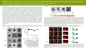 科学海报STEMdiff™ Cerebral Organoid Kit: A New Tool for the Culture of 3D Brain Organoids Derived from hPSCs
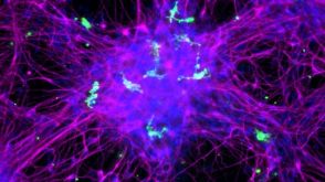 实验方案How to Co-Culture Human Pluripotent Stem Cell (hPSC)-Derived Forebrain Neurons and Microglia
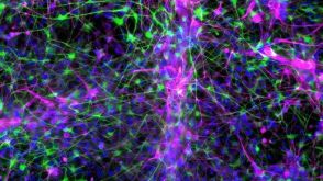 实验方案How to Co-Culture Astrocytes and NGN2 mRNA-Driven Induced Forebrain Neurons Derived from Human Pluripotent Stem Cells
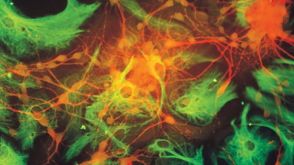


 沪公网安备31010102008431号