Heterotopically transplanted CVO neural stem cells generate neurons and migrate with SVZ cells in the adult mouse brain.
Production of new neurons throughout adulthood has been well characterized in two brain regions,the subventricular zone (SVZ) of the anterolateral ventricle and the subgranular zone (SGZ) of the hippocampus. The neurons produced from these regions arise from neural stem cells (NSCs) found in highly regulated stem cell niches. We recently showed that midline structures called circumventricular organs (CVOs) also contain NSCs capable of neurogenesis and/or astrogliogenesis in vitro and in situ (Bennett et al.). The present study demonstrates that NSCs derived from two astrogliogenic CVOs,the median eminence and organum vasculosum of the lamina terminalis of the nestin-GFP mouse,possess the potential to integrate into the SVZ and differentiate into cells with a neuronal phenotype. These NSCs,following expansion and BrdU-labeling in culture and heterotopic transplantation into a region proximal to the SVZ in adult mice,migrate caudally to the SVZ and express early neuronal markers (TUC-4,PSA-NCAM) as they migrate along the rostral migratory stream. CVO-derived BrdU(+) cells ultimately reach the olfactory bulb where they express early (PSA-NCAM) and mature (NeuN) neuronal markers. Collectively,these data suggest that although NSCs derived from the ME and OVLT CVOs are astrogliogenic in situ,they produce cells phenotypic of neurons in vivo when placed in a neurogenic environment. These findings may have implications for neural repair in the adult brain.
View Publication
Fernando P et al. (OCT 2005)
FASEB journal : official publication of the Federation of American Societies for Experimental Biology 19 12 1671--3
Neural stem cell differentiation is dependent upon endogenous caspase 3 activity.
Caspase proteases have become the focal point for the development and application of anti-apoptotic therapies in a variety of central nervous system diseases. However,this approach is based on the premise that caspase function is limited to invoking cell death signals. Here,we show that caspase-3 activity is elevated in nonapoptotic differentiating neuronal cell populations. Moreover,peptide inhibition of protease activity effectively inhibits the differentiation process in a cultured neurosphere model. These results implicate caspase-3 activation as a conserved feature of neuronal differentiation and suggest that targeted inhibition of this protease in neural cell populations may have unintended consequences.
View Publication
Sox2 expression defines a heterogeneous population of neurosphere-forming cells in the adult murine brain.
The identification of neural stem cells (NSCs) in situ has been prevented by the inability to identify a marker consistently expressed in all adult NSCs and is thus generally accomplished using the in vitro neurosphere-forming assay. The high-mobility group transcription factor Sox2 is expressed in embryonic neural epithelial stem cells; because these cells are thought to give rise to the adult NSC population,we hypothesized that Sox2 may continue to be expressed in adult NSCs. Using Sox2:EGFP transgenic mice,we show that Sox2 is expressed in neurogenic regions along the rostral-caudal axis of the central nervous system throughout life. Furthermore,all neurospheres derived from these neurogenic regions express Sox2,suggesting that Sox2 is indeed expressed in adult NSCs. We demonstrate that NSCs are heterogeneous within the adult brain,with differing capacities for cell production. In vitro,all neurospheres express Sox2,but the expression of markers common to early progenitor cells within individual neurospheres varies; this heterogeneity of NSCs is mirrored in vivo. For example,both glial fibrillary acidic protein and NG2 are expressed within individual neurospheres,but their expression is mutually exclusive; likewise,these two markers show distinct staining patterns within the Sox2+ regions of the brain's neurogenic regions. Thus,we propose that the expression of Sox2 is a unifying characteristic of NSCs in the adult brain,but that not all NSCs maintain the ability to form all neural cell types in vivo.
View Publication
Guillou L et al. (NOV 2016)
Biophysical journal 111 9 2039--2050
Measuring Cell Viscoelastic Properties Using a Microfluidic Extensional Flow Device.
The quantification of cellular mechanical properties is of tremendous interest in biology and medicine. Recent microfluidic technologies that infer cellular mechanical properties based on analysis of cellular deformations during microchannel traversal have dramatically improved throughput over traditional single-cell rheological tools,yet the extraction of material parameters from these measurements remains quite complex due to challenges such as confinement by channel walls and the domination of complex inertial forces. Here,we describe a simple microfluidic platform that uses hydrodynamic forces at low Reynolds number and low confinement to elongate single cells near the stagnation point of a planar extensional flow. In tandem,we present,to our knowledge,a novel analytical framework that enables determination of cellular viscoelastic properties (stiffness and fluidity) from these measurements. We validated our system and analysis by measuring the stiffness of cross-linked dextran microparticles,which yielded reasonable agreement with previously reported values and our micropipette aspiration measurements. We then measured viscoelastic properties of 3T3 fibroblasts and glioblastoma tumor initiating cells. Our system captures the expected changes in elastic modulus induced in 3T3 fibroblasts and tumor initiating cells in response to agents that soften (cytochalasin D) or stiffen (paraformaldehyde) the cytoskeleton. The simplicity of the device coupled with our analytical model allows straightforward measurement of the viscoelastic properties of cells and soft,spherical objects.
View Publication
产品号#:
05750
05751
产品名:
NeuroCult™ NS-A 基础培养基(人)
NeuroCult™ NS-A 扩增试剂盒(人)
Hackett C et al. ( 2014)
American journal of translational research 6 2 119--28
Transplantation of Fas-deficient or wild-type neural stem/progenitor cells (NPCs) is equally efficient in treating experimental autoimmune encephalomyelitis (EAE).
Studies have shown that neural stem/progenitor cell (NPC) transplantation is beneficial in experimental autoimmune encephalomyelitis (EAE),an established animal model of multiple sclerosis (MS). It is unclear whether NPCs have the ability to integrate into the host CNS to replace lost cells or if their main mechanism of action is via bystander immunomodulation. Understanding the mechanisms by which NPCs exert their beneficial effects as well as exploring methods to increase post-transplantation survival and differentiation is critical to advancing this treatment strategy. Using the EAE model and Fas-deficient (lpr) NPCs,we investigated the effects of altering the Fas system in NPC transplantation therapy. We show that transplantation of NPCs into EAE mice ameliorates clinical symptoms with greater efficacy than sham treatments regardless of cell type (wt or lpr). NPC transplantation via retro-orbital injections significantly decreased inflammatory infiltrates at the acute time point,with a similar trend at the chronic time point. Both wt and lpr NPCs injected into mice with EAE were able to home to sites of CNS inflammation in the periventricular brain and lumbar spinal cord. Both wt and lpr NPCs have the same capacity for inducing apoptosis of Th1 and Th17 cells,and minimal numbers of NPCs entered the CNS. These cells did not express terminal differentiation markers,suggesting that NPCs exert their effects mainly via bystander peripheral immunomodulation.
View Publication
Maynard KR and Stein E (NOV 2012)
The Journal of neuroscience : the official journal of the Society for Neuroscience 32 47 16637--50
DSCAM contributes to dendrite arborization and spine formation in the developing cerebral cortex.
Down syndrome cell adhesion molecule,or DSCAM,has been implicated in many neurodevelopmental processes including axon guidance,dendrite arborization,and synapse formation. Here we show that DSCAM plays an important role in regulating the morphogenesis of cortical pyramidal neurons in the mouse. We report that DSCAM expression is developmentally regulated and localizes to synaptic plasma membranes during a time of robust cortical dendrite arborization and spine formation. Analysis of mice that carry a spontaneous mutation in DSCAM (DSCAM(del17)) revealed gross morphological changes in brain size and shape in addition to subtle changes in cortical organization,volume,and lamination. Early postnatal mutant mice displayed a transient decrease in cortical thickness,but these reductions could not be attributed to changes in neuron production or cell death. DSCAM(del17) mutants showed temporary impairments in the branching of layer V pyramidal neuron dendrites at P10 and P17 that recovered to normal by adulthood. Defects in DSCAM(del17) dendrite branching correlated with a temporal increase in apical branch spine density and lasting changes in spine morphology. At P15 and P42,mutant mice displayed a decrease in the percentage of large,stable spines and an increase in the percentage of small,immature spines. Together,our findings suggest that DSCAM contributes to pyramidal neuron morphogenesis by regulating dendrite arborization and spine formation during cortical circuit development.
View Publication

 EasySep™小鼠TIL(CD45)正选试剂盒


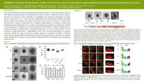 科学海报STEMdiff™ Cerebral Organoid Kit: A New Tool for the Culture of 3D Brain Organoids Derived from hPSCs
 科学海报Single-Cell RNA Sequencing Analysis of Regionally Patterned Human Pluripotent Stem Cell-Derived Neural Organoids


 沪公网安备31010102008431号