P. Petrov et al. (mar 2019)
Scientific reports 9 1 4155
Computational analysis of the evolutionarily conserved Missing In Metastasis/Metastasis Suppressor 1 gene predicts novel interactions, regulatory regions and transcriptional control.
Missing in Metastasis (MIM),or Metastasis Suppressor 1 (MTSS1),is a highly conserved protein,which links the plasma membrane to the actin cytoskeleton. MIM has been implicated in various cancers,however,its modes of action remain largely enigmatic. Here,we performed an extensive in silico characterisation of MIM to gain better understanding of its function. We detected previously unappreciated functional motifs including adaptor protein (AP) complex interaction site and a C-helix,pointing to a role in endocytosis and regulation of actin dynamics,respectively. We also identified new functional regions,characterised with phosphorylation sites or distinct hydrophilic properties. Strong negative selection during evolution,yielding high conservation of MIM,has been combined with positive selection at key sites. Interestingly,our analysis of intra-molecular co-evolution revealed potential regulatory hotspots that coincided with reduced potentially pathogenic polymorphisms. We explored databases for the mutations and expression levels of MIM in cancer. Experimentally,we focused on chronic lymphocytic leukaemia (CLL),where MIM showed high overall expression,however,downregulation on poor prognosis samples. Finally,we propose strong conservation of MTSS1 also on the transcriptional level and predict novel transcriptional regulators. Our data highlight important targets for future studies on the role of MIM in different tissues and cancers.
View Publication
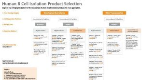
产品号#:
15024
15064
产品名:
RosetteSep™人B细胞富集抗体混合物
RosetteSep™人B细胞富集抗体混合物
A. A. Titov et al. (jul 2019)
Journal of immunology (Baltimore,Md. : 1950) 203 2 338--348
Metformin Inhibits the Type 1 IFN Response in Human CD4+ T Cells.
In systemic lupus erythematosus,defective clearance of apoptotic debris and activation of innate cells result in a chronically activated type 1 IFN response,which can be measured in PBMCs of most patients. Metformin,a widely used prescription drug for Type 2 diabetes,has a therapeutic effect in several mouse models of lupus through mechanisms involving inhibition of oxidative phosphorylation and a decrease in CD4+ T cell activation. In this study,we report that in CD4+ T cells from human healthy controls and human systemic lupus erythematosus patients,metformin inhibits the transcription of IFN-stimulated genes (ISGs) after IFN-alpha treatment. Accordingly,metformin inhibited the phosphorylation of pSTAT1 (Y701) and its binding to IFN-stimulated response elements that control ISG expression. These effects were independent of AMPK activation or mTORC1 inhibition but were replicated using inhibitors of the electron transport chain respiratory complexes I,III,and IV. This indicates that mitochondrial respiration is required for ISG expression in CD4+ T cells and provides a novel mechanism by which metformin may exert a therapeutic effect in autoimmune diseases.
View Publication
J. U. Hermansen et al. (dec 2018)
Scientific reports 8 1 17651
Cryopreservation of primary B cells minimally influences their signaling responses.
Phospho flow is a powerful approach to detect cell signaling aberrations,identify biomarkers and assess pharmacodynamics,and can be performed using cryopreserved samples. The effects of cryopreservation on signaling responses and the reproducibility of phospho flow measurements are however unknown in many cell systems. Here,B lymphocytes were isolated from healthy donors and patients with the B cell malignancy chronic lymphocytic leukemia and analyzed by phospho flow using phospho-specific antibodies targeting 20 different protein epitopes. Cells were analyzed both at basal conditions and after activation of cluster of differentiation 40 (CD40) or the B cell receptor. Pharmacodynamics of the novel pathway inhibitor ibrutinib was also assessed. At all conditions,fresh cells were compared to cryopreserved cells. Minimal variation between fresh and frozen samples was detected. Reproducibility was tested by running samples from the same donors in different experiments. The results demonstrate reproducibility across different phospho flow runs and support the use of cryopreserved samples in future phospho flow studies of B lymphocytes.
View Publication
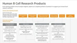
产品号#:
15024
15064
产品名:
RosetteSep™人B细胞富集抗体混合物
RosetteSep™人B细胞富集抗体混合物
Thompson EA et al. (APR 2016)
Journal of Immunology 196 7 3054--63
Shortened Intervals during Heterologous Boosting Preserve Memory CD8 T Cell Function but Compromise Longevity.
Developing vaccine strategies to generate high numbers of Ag-specific CD8 T cells may be necessary for protection against recalcitrant pathogens. Heterologous prime-boost-boost immunization has been shown to result in large quantities of functional memory CD8 T cells with protective capacities and long-term stability. Completing the serial immunization steps for heterologous prime-boost-boost can be lengthy,leaving the host vulnerable for an extensive period of time during the vaccination process. We show in this study that shortening the intervals between boosting events to 2 wk results in high numbers of functional and protective Ag-specific CD8 T cells. This protection is comparable to that achieved with long-term boosting intervals. Short-boosted Ag-specific CD8 T cells display a canonical memory T cell signature associated with long-lived memory and have identical proliferative potential to long-boosted T cells Both populations robustly respond to antigenic re-exposure. Despite this,short-boosted Ag-specific CD8 T cells continue to contract gradually over time,which correlates to metabolic differences between short- and long-boosted CD8 T cells at early memory time points. Our studies indicate that shortening the interval between boosts can yield abundant,functional Ag-specific CD8 T cells that are poised for immediate protection; however,this is at the expense of forming stable long-term memory.
View Publication
产品号#:
17951
17951RF
17952
17952RF
19254
19254RF
19853
19853RF
100-0695
100-0696
产品名:
EasySep™人T细胞分选试剂盒
RoboSep™ 人T细胞分选试剂盒
EasySep™人CD4+ T细胞分选试剂盒
RoboSep™ 人CD4+ T细胞分选试剂盒
EasySep™人Naïve B细胞富集试剂盒
RoboSep™ 人Naïve B细胞富集试剂盒含滤芯吸头
EasySep™小鼠CD8+ T细胞分选试剂盒
RoboSep™ 小鼠CD8+ T细胞分选试剂盒
EasySep™人T细胞分选试剂盒
EasySep™人CD4+ T细胞分离试剂盒
Flach A-C et al. (MAR 2016)
Proceedings of the National Academy of Sciences of the United States of America 113 12 3323--8
Autoantibody-boosted T-cell reactivation in the target organ triggers manifestation of autoimmune CNS disease.
Multiple sclerosis (MS) is caused by T cells that are reactive for brain antigens. In experimental autoimmune encephalomyelitis,the animal model for MS,myelin-reactive T cells initiate the autoimmune process when entering the nervous tissue and become reactivated upon local encounter of their cognate CNS antigen. Thereby,the strength of the T-cellular reactivation process within the CNS tissue is crucial for the manifestation and the severity of the clinical disease. Recently,B cells were found to participate in the pathogenesis of CNS autoimmunity,with several diverse underlying mechanisms being under discussion. We here report that B cells play an important role in promoting the initiation process of CNS autoimmunity. Myelin-specific antibodies produced by autoreactive B cells after activation in the periphery diffused into the CNS together with the first invading pathogenic T cells. The antibodies accumulated in resident antigen-presenting phagocytes and significantly enhanced the activation of the incoming effector T cells. The ensuing strong blood-brain barrier disruption and immune cell recruitment resulted in rapid manifestation of clinical disease. Therefore,myelin oligodendrocyte glycoprotein (MOG)-specific autoantibodies can initiate disease bouts by cooperating with the autoreactive T cells in helping them to recognize their autoantigen and become efficiently reactivated within the immune-deprived nervous tissue.
View Publication
产品号#:
19851
19851RF
19854
19854RF
产品名:
EasySep™小鼠T细胞分选试剂盒
RoboSep™ 小鼠T细胞分选试剂盒
EasySep™小鼠B细胞分选试剂盒
RoboSep™ 小鼠B细胞分选试剂盒
de Valle E et al. (APR 2016)
The Journal of Experimental Medicine 213 4 621--41
NFκB1 is essential to prevent the development of multiorgan autoimmunity by limiting IL-6 production in follicular B cells.
We examined the role of NFκB1 in the homeostasis and function of peripheral follicular (Fo) B cells. Aging mice lacking NFκB1 (Nfκb1(-/-)) develop lymphoproliferative and multiorgan autoimmune disease attributed in large part to the deregulated activity ofNfκb1(-/-)Fo B cells that produce excessive levels of the proinflammatory cytokine interleukin 6 (IL-6). Despite enhanced germinal center (GC) B cell differentiation,the formation of GC structures was severely disrupted in theNfκb1(-/-)mice. Bone marrow chimeric mice revealed that the Fo B cell-intrinsic loss of NFκB1 led to the spontaneous generation of GC B cells. This was primarily the result of an increase in IL-6 levels,which promotes the differentiation of Fo helper CD4(+)T cells and acts in an autocrine manner to reduce antigen receptor and toll-like receptor activation thresholds in a population of proliferating IgM(+)Nfκb1(-/-)Fo B cells. We demonstrate that p50-NFκB1 repressesIl-6transcription in Fo B cells,with the loss of NFκB1 also resulting in the uncontrolled RELA-driven transcription ofIl-6.Collectively,our findings identify a previously unrecognized role for NFκB1 in preventing multiorgan autoimmunity through its negative regulation ofIl-6gene expression in Fo B cells.
View Publication
产品号#:
19854
19854RF
产品名:
EasySep™小鼠B细胞分选试剂盒
RoboSep™ 小鼠B细胞分选试剂盒
Valsecchi R et al. (APR 2016)
Blood 127 16 1987--97
HIF-1α regulates the interaction of chronic lymphocytic leukemia cells with the tumor microenvironment.
Hypoxia-inducible transcription factors (HIFs) regulate a wide array of adaptive responses to hypoxia and are often activated in solid tumors and hematologic malignancies due to intratumoral hypoxia and emerging new layers of regulation. We found that in chronic lymphocytic leukemia (CLL),HIF-1α is a novel regulator of the interaction of CLL cells with protective leukemia microenvironments and,in turn,is regulated by this interaction in a positive feedback loop that promotes leukemia survival and propagation. Through unbiased microarray analysis,we found that in CLL cells,HIF-1α regulates the expression of important chemokine receptors and cell adhesion molecules that control the interaction of leukemic cells with bone marrow and spleen microenvironments. Inactivation of HIF-1α impairs chemotaxis and cell adhesion to stroma,reduces bone marrow and spleen colonization in xenograft and allograft CLL mouse models,and prolongs survival in mice. Of interest,we found that in CLL cells,HIF-1α is transcriptionally regulated after coculture with stromal cells. Furthermore,HIF-1α messenger RNA levels vary significantly within CLL patients and correlate with the expression of HIF-1α target genes,including CXCR4,thus further emphasizing the relevance of HIF-1α expression to CLL pathogenesis.
View Publication

 EasySep™小鼠TIL(CD45)正选试剂盒


 实验方案Optimizing Delivery Efficiency with Fluorescent Dextran Using the CellPore™ Transfection System

 沪公网安备31010102008431号